


To report reference values for the horizontal rectus muscles thickness using Spectral Domain optical coherence tomography (SD-OCT), and to evaluate whether there are any correlations between the muscle thickness and gender, age, or axial length (AL).
Materials and methodsA cross-sectional study was conducted on 131 right eyes of healthy subjects. The gender and age were recorded, and axial length was measured using an optical biometer. The medial rectus (MR) muscle thickness was measured at 7.2 and 9.2mm from the limbus, and the lateral rectus (LR) at 8.5 and 10.5mm from the limbus using OCT. A multivariate model was adjusted to determine whether gender, age, and axial length could have an impact on the muscle thickness.
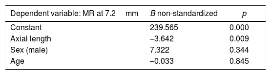
ResultsMean age was 43.3±20.9 years (range 6–86), and 59% were women. Mean AL was 24.9±2.7mm (range: 20.4–33.8). Mean thickness was 188.5±51.2μm (range 69–342) for the LR at 8.5 and 186.5±45.9μm (range 75–269) at 10.5mm, and for the MR, 158.1±39.1μm (range 69–273) at 7.2mm and 193.7±55.9μm (range 105–386) at 9.2mm. A correlation was observed between the AL and MR thickness (R=−0.255; p=0.023) while no correlation was observed for the LR (p≥0.203). No correlations were found between thickness and gender or thickness and age (p≥0.125).
ConclusionsThe reference ranges of the horizontal rectus muscles thickness was described using SD-OCT, observing an association between the AL and the MR thickness.
Describir los valores de normalidad del grosor de los músculos rectos horizontales mediante tomografía de coherencia óptica de dominio espectral (SD-OCT) y valorar si existe correlación entre el grosor muscular y el sexo, la edad o la longitud axial (LA).
Material y métodosEstudio transversal de 131 ojos derechos de pacientes sin enfermedad oftalmológica. Se recogieron el sexo y la edad y se midió la LA utilizando un biómetro óptico Lenstar LS 900 (Haag-Streit AG, Koeniz, Suiza). El grosor del recto medio (RM) se midió a 7,2 y 9,2mm desde el limbo y el recto lateral (RL) a 8,5 y 10,5mm utilizando la OCT. Se ajustó un modelo multivariable para analizar si el sexo, la edad y la LA podrían influir sobre el grosor muscular.
ResultadosLa edad media fue 43,3±20,9 años (rango 6-86), siendo 59% mujeres. La LA media fue 24,9±2,7mm (rango: 20,4-33,8). El grosor medio del RL a 8,5mm fue 188,5±51,2μm (rango 69-342) y 186,5±45,9μm (rango 75-269) a 10,5mm. El grosor del RM fue 158,1±39,1μm (rango 69-273) a 7,2mm y 193,7±55,9μm (rango 105-386) a 9,2mm. Se observó correlación entre el grosor del RM y la LA (R=–0,255; p=0,023), no hallándose correlación para el RL (p≥0,203). Tampoco se encontró asociación entre el grosor y el sexo o la edad (p≥0,125).
ConclusionesLa OCT permite medir el grosor de los músculos rectos horizontales, observándose una asociación entre el grosor del RM y la LA.
