Buscar en
Archivos de la Sociedad Española de Oftalmología (English Edition)
Toda la web
Inicio Archivos de la Sociedad Española de Oftalmología (English Edition) Assessment of changes in the macula and optic nerve head using optical coherence...
Journal Information
Vol. 95. Issue 6.
Pages 271-278 (June 2020)
Share
Share
Download PDF
More article options
Visits
4
Vol. 95. Issue 6.
Pages 271-278 (June 2020)
Original article
Assessment of changes in the macula and optic nerve head using optical coherence tomography in patients with attention deficit hyperactivity disorder
Evaluación de los cambios en la mácula y en el nervio óptico mediante tomografía de coherencia óptica en pacientes con trastorno por déficit de atención e hiperactividad
Visits
4
I. Sánchez-Guilléna,b,
Corresponding author
inessanchezguillen@gmail.com

Corresponding author.
, I. Almorín-Fernández-Vigoa, J.I. Fernández-Vigoc,d, L. de-Pablo-Gómez-de-Liañoc,e, B. Kudsiehc,f, J.A. Fernández-Vigoa,c,g
a Centro Internacional de Oftalmología Avanzada, Badajoz, Spain
b Departamento de Oftalmología, Hospital de Zafra, Zafra, Badajoz, Spain
c Centro Internacional de Oftalmología Avanzada, Madrid, Spain
d Departamento de Oftalmología, Hospital Clínico San Carlos, Instituto de Investigación Sanitaria del Hospital Clínico San Carlos (IdISSC), Madrid, Spain
e Departamento de Oftalmología, Hospital 12 de Octubre, Madrid, Spain
f Departamento de Oftalmología, Hospital Puerta de Hierro, Majadahonda, Madrid, Spain
g Facultad de Medicina, Universidad de Extremadura, Badajoz, Spain
This item has received
Article information
Abstract
Full Text
Bibliography
Download PDF
Statistics
Figures (2)
Tables (3)
Table 1. Results of macular thickness measurements in control subjects compared to those with attention deficits and hyper timid to disorder (ADHD), and between patients with ADHD with and without treatment.
Table 2. Results of retina ganglion cell complex (GCC) measurements in control subjects compared to those with attention deficit and hyperactivity disorder (ADHD) and between patients with ADHD with and without treatment.
Table 3. Results of retinal nerve fiber layer (RNFL) measurements in control subjects compared to those with attention deficit and hyperactivity disorder (ADHD) and between patients with ADHD with and without treatment.
Show moreShow less
Abstract
Introduction

To assess if there are any differences in macular and papillary thickness using optical coherence tomography (OCT) in patients with attention deficit hyperactivity disorder (ADHD) compared with a control group, including if there are differences between ADHD patients with and without treatment.

Methods

Prospective observational study including 92 eyes of 46 patients divided into 2 groups: 46 eyes of 23 patients with ADHD, and a control group of 46 eyes of 23 healthy patients. The group of patients with ADHD was subdivided into those on treatment with methylphenidate (n=28) and those not on treatment (n=18). The macular thickness, the ganglion cell complex (GCC), and the retinal nerve fiber layer (RNFL) at the papillary level were measured in 12 sectors.
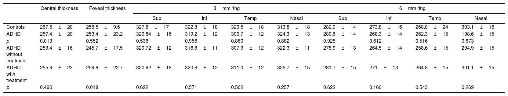
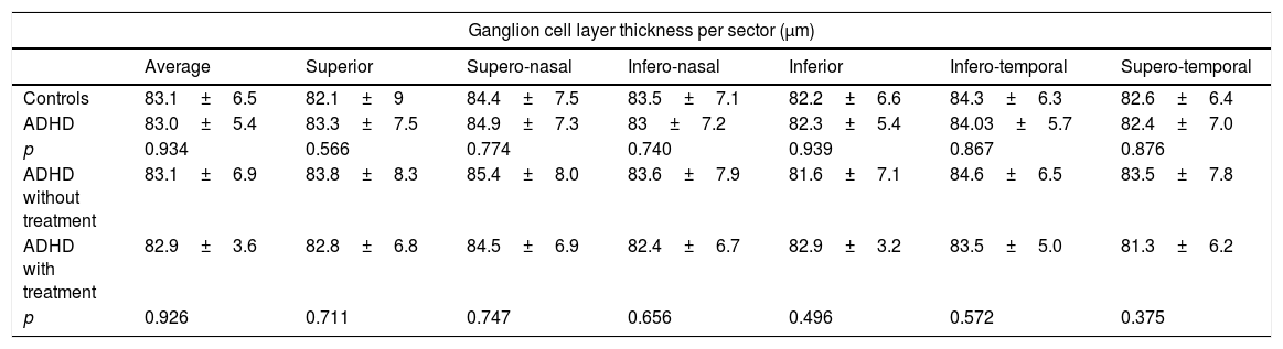
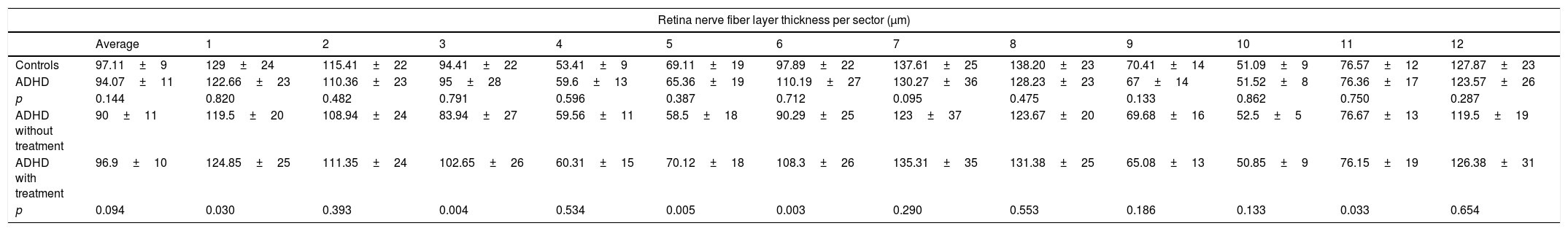
Results

A lower central macular thickness was observed in the ADHD patients than in the controls (257.4±20μm versus 267.5±20μm, p=0.013), with no differences observed in the GCC (p=0.566), or in the RNFL (p=0.095). There were no differences in the patients with ADHD with and without treatment, as regards macular thickness and the GCC (p=0.160 and p=0.375 respectively), but a lower foveal thickness (p=0.018) and RNFL in 5/12 sectors at the papillary level (p=0.033) were observed in those without treatment.

Conclusions

A lower macular thickness was observed in patients with ADHD than in controls. In addition, patients with ADHD without treatment had a lower thickness of the fovea and RNFL than those patients on treatment.

Keywords:
Attention deficit hyperactivity disorder
Macular thickness
Retina nerve fiber layer
Optical coherence tomography
Methylphenidate
Resumen
Introducción

Determinar si existen diferencias en el grosor macular y papilar mediante tomografía de coherencia óptica (OCT) en pacientes con trastorno por déficit de atención e hiperactividad (TDAH) comparando con un grupo control, evaluando además si existen diferencias entre pacientes con TDAH con y sin tratamiento.

Métodos

Estudio transversal en el que se incluyó a 92 ojos de 46 pacientes, divididos en 2 grupos: 46 ojos de 23 pacientes con TDAH y un grupo control de 46 ojos de 23 sujetos. El grupo de pacientes con TDAH se subdividió en aquellos con tratamiento con metilfenidato o derivados (n=28) y aquellos sin tratamiento (n=18). Se midió el grosor macular, el complejo de células ganglionares (CCG) y la capa de fibras nerviosas de la retina (CFNR) a nivel papilar en 12 sectores.

Resultados

Se observó un menor grosor macular central en los TDAH que en controles (257,4±20μm vs. 267,5±20μm; p=0,013), no observándose diferencias en el CCG (p ≥ 0,566), ni en la CFNR (p ≥ 0,095). En los pacientes con TDAH con y sin tratamiento no se observaron diferencias en el grosor macular ni en el CCG (p ≥ 0,160 y 0,375, respectivamente), pero se objetivó un menor grosor foveal (p=0,018) y de la CFNR en 5/12 sectores a nivel papilar (p ≤ 0,033) en aquellos sin tratamiento.

Conclusiones

Se observó un menor grosor macular en los pacientes con TDAH que en controles. Además, los pacientes con TDAH sin tratamiento presentaron un menor grosor foveal y de la CFNR que aquellos pacientes en tratamiento.

Palabras clave:
Trastorno por déficit de atención e hiperactividad
Grosor macular
Capa fibras nerviosas retina
Tomografía de coherencia óptica
Metilfenidato

Article

These are the options to access the full texts of the publication Archivos de la Sociedad Española de Oftalmología (English Edition)
Subscriber
Subscriber

If you already have your login data, please click here .

If you have forgotten your password you can you can recover it by clicking here and selecting the option “I have forgotten my password”
Subscribe
Subscribe to

Archivos de la Sociedad Española de Oftalmología (English Edition)

Purchase
Purchase article

Purchasing article the PDF version will be downloaded

Price 19.34 €

Purchase now
Contact
Phone for subscriptions and reporting of errors
From Monday to Friday from 9 a.m. to 6 p.m. (GMT + 1) except for the months of July and August which will be from 9 a.m. to 3 p.m.
Calls from Spain
932 415 960
Calls from outside Spain
+34 932 415 960
E-mail
Article options
Tools
es en pt

¿Es usted profesional sanitario apto para prescribir o dispensar medicamentos?

Are you a health professional able to prescribe or dispense drugs?

Você é um profissional de saúde habilitado a prescrever ou dispensar medicamentos