


The frontal sinus is the paranasal cavity with the greatest anatomical variability. Since the level of difficulty in dissecting the frontal recess during surgery is directly related to these anatomical variations, identifying them in the preoperative stage is crucial. This study aims to determine the most prevalent tomographic characteristics of the frontal sinus in paranasal sinus CT scans performed at a tertiary-level hospital in Lima, Peru, during the period 2023–2024.
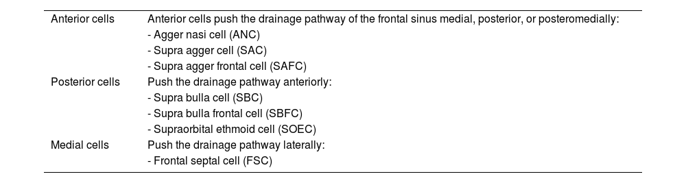
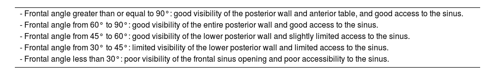
Materials and methodsThis is an observational, descriptive, cross-sectional study. Paranasal sinus CT scans obtained between July 2023 and June 2024 were analyzed. Sinus morphology, anteroposterior diameter of the frontal ostium, frontal angle, and frontal ostium grade were evaluated. Identification of ethmoidal cells and measurement of the distance from the columella to the frontal beak and to the anterior skull base were also included. Data was collected using Excel spreadsheets, and frequency tabulations were performed using IBM SPSS Statistics software.
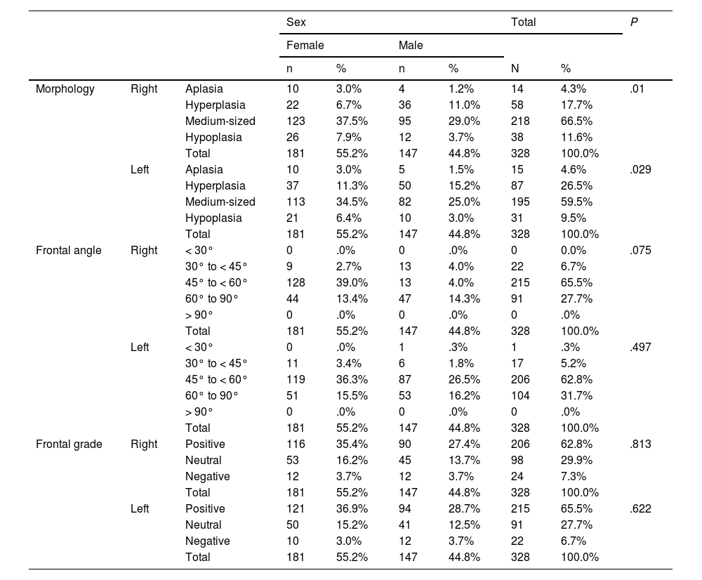
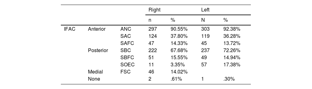
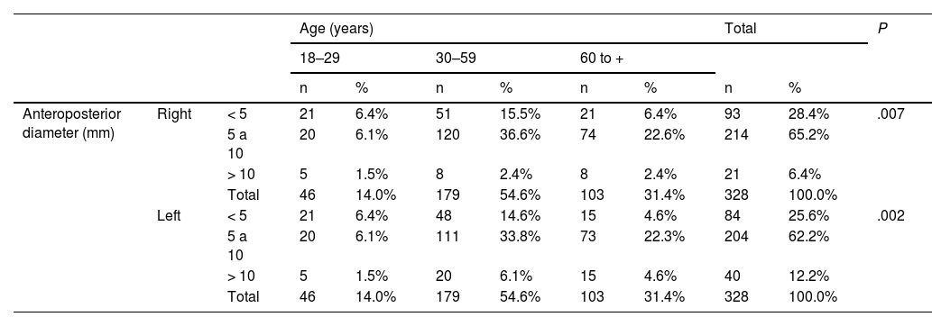
ResultsA total of 328 CT scans were included. In our sample, the most frequent age group was 30–59 years (54.6%), and the female sex predominated (55.2%). The most frequent morphology of the frontal sinus was medium size (66.5% on the right side and 59.5% on the left side). Hyperplasia predominated in males. More than 60% of the analyzed CT scans presented an anteroposterior distance of the frontal ostium between 5–10 mm (64.63% on the right side and 60.98% on the left). Adults over 30 years of age showed a diameter >10 mm more frequently than those aged 18–29 years. The frontal angle between 45° and <60° was the most frequent (65.55% on the right side and 62.80% on the left), and the most common grade was positive (62.80% on the right side and 65.55% on the left). The mean D-CFB was approximately 5.6 mm on the right side and 5.7 mm on the left side. Males presented greater measurements (approximately 0.5 mm more) than females. The mean D-CASB was 6.39 mm (right) and 6.53 mm (left). Males presented a significantly greater distance (∼0.5–0.6 mm more) than females (P < .001). The most prevalent cells were the agger nasi cell (90.55% on the right side and 92.38% on the left side) and the suprabullar cell (67.68% on the right side and 72.26% on the left side).
ConclusionsThis study provides a detailed tomographic characterization of the frontal sinus in a Peruvian population, serving as a theoretical basis to optimize surgical planning and anticipate potential challenges in the endoscopic approach to this anatomical region. The IFAC classification, frontal ostium diameter, and frontal angle are key factors to consider in surgical decision-making.
El seno frontal es la cavidad paranasal con mayor variabilidad anatómica. Dado que el nivel de dificultad de la disección del receso frontal durante la cirugía está directamente relacionado a estas variantes, resulta crucial identificarlas en la etapa preoperatoria. Este estudio se propone determinar las características tomográficas de mayor prevalencia del seno frontal en tomografías de senos paranasales realizadas en un hospital de tercer nivel en Lima, Perú, durante el año 2023–2024.
Materiales y métodosEstudio observacional, descriptivo, de corte transversal. Se analizaron tomografías de senos paranasales obtenidas durante el periodo julio 2023 a junio 2024, determinándose morfología, diámetro anteroposterior del ostium frontal, ángulo frontal y grado del ostium frontal, así como la identificación de celdillas etmoidales y medición de la distancia desde la columela al pico frontal y a la base del cráneo anterior. Los datos fueron recolectados en tablas de Excel y se realizaron tabulaciones de frecuencias utilizando el programa IBM SPSS statistics.
ResultadosSe incluyeron 328 tomografías. En nuestra muestra, el grupo etario más frecuente fue el de 30–59 años (54.6%) y predominó el sexo femenino (55.2%). La morfología más frecuente del seno frontal fue el tamaño medio (66.5% en el lado derecho y 59.5% en el lado izquierdo). La hiperplasia predominó en varones. Más del 60% de tomografías analizadas presentaron una distancia anteroposterior del ostium frontal entre 5–10 mm (64.63% en el lado derecho y 60.98% en el izquierdo). Los adultos mayores de 30 años presentaron un diámetro >10 mm con mayor frecuencia que el grupo etario comprendido entre los 18 y 29 años. El ángulo frontal entre 45° y <60° fue el más frecuente (65.55% en el lado derecho y 62.80% en el izquierdo) y el grado más común fue el positivo (62.80% en el lado derecho y 65.55% en el izquierdo). La D-CFB promedio fue de ∼5,6 mm en el lado derecho y ∼5,7 mm en el izquierdo. El sexo masculino presentó medidas mayores (aproximadamente 0,5 mm) más que las mujeres. La D-CASB promedio fue 6,39 mm (derecho) y 6,53 mm (izquierdo). El sexo masculino presentó significativamente una distancia mayor (∼0,5–0,6 mm más) que las mujeres (P < ,001). Las celdillas más prevalentes fueron la celdilla agger nassi (90.55% en el lado derecho y 92.38% en el lado izquierdo) y la celdilla suprabullar (67.68% en el lado derecho y 72.26% en el lado izquierdo).
ConclusionesEste estudio aporta información tomográfica detallada sobre el seno frontal en una población peruana, sirviendo de sustento teórico para optimizar la planificación quirúrgica y prever posibles dificultades en el abordaje endoscópico de dicha región anatómica. La clasificación IFAC, el diámetro del ostium frontal y el ángulo frontal son factores clave a considerar en la toma de decisiones quirúrgicas.