


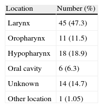
The aim of this study was to determine the predictive value of computed tomography (CT) i.e., its sensitivity and specificity in detecting metastatic lymph nodes of head and neck tumors. We also studied the capacity of CT in correct nodal lymph staging.
Patients and methodsA CT was performed on 95 patients diagnosed with neoplastic disease of the pharynx and/or larynx. All patients subsequently underwent cervical lymph node dissections. In the imaging study, the following parameters were considered for suspected radiological nodal involvement: lymph node diameter greater than 10mm, lesion margins poorly defined, capsule enhancement after contrast administration and lymph nodes that, despite their size, had signs of central necrosis.
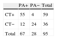
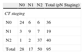
ResultsIn the dissections, 70.53% resulted N+ in the histological study. The sensitivity of CT was 82.09% and the specificity, 85.71%. The CT detected positivity in 55 of the 67 histologically pathological dissections, while the CT detected negativity in 24 of the 28 dissections histologically negative. The weighted kappa index value was 0.6408, indicating limited capacity for appropriate staging of the lymph nodes.
ConclusionsWhile the ability of CT to detect metastatic lymph nodes in head and neck tumors is quite acceptable, it is less so for correctly staging them. It is therefore necessary to look for other imaging tests that provide greater accuracy to avoid unnecessary elective neck dissections and to reduce morbidity and mortality from them. We must now pay attention to new imaging techniques such as PET and PET/CT.
Este estudio tiene como objetivo determinar el valor predictivo de la Tomografía Computerizada (TC), es decir, su sensibilidad y especificidad, en la detección de adenopatías metastásicas de tumores de cabeza y cuello. Además se estudia la capacidad de la TC para estadificar correctamente la afectación ganglionar.
Pacientes y métodoLa TC se efectuó a 95 pacientes diagnosticados de neoplasia de faringe y/o laringe que posteriormente fueron intervenidos de vaciamientos cervicales ganglionares. En el estudio de imagen se consideraron los siguientes parámetros radiológicos para sospechar afección ganglionar; diámetro de la adenopatía superior a 10mm, márgenes de la lesión mal definidos, realce de la cápsula tras la administración de contraste y adenopatías que independientemente del tamaño tuviesen signos de necrosis central.
ResultadosEl 70,53% de los vaciamientos resultó N+ en el estudio histopatológico. La Sensibilidad de la TC fue de 82,09% y la Especificidad de 85,71%. De los 67 vaciamientos histológicamente patológicos, la TC detectó positividad en 55, mientras que de los 28 vaciamientos histológicamente negativos, la TC detectó como negativos 24. El valor índece kappa ponderado fue de 0,6408, que indica limitada capacidad para la correcta estadificación de las adenopatías.
ConclusionesSi bien la capacidad de la TC para detectar adenopatias metastásicas en los tumores de cabeza y cuello es aceptale, no lo es tanto para realizar una correcta estadificación de las mismas. Por tanto es necesaria la búsqueda de otras pruebas de imagen que nos aporten una mayor precisión para así evitar vaciamientos electivos innecesarios y reducir la morbimortalidad de los mismos, debiendo actualmente prestar atención en las nuevas técnicas de imagen como son la PETy la PET/TC.