


Migrainous dizziness is one of the most frequent complaints. Dizziness associated with migraine may be the result of abnormal eye movements. Brain imaging and changes in eye movements may explain the dizziness and highlight possible pathophysiological substrates in migraine dizziness. Our aim is to evaluate eye movement using videonystagmography (VNG) and video head impulse test (vHIT) and to study the occipital lobe metabolic profile in vestibular migraine patients (VM).
Materials and methodsThere were 2 groups enrolled in the study; the first group consisted of 25 vestibular migraine patients (VM) according to the recent criteria of Barany society. The second group consisted of 20 age matched healthy subjects. Both groups underwent the following: (1) A detailed history, VNG test protocol, vHIT in three planes. (2) Magnetic resonance imaging (MRI) for the brain and inner ear using 1.5T magnet and proton magnetic resonance spectroscopy (H1-MRS).
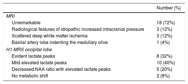
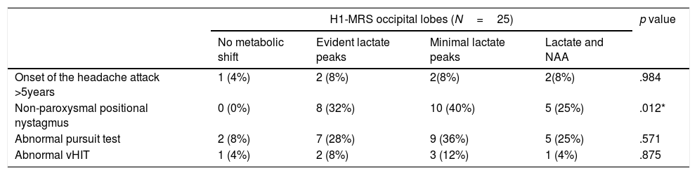
ResultsSixty eight percent of the patients complained of spontaneous vertigo and 28% complained of positional vertigo. Non-paroxysmal positional nystagmus was recorded in 92% during their dizzy spell. The brain MRI was unremarkable in 72% of the cases. Chemical shift in the occipital lobe was found in 92% of VM. Lactate peaks were statistically significant related with the presence of non-paroxysmal positional nystagmus.
ConclusionsA statistically significant relationship exists between non-paroxysmal positional nystagmus and presence of lactate peaks in the occipital lobe in VM patients.
El mareo migrañoso es una de las quejas más frecuentes. Las pruebas de imagen del cerebro y los cambios en los movimientos oculares pueden explicar los mareos y destacar los posibles sustratos fisiopatológicos en la migraña vestibular. Nuestro objetivo fue evaluar el movimiento ocular utilizando videonistagmografía (VNG) y la prueba de impulso cefálico por vídeo (vHIT), y estudiar el perfil metabólico del lóbulo occipital en pacientes con migraña vestibular (VM).
Materiales y métodosSe incluyeron dos grupos en el estudio; el primer grupo consistió en 25 pacientes con VM según los criterios recientes de la sociedad Bárány. El segundo grupo consistió en 20 sujetos sanos emparejados por edad. Ambos grupos se sometieron a lo siguiente: 1) Una historia detallada, protocolo de prueba de VNG y vHIT en 3 planos, y 2) Imágenes de resonancia magnética (IRM) para el cerebro y el oído interno con el imán de 1,5 tesla y la espectroscopía de resonancia magnética de protones (H1-MRS).
ResultadosEl 68% de los pacientes se quejó de vértigo espontáneo, y el 28% de vértigo posicional. El nistagmo posicional no paroxístico se registró en el 60% de los pacientes durante su mareo. La resonancia magnética cerebral no mostró alteraciones en el 72% de los casos. El cambio químico en el lóbulo occipital se encontró en el 92% de los casos de VM. Los picos de lactato fueron estadísticamente significativos con relación a la presencia de nistagmo posicional no paroxístico.
ConclusionesExiste una relación estadísticamente significativa entre el nistagmo posicional no paroxístico y la presencia de picos de lactato en el lóbulo occipital en pacientes con VM.