


The enlarged vestibular aqueduct (EVA) is the most frequent malformation of the inner ear associated with sensorineural hearing loss (5–15%). It exists when the diameter in imaging tests is greater than 1.5 mm at its midpoint. The association between hearing loss and EVA has been described in a syndromic and non-syndromic manner. It can appear as a familial or isolated form and the audiological profile is highly variable. The gene responsible for sensorineural hearing loss associated with EVA is located in the same region described for Pendred syndrome, where the SCL26A4 gene is located.
ObjectiveTo describe a series of children diagnosed with EVA in order to study their clinical and audiological characteristics, as well as the associated genetic and vestibular alterations.
MethodRetrospective study of data collection of children diagnosed with EVA, from April 2014 to February 2023.
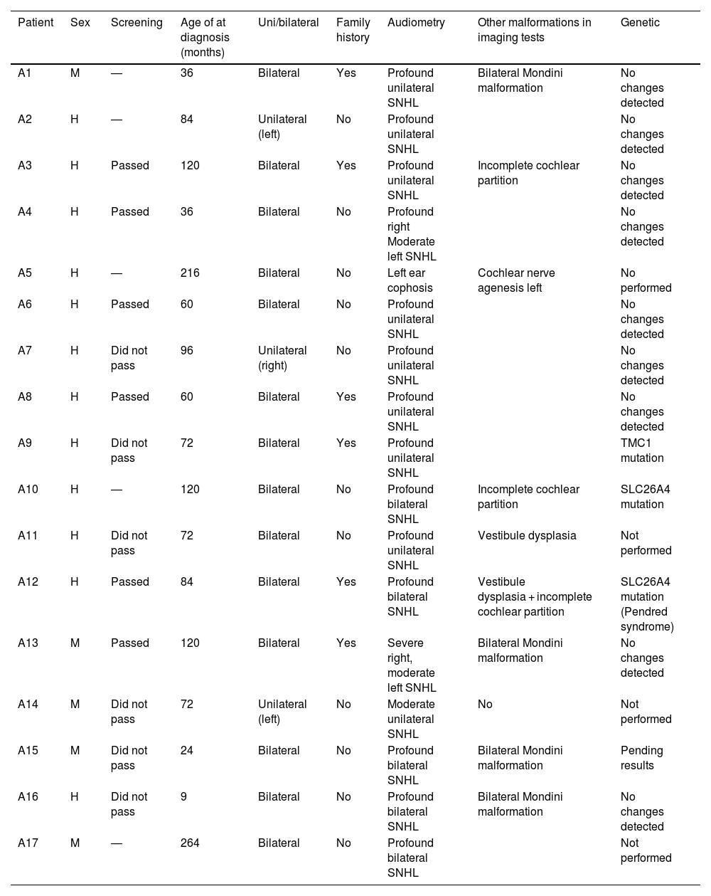
ResultsOf the 17 cases, 12 were male and 5 were female. 5 of them were unilateral and 12 bilateral. In 5 cases, a cranial traumatism triggered the hearing loss. Genetic alterations were detected in 3 cases: 2 mutations in the SCL26A4 gene and 1 mutation in the MCT1 gene. 13 patients (76.5%) were rehabilitated with hearing aids and 9 of them required cochlear implantation.
DiscussionThe clinical importance of AVD lies in the fact that it is a frequent finding in the context of postneonatal hearing loss. It is convenient to have a high suspicion to diagnose it with imaging tests, to monitor its evolution, and to rehabilitate early.
El acueducto vestibular dilatado (AVD) constituye la malformación más frecuente del oído interno asociada con hipoacusia neurosensorial (5–15%). Existe cuando el diámetro en las pruebas de imagen es superior 1,5 mm en su punto medio. La asociación entre hipoacusia y AVD se ha descrito de forma sindrómica y no sindrómica. Puede aparecer como una forma familiar o aislada y el perfil audiológico es muy variable. El gen responsable de la hipoacusia neurosensorial asociada al AVD está localizado en la misma región descrita para el síndrome de Pendred, donde se localiza el gen SCL26A4.
ObjetivoDescribir una serie de niños diagnosticados de AVD con el fin de estudiar sus características clínicas y audiológicas, así como las alteraciones genéticas y vestibulares asociadas.
MétodoEstudio retrospectivo de recogida de datos de niños diagnosticados de AVD, desde abril de 2014 hasta febrero 2023.
ResultadosDe los 17 casos, 12 eran del sexo masculino y 5 del sexo femenino. 5 de los fueron unilaterales y 12 bilaterales. En 5 casos, un traumatismo craneoencefálico desencadenó la hipoacusia. Se detectaron alteraciones genéticas en 3 casos: 2 mutaciones en el gen SCL26A4 y 1 mutación en el gen MCT1. 13 pacientes (76.5%) fueron rehabilitados con audioprótesis y 9 de ellos requirieron una implantación coclear.
DiscusiónLa importancia clínica del AVD radica en que es un hallazgo frecuente en el contexto de una hipoacusia postneonatal. Conviene tener una elevada sospecha para diagnosticarlo con pruebas de imagen, para de vigilar su evolución, y rehabilitar de manera precoz.