Buscar en
Acta Otorrinolaringológica Española
Toda la web
Inicio Acta Otorrinolaringológica Española Diagnostic Utility of Labyrinth Capsule Bone Density in the Diagnosis of Otoscle...
Journal Information
Vol. 71. Issue 4.
Pages 242-248 (July - August 2020)
Share
Share
Download PDF
More article options
Visits
5
Vol. 71. Issue 4.
Pages 242-248 (July - August 2020)
Original article
Diagnostic Utility of Labyrinth Capsule Bone Density in the Diagnosis of Otosclerosis With High Resolution Tomography
Utilidad diagnóstica de la densidad ósea de la cápsula laberíntica en el diagnóstico de la otosclerosis por tomografía de alta resolución
Visits
5
Ignacio Viza Puiggrósa,
Corresponding author
ignacioviza@gmail.com

Corresponding author.
, Esther Granell Morenob, Carlos Calvo Navarroa, Mercè Bohé Roviraa, César Orús Dotuc, Miquel Quer i Agustíc
a Servicio ORL, Hospital Plató, Barcelona, Spain
b Servicio de Radiología, Hospital de la Santa Creu i Sant Pau, Barcelona, Spain
c Servicio de ORL, Hospital de la Santa Creu i Sant Pau, Barcelona, Spain
This item has received
Article information
Abstract
Full Text
Bibliography
Download PDF
Statistics
Figures (2)
Tables (3)
Table 1. Densities in Hounsfield Units (HU) of Each Region of Interest (ROI).
Table 2. Average of Mean Densities in Hounsfield Units (HU).
Table 3. Diagnostic Yield. Assessed Using the Area Under the ROC Curve of Each Area of Interest (ROI).
Show moreShow less
Abstract
Objective

The aim of this study is to compare the bone density around the otic capsule in otosclerotic patients with a control group, and find the cut-off values of bone density from which we can diagnose the disease.
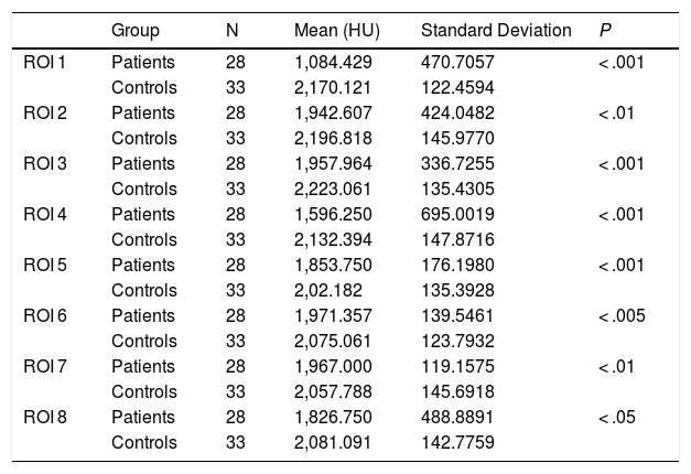
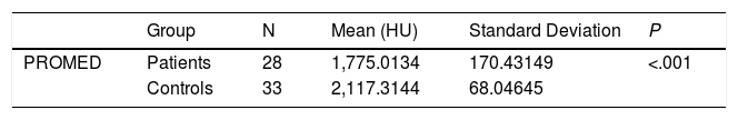
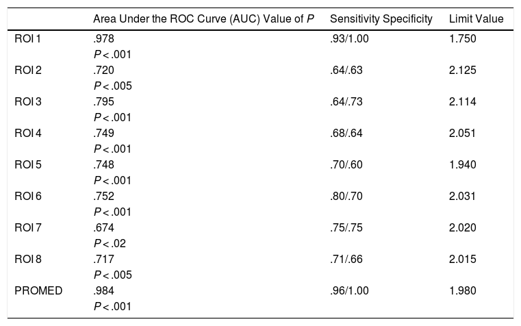
Material and methods

A retrospective case-control study was performed. Bone densities in Houndfield units (HU) from 28 otosclerotic ears were compared to the densities of 33 non otosclerotic capsules. These densities were measured in eight regions of interest (ROI) where the otosclerotic foci are usually found. The mean density of these regions (PROMED) was taken. Furthermore, the ROC curves of each ROI and the mean density (PROMED) were calculated.

Results

All radiological densities in HU of each ROI and the mean density in otosclerotic patients were lower compared to non otosclerotic ears. The Area Under the ROC curve of each ROI and the mean density, showed that the areas with greater accuracy for the diagnosis of otosclerosis were mean density, the fissula ante fenestram, and precochlear region, with cut-off values of 1980HU, 1750HU and 2114HU respectively.

Conclusion

The mean density of the otic capsule (PROMED), the density in the fissula ante fenestram ( ROI1) and in the precochlear region (ROI3) seem to be the most useful parameters to make a diagnosis of otosclerosis.

Keywords:
Otosclerosis
Otospongiosis
Densitometry
High resolution tomography
Hounsfield units
Resumen
Objetivo

El objetivo de este estudio es comparar la densidad ósea alrededor de la cápsula ótica en pacientes otosclerosos con un grupo control y encontrar el límite de densidad ósea a partir del cual podemos diagnosticar la enfermedad.

Material y métodos

se realizó un estudio retrospectivo de casos y controles. La densidad ósea en unidades de Houndfield (HU) de 28 oídos otosclerosos fue comparada con la densidad de 33 cápsulas no otoscleróticas. La densidad fue medida en ocho áreas de interés (ROI) donde normalmente se encuentran los focos otoscleróticos. Adicionalmente se realizó la densidad media de estas regiones (PROMED). Además, se calcularon las curvas ROC de cada ROI y la densidad media (PROMED).

Resultados

Todas las densidades radiológicas en HU de cada ROI y la densidad media en pacientes otosclerosos fueron menores en comparación con los oídos no otosclerosos. El área bajo la curva ROC de cada ROI y la densidad media, mostraron que las áreas con mayor rendimiento diagnóstico fueron la densidad media, la fissula antefenestram y la región precoclear, con valores de corte de 1980HU, 1750HU y 2114HU respectivamente..

Conclusión

La densidad media de la cápsula ótica (PROMED), la densidad en Fissula antefenestram (ROI1) y en la región precoclear (ROI3) parecen ser los parámetros más útiles para realizar el diagnóstico de otosclerosis.

Palabras clave:
Otosclerosis
Otospongiosis
Densitometría
Tomografía de alta resolución
Unidades Hounsfield

Article

These are the options to access the full texts of the publication Acta Otorrinolaringológica Española
Subscriber
Subscriber

If you already have your login data, please click here .

If you have forgotten your password you can you can recover it by clicking here and selecting the option “I have forgotten my password”
Subscribe
Subscribe to

Acta Otorrinolaringológica Española

Purchase
Purchase article

Purchasing article the PDF version will be downloaded

Price 19.34 €

Purchase now
Contact
Phone for subscriptions and reporting of errors
From Monday to Friday from 9 a.m. to 6 p.m. (GMT + 1) except for the months of July and August which will be from 9 a.m. to 3 p.m.
Calls from Spain
932 415 960
Calls from outside Spain
+34 932 415 960
E-mail
Article options
Tools
es en pt

¿Es usted profesional sanitario apto para prescribir o dispensar medicamentos?

Are you a health professional able to prescribe or dispense drugs?

Você é um profissional de saúde habilitado a prescrever ou dispensar medicamentos

Quizás le interese:
10.1016/j.otoeng.2021.04.003
No mostrar más