Buscar en
Gastroenterología y Hepatología (English Edition)
Toda la web
Inicio Gastroenterología y Hepatología (English Edition) Enteric duplication cyst as a rare presentation of adult acute abdomen
Journal Information
Image of the month
Full text access
Enteric duplication cyst as a rare presentation of adult acute abdomen
Quiste de duplicación entérica como presentación poco frecuente de abdomen agudo en adulto
Visits
17
Gerard Rafarta,
Corresponding author
, Clara Bassaganyasa, Xavier Moralesb, Míriam Cuatrecasasc,d, Jordi Rimolaa,e
a Servicio de Radiodiagnóstico, Hospital Clínic de Barcelona, Barcelona, Spain
b Servicio de Cirugía General, Hospital Clínic de Barcelona, Barcelona, Spain
c Servicio de Anatomía Patológica, Hospital Clínic de Barcelona, Barcelona, Spain
d Universitat de Barcelona, Barcelona, Spain
e IDIBAPS, Barcelona, Spain
This item has received
Article information
Full Text
Bibliography
Download PDF
Statistics
Figures (2)
Full Text

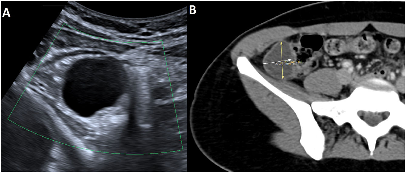
This was a 24-year-old woman, who attended Accident and Emergency for right iliac fossa (RIF) pain and nausea. On physical examination, the patient showed guarding in RIF, with no analytical abnormalities. Abdominal ultrasound showed a 27 mm peri-caecal cystic lesion1 which was confirmed by computed tomography (CT). The CT scan showed the lesion to be located alongside the caecum, not attached to the vermiform appendix, having thin, well-defined walls, with no signs of inflammation (Fig. 1). Diagnostic imaging suggested enteric duplication cyst (EDC) as a first choice, with appendiceal mucocele or endometrioma being less likely.

Figure 1.

Image A: abdominal ultrasound showing a well-defined, anechogenic lesion lateral to the caecum, with thin walls and no vascularisation on colour Doppler study. Image B: Contrast-enhanced CT scan showing a lesion located in the RIF, adjacent to the caecum and independent of the vermiform appendix, with well-defined walls.

(0.09MB).

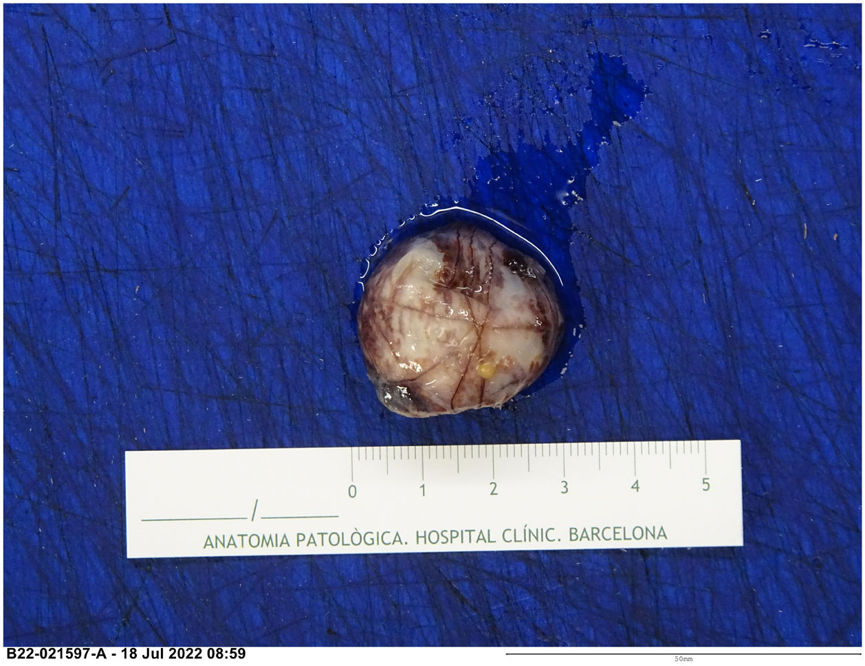
Laparoscopy identified a cystic lesion sharing serous membrane with the caecum and excision was performed. Pathology described a cystic lesion with fibrous walls lined by an epithelium of mesothelial cells without atypia, compatible with EDC (Fig. 2).

Figure 2.

Well-defined cystic lesion consisting of fibrous walls and presence of congestive vessels.

(0.21MB).

EDC is a rare congenital lesion diagnosed in the paediatric age group.2 It is often asymptomatic or presents with very non-specific symptoms, including abdominal pain, obstruction or intussusception.3 It can be found anywhere in the gastrointestinal tract, the ileum being the most common location and the colon the least.4 Given its low incidence in adults, it is important to be aware of this condition as a rare cause of acute abdomen due to its difficult diagnosis.

References
[1]
M. Di Serafino, C. Mercogliano, G. Vallone.
Ultrasound evaluation of the enteric duplication cyst: the gut signature.
J Ultrasound, 19 (2016), pp. 131-133
[2]
P.S. Puligandla, L.T. Nguyen, D. St-Vil, H. Flageole, A.L. Bensoussan, V.H. Nguyen, J.M. Laberge.
Gastrointestinal duplications.
J Pediatr Surg, 38 (2003), pp. 740-744
[3]
K. Kyo, M. Azuma, K. Okamoto, M. Nishiyama, T. Shimamura, A. Maema, et al.
Laparoscopic resection of adult colon duplication causing intussusception.
World J Gastroenterol, 22 (2016), pp. 2398-2402
[4]
S.C. Tong, M. Pitman, S.A. Anupindi.
Best cases from the AFIP. Ileocecal Enteric Duplication Cyst: Radiologic-Pathologic Correlation.
Radiographics, 22 (2002), pp. 1217-1222
Copyright © 2023. The Author(s)
Article options
Tools
es en pt

¿Es usted profesional sanitario apto para prescribir o dispensar medicamentos?

Are you a health professional able to prescribe or dispense drugs?

Você é um profissional de saúde habilitado a prescrever ou dispensar medicamentos