Buscar en
Archivos de la Sociedad Española de Oftalmología (English Edition)
Toda la web
Inicio Archivos de la Sociedad Española de Oftalmología (English Edition) Ocular surface analysis in patients diagnosed with X-linked ichthyosis
Journal Information
Vol. 95. Issue 11.
Pages 565-568 (November 2020)
Share
Share
Download PDF
More article options
Visits
7
Vol. 95. Issue 11.
Pages 565-568 (November 2020)
Short communication
Ocular surface analysis in patients diagnosed with X-linked ichthyosis
Análisis de superficie ocular en pacientes diagnosticados de ictiosis X
Visits
7
F.B. Domínguez-Serranoa, M. Caro-Magdalenoa,b,c,
Corresponding author
drmanuelcaro@gmail.com

Corresponding author.
, B. Mataix-Alberta, P. Molina-Solanaa, J. Montero-Iruzubietaa, E. Rodríguez-de-la-Rúaa,b,c
a UGC Oftalmología, Hospital Universitario Virgen Macarena, Sevilla, Spain
b Departamento de Cirugía, Universidad de Sevilla, Sevilla, Spain
c RETICS-OFTARED, Spain
This item has received
Article information
Abstract
Full Text
Bibliography
Download PDF
Statistics
Figures (2)
Abstract

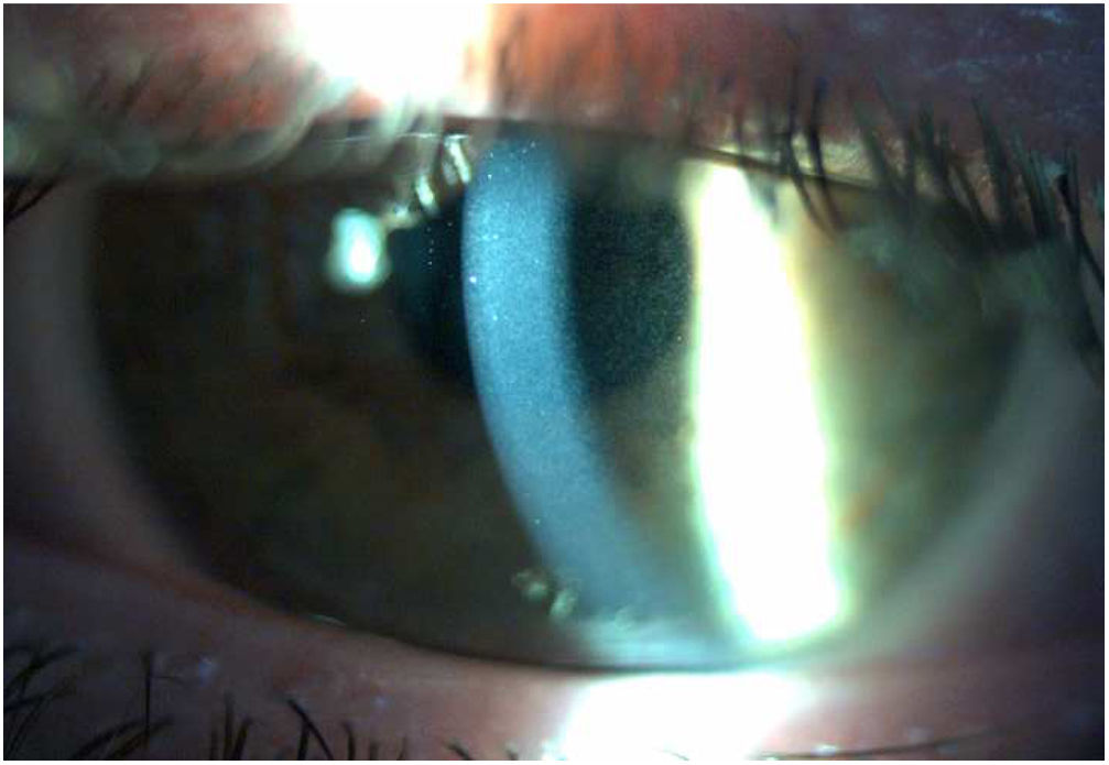
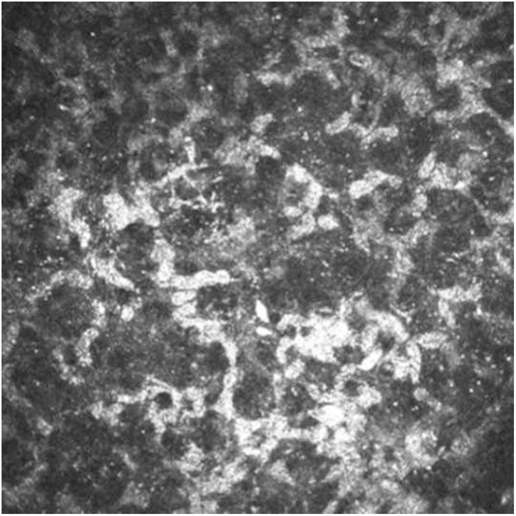
Seven patients (14 eyes) diagnosed with X-linked ichthyosis were studied using the Schirmer test, biomicroscopy, tonometry, endothelial count, optical coherence tomography, Pentacam®, ocular surface analyser, and confocal microscopy. The mean age was 33.83±20.17 years (range: 7–64 years). The most frequent findings in biomicroscopy were Meibomian glands dysfunction (83.3%) and stromal corneal opacities (33%). The tear break-up time was found shortened in 25% of the eyes. Confocal microscopy (both eyes) revealed activated keratocytes with hyper-reflective particles inside them in the anterior stroma and outside them in the posterior stroma. It is believed that the inclusion of the use of confocal microscopy will help in a better understanding of the corneal pathology associated with ichthyosis X, as well as new characteristics of these patients.

Keywords:
X ichthyosis
Confocal microscopy
Corneal opacities
Keratocytes
Hyper-reflective deposits
Meibomian gland dysfunction
Resumen

Se estudió a 7 pacientes (14 ojos) diagnosticados de ictiosis X mediante test de Schirmer, biomicroscopia, tonometría, recuento endotelial, tomografía de coherencia óptica, Pentacam, analizador de superficie ocular y microscopia confocal. La edad media fue 33,83±20,17 años (rango: 7-64 años). Los hallazgos más frecuentes en biomicrocoscopia fueron disfunción de glándulas de Meibomio (83,3%) y opacidades corneales estromales (33%). El tiempo de rotura de la película lagrimal se encontró acortado en el 25% de los ojos. La microscopia confocal (2 ojos) reveló queratocitos activados con partículas hiperreflectivas en su interior en estroma anterior y fuera de ellos en estroma posterior. Creemos que la extensión del uso de la microscopia confocal permitirá conocer mejor la enfermedad corneal asociada a ictiosis X y nuevas características de estos pacientes.

Palabras clave:
Ictiosis X
Microscopia confocal
Opacidades corneales
Queratocitos
Depósitos hiperreflectivos
Disfunción de glándulas de Meibomio

Article

These are the options to access the full texts of the publication Archivos de la Sociedad Española de Oftalmología (English Edition)
Subscriber
Subscriber

If you already have your login data, please click here .

If you have forgotten your password you can you can recover it by clicking here and selecting the option “I have forgotten my password”
Subscribe
Subscribe to

Archivos de la Sociedad Española de Oftalmología (English Edition)

Purchase
Purchase article

Purchasing article the PDF version will be downloaded

Price 19.34 €

Purchase now
Contact
Phone for subscriptions and reporting of errors
From Monday to Friday from 9 a.m. to 6 p.m. (GMT + 1) except for the months of July and August which will be from 9 a.m. to 3 p.m.
Calls from Spain
932 415 960
Calls from outside Spain
+34 932 415 960
E-mail
Article options
Tools
es en pt

¿Es usted profesional sanitario apto para prescribir o dispensar medicamentos?

Are you a health professional able to prescribe or dispense drugs?

Você é um profissional de saúde habilitado a prescrever ou dispensar medicamentos

Quizás le interese:
10.1016/j.oftale.2019.03.003
No mostrar más