Journal Information
Vol. 37. Issue 5.
Pages 453-454 (May 2018)
Share
Share
Download PDF
More article options
Visits
3876
Vol. 37. Issue 5.
Pages 453-454 (May 2018)
Image in Cardiology
Open Access
Rare forms of pediatric congenital right ventricular fistulae
Fístulas congénitas pediátricas raras do ventrículo direito
Visits
3876
Natália Noronha
Corresponding author
natalianoronhaf@gmail.com

Corresponding author.
, Pedro Pires Epifânio, Paula Martins, António Pires, António Marinho, Eduardo Castela
Serviço de Cardiologia Pediátrica, Hospital Pediátrico de Coimbra, Centro Hospitalar e Universitário de Coimbra, Coimbra, Portugal
This item has received

Under a Creative Commons license
Article information
Full Text
Download PDF
Statistics
Figures (3)
Show moreShow less
Full Text

Abnormal vascular connections to the right ventricle (RV) are rare congenital malformations. The majority of cases are diagnosed incidentally, and most patients are asymptomatic. The authors present three cases of right ventricular fistulae of non-coronary origin.
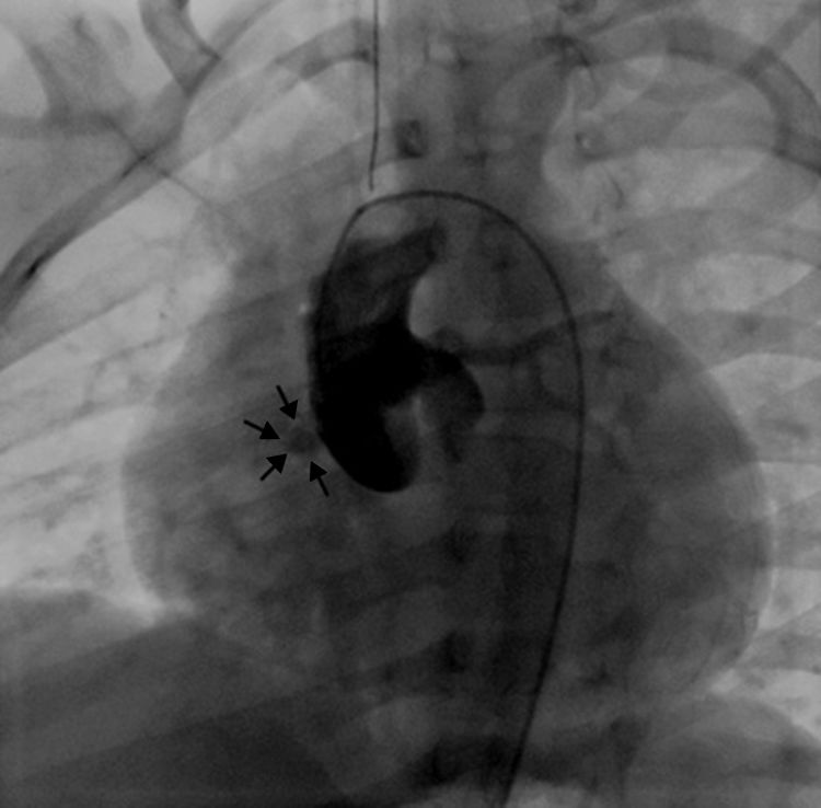
Case 1

A 2-year-old asymptomatic boy with a heart murmur underwent an echocardiogram which showed turbulent flow in the subaortic region. He was catheterized and the aortic angiogram revealed a hemodynamically insignificant fistulous tract between the right sinus of Valsalva and the RV (Figure 1).

Figure 1.

Aortography showing a fistula between the right sinus of Valsalva and the right ventricle.

(0.05MB).
Case 2

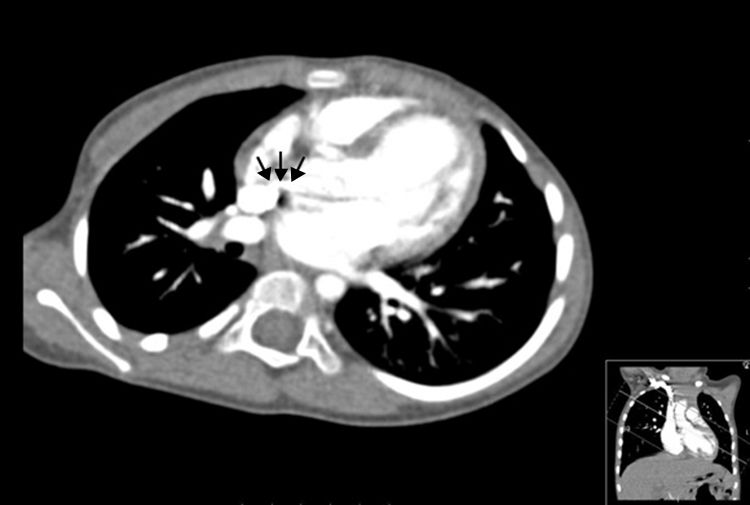
A 3-year-old asymptomatic boy was referred due to a murmur suggestive of a ventricular septal defect. In the echocardiogram, a turbulent flow was evident in the proximal ascending aorta, as well as in the right ventricular wall. In order to clarify the morphology of this apparent fistulous tract, he underwent a cardiac computerized tomography scan, which confirmed the presence of a fistula between the ascending aorta and the RV (Figure 2).

Figure 2.

Cardiac computerized tomography scan showing a fistula between the ascending aorta and the right ventricle.

(0.04MB).
Case 3

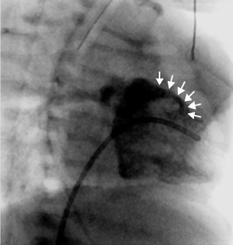
A 19-day-old baby boy was admitted to a Pediatric Intensive Care Unit following cardiorespiratory arrest of unknown cause. Cardiac catheterization to rule out anomalous coronary artery anatomy was performed and the right ventricular angiogram revealed a fistulous tract between the RV and the pulmonary artery (Figure 3).

Figure 3.

Ventriculography showing a fistula between the RV and the main pulmonary artery.

(0.05MB).
Conclusions

The majority of congenital fistulae to the RV originate in the coronary artery bed. Other sites of origin, such as those demonstrated in these cases, are exceedingly rare. To the best of our knowledge, few have been reported during infancy. As these fistulae were found accidentally, we opted for a conservative approach.

Conflicts of interest

The authors have no conflicts of interest to declare.

Copyright © 2018. Sociedade Portuguesa de Cardiologia
Idiomas
Revista Portuguesa de Cardiologia (English edition)
Article options
Tools
en pt

Are you a health professional able to prescribe or dispense drugs?

Você é um profissional de saúde habilitado a prescrever ou dispensar medicamentos

By checking that you are a health professional, you are stating that you are aware and accept that the Portuguese Journal of Cardiology (RPC) is the Data Controller that processes the personal information of users of its website, with its registered office at Campo Grande, n.º 28, 13.º, 1700-093 Lisbon, telephone 217 970 685 and 217 817 630, fax 217 931 095, and email revista@spc.pt. I declare for all purposes that the information provided herein is accurate and correct.