


An observational, ambispective study comparing the long-term clinical and radiological outcomes of two surface coatings (Porocoat® and Gription®) in Pinnacle® acetabular cups used for primary total hip arthroplasty.
Materials and methodsA total of 50 consecutively implanted cups between January 2012 and July 2013 were analyzed, categorized into Group A (n=25 Pinnacle®-Porocoat® cups) and Group B (n=25 Pinnacle®-Gription® cups). Clinical and radiological outcomes of both groups, including the need for surgical revision and time to revision, were compared over long-term follow-up, during the decade following the index procedure.
ResultsThe study population had a median age of 70 [61–73] years and was predominantly male (64%, n=32). Preoperative BMI and functional evaluation were similar for both coating types. Revision-free survival was 96% in Group A and 92% in Group B (p=.591). Both groups showed significant and comparable clinical improvement, with no cases of aseptic loosening. Only two Pinnacle®-Gription® cups presented with non-progressive radiolucent lines.
ConclusionsBoth Porocoat® and Gription® demonstrated stable fixation and favorable long-term clinical and radiological outcomes, with no significant differences observed during follow-up after primary total hip arthroplasty.
Estudio observacional y ambispectivo que comparó los resultados clínicos y radiológicos a largo plazo de 2 recubrimientos de superficie (Porocoat® y Gription®) en cotilos Pinnacle® en artroplastia total primaria de cadera.
Material y métodosSe analizaron 50 cotilos implantados consecutivamente entre enero de 2012 y julio de 2013, categorizados en Grupo A (n=25 cotilos Pinnacle®-Porocoat®) y Grupo B (n=25 cotilos Pinnacle®-Gription®). Los resultados clínicos y radiológicos de ambos grupos, incluyendo la necesidad de revisión quirúrgica y el tiempo hasta la misma, fueron comparados a largo plazo, durante la década posterior a la intervención inicial.
ResultadosLa población de estudio se caracterizó por una mediana de edad global de 70 [61-73] años, siendo esta predominantemente masculina (64%, n=32). El IMC y la evaluación funcional preoperatorias fueron similares para ambos tipos de recubrimiento. La supervivencia libre de revisión quirúrgica fue del 96% en el Grupo A y del 92% en el Grupo B (p=0,591). Ambos grupos mostraron mejoría clínica significativas y similares, no registrándose aflojamientos asépticos. Únicamente 2 cotilos Pinnacle®-Gription® presentaron líneas radiolucentes no progresivas.
ConclusionesTanto Porocoat® como Gription® ofrecieron una fijación estable, así como resultados clínicos y radiológicos positivos y duraderos, sin diferencias durante el seguimiento a largo plazo tras artroplastia total primaria de cadera.
In recent decades, tantalum has become an increasingly popular material for acetabular components. Its porosity, elasticity, and high coefficient of friction enable excellent osseointegration, even in cases where bone quality is compromised, for example in revision surgery.1,2 These properties have led to improved clinical and radiological outcomes compared with titanium.3,4 Despite these advantages, tantalum's high cost and limited availability have driven the development of porous titanium alternatives, which aim to replicate its results in a more cost-effective manner.5–7
The Pinnacle® cup (DePuy, Warsaw, IN, USA), a third-generation cup with a Porocoat® coating, was introduced in 2001. This has a porosity of 20%–60% and a pore size of 250μm.8 In 2007, the Gription® coating was introduced, increasing porosity to approximately 80% and pore size to 300μm. The transition from one coating to the other resulted in an increase in the coefficient of friction from .8 to 1.2.9
Despite the theoretical advantages, the available evidence is mixed. While some studies have reported higher rates of radiolucent lines and suboptimal short-term outcomes with new, highly porous coatings,10–12 others have demonstrated similar results.13 Few studies have evaluated the long-term outcomes of the Pinnacle® cup with different coatings.
This study aims to compare the long-term clinical and radiological outcomes of the same titanium cup with two different surface coatings: Porocoat® and Gription®.
Material and methodDesign and study populationThis was a single-centre, observational, non-randomised, retrospective study with prospective data collection. It was designed to compare the clinical and radiological outcomes of Pinnacle® acetabular cups with two different coatings under real-world clinical conditions. The study included all acetabular cups implanted consecutively by the same surgical team between January 2012 and July 2013, following approval by the centre's Ethics Committee (PI-2626). Cases in which the Pinnacle® acetabular cup was implanted outside of the study period, or where there was insufficient clinical information or follow-up, were excluded.
During the study period, 51 Pinnacle® acetabular cups were implanted. One patient was lost to follow-up and excluded, resulting in a final sample of 50 cups corresponding to 48 patients. The cases were grouped according to the type of coating: Group A (n=25, Pinnacle®-Porocoat® cups) and Group B (n=25, Pinnacle®-Gription® cups). Porocoat® cups were implanted between January and March 2012, and Gription® cups were implanted from April 2012 onwards.
The choice of new implant type was not the responsibility of the treating medical team, but was instead an administrative decision made by the hospital's procurement department. All patients who received at least one Pinnacle® cup during the study period were eligible, and there were no specific exclusion criteria. The analysis was conducted using the acetabular cup as the unit of analysis. It should be noted that 3 patients had bilateral prostheses with two different surface coatings, one of which had two different coatings.
The median age was 70 [61–73] years, and 64% were male (n=32). In terms of aetiology, the main diagnosis leading to surgery was primary osteoarthritis, present in 35 cases (70%), followed by avascular necrosis (n=10), rheumatoid arthritis (n=3) and, in isolated cases, femoral neck fracture (n=1) and Perthes’ disease (n=1).
Surgical procedureForty of the arthroplasties were performed via a direct lateral approach and 10 via a posterolateral approach. There were no significant differences in the type of surgical approach between the groups, with the direct lateral approach being predominantly used (40 cases) and, to a lesser extent, the posterolateral approach (10 cases), distributed similarly between both groups (p=1.000). All procedures were performed under spinal anaesthesia and in the lateral decubitus position.
The acetabular cups were reamed 1mm smaller than the size of the implanted cup to achieve good peripheral press-fit fixation. In all cases, supplementary fixation with screws was employed and the Summit® uncemented stem (DePuy, Warsaw, IN, USA) was implanted. The bearing pair was chosen based on the patient's age and baseline characteristics: metal-polyethylene (Articul/EZE® Metal Head, DePuy, Warsaw, IN, USA) was used in 33 patients, ceramic-ceramic (Biolox delta®, Ceramtec, Germany) in 13 patients and ceramic polyethylene in 4 patients.
A highly cross-linked polyethylene liner (Marathon®, DePuy, Warsaw, IN, USA) was used. The size of the femoral head was determined based on the bearing pair and the size of the acetabular cup, with 35 heads measuring 28mm and 15 heads measuring 32mm.
Clinical and radiological assessmentPatients were assessed before and after surgery with periodic follow-ups at six weeks, 12 weeks, six months, and annually throughout the postoperative follow-up period. For the purposes of comparison, however, the analyses in this study focused on baseline results and the latest available follow-up. Body mass index (BMI) was calculated as the ratio of weight to height obtained at the initial preoperative assessment. Functional capacity and patient-reported quality of life were assessed prospectively using the Harris Hip Score (HHS) and the Merle d’Aubigné and Postel (MAP) scales,14,15 during the preoperative clinical interview and at the final follow-up visit.
For the radiographic analysis, standardised anteroposterior pelvic radiographs were evaluated, using the diameter of the femoral head for correct calibration. All radiographs were reviewed by a single experienced assessor, who was blinded to the type of coating. Measurements included the acetabular abduction angle, the horizontal distance to the Köhler line, the vertical distance to the base of the teardrop, and the distance to the centre of rotation according to Ranawat,16 expressed in millimetres.
The presence of radiolucent lines was analysed, as well as their pattern, size, and possible osteolysis in the DeLee and Charnley zones.17 It was assessed whether the radiolucent lines were progressive, defining progression as an increase in radiolucency of at least 1mm between two radiological measurements and maintained over time.
Liner wear was assessed using Kim's method,18 and component integration was analysed using Engh's criteria.19 Unlike the prospective clinical assessment, the radiological analysis was conducted retrospectively; for this reason, the design is considered retrospective with prospective data collection.
Finally, intraoperative (intraoperative fracture) and postoperative (dislocation, aseptic or septic loosening, polyethylene wear, and periprosthetic fracture) complications were recorded during follow-up.
Statistical analysisCategorical variables were expressed as counts and percentages, while quantitative variables were expressed as the mean±standard deviation or the median (interquartile range), depending on their distribution. χ2 or Fisher's exact tests were used to compare the distributions of the categorical variables between the study groups, as appropriate. The normality of quantitative datasets was assessed using the Shapiro–Wilk test, with Student's t-tests or Wilcoxon rank-sum tests applied as appropriate. Time to surgical revision was assessed using Kaplan–Meier survival analysis, with the two groups being compared using the log-rank test. A p-value of less than .05 was considered statistically significant, and all analyses were performed using StataStata® BE 18.0 statistical software (StataCorp).
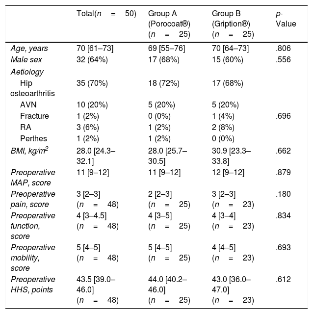
ResultsAs shown in Table 1, both groups had similar baseline characteristics. The median follow-up period was 10.6 [8.9–11.8] years.
Baseline characteristics.
| Total(n=50) | Group A (Porocoat®)(n=25) | Group B (Gription®)(n=25) | p-Value | |
|---|---|---|---|---|
| Age, years | 70 [61–73] | 69 [55–76] | 70 [64–73] | .806 |
| Male sex | 32 (64%) | 17 (68%) | 15 (60%) | .556 |
| Aetiology | ||||
| Hip osteoarthritis | 35 (70%) | 18 (72%) | 17 (68%) | |
| AVN | 10 (20%) | 5 (20%) | 5 (20%) | |
| Fracture | 1 (2%) | 0 (0%) | 1 (4%) | .696 |
| RA | 3 (6%) | 1 (2%) | 2 (8%) | |
| Perthes | 1 (2%) | 1 (2%) | 0 (0%) | |
| BMI, kg/m2 | 28.0 [24.3–32.1] | 28.0 [25.7–30.5] | 30.9 [23.3–33.8] | .662 |
| Preoperative MAP, score | 11 [9–12] | 11 [9–12] | 12 [9–12] | .879 |
| Preoperative pain, score | 3 [2–3] (n=48) | 2 [2–3] (n=25) | 3 [2–3] (n=23) | .180 |
| Preoperative function, score | 4 [3–4.5] (n=48) | 4 [3–5] (n=25) | 4 [3–4] (n=23) | .834 |
| Preoperative mobility, score | 5 [4–5] (n=48) | 5 [4–5] (n=25) | 4 [4–5] (n=23) | .693 |
| Preoperative HHS, points | 43.5 [39.0–46.0] (n=48) | 44.0 [40.2–46.0] (n=25) | 43.0 [36.0–47.0] (n=23) | .612 |
Categorical variables expressed as a percentage (%) and quantitative variables as a median [P25–P75].
AVN: avascular necrosis; BMI: body mass index; HHS: Harris Hip Score; MAP: Merle D’Aubigné and Postel; RA: rheumatoid arthritis.
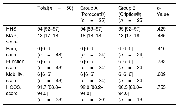
Compared with baseline (Tables 1 and 2), the change in HHS score between baseline and final follow-up did not differ between groups, with a median HHS delta score of +51 [45.1–54.0] in Group A and +53.0 [48.6–57.0] in Group B (p=.130). Similarly, the median MAP delta score was +7 [5–8] and +6 [6–8] for each group, respectively (p=.743).
Clinical results at last follow-up visit.
| Total(n=50) | Group A (Porocoat®)(n=25) | Group B (Gription®)(n=25) | p-Value | |
|---|---|---|---|---|
| HHS | 94 [92–97] | 94 [89–97] | 95 [92–97] | .429 |
| MAP, score | 18 [17–18] | 18 [18–18] | 18 [17–18] | .485 |
| Pain, score | 6 [6–6] (n=48) | 6 [6–6] (n=24) | 6 [6–6] (n=24) | .416 |
| Function, score | 6 [6–6] (n=48) | 6 [6–6] (n=24) | 6 [6–6] (n=24) | .783 |
| Mobility, score | 6 [6–6] (n=48) | 6 [6–6] (n=24) | 6 [6–6] (n=24) | .609 |
| HOOS, score | 91.7 [88.8–94.0] (n=38) | 92.0 [88.2–94.0] (n=20) | 90.5 [89.0–94.0] (n=18) | .755 |
Quantitative variables are expressed as the median [P25–P75].
BMI: body mass index; HHS: Harris Hip Score; MAP: Merle D’Aubigné and Postel.
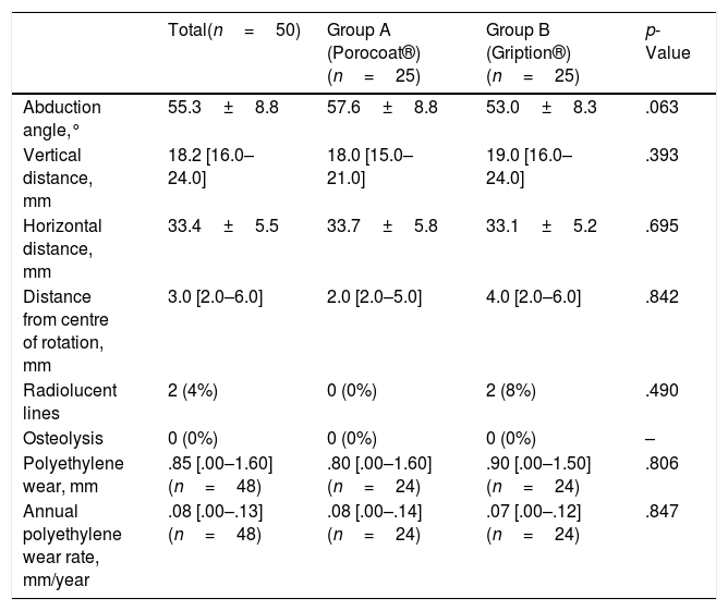
No differences were observed in the orientation or positioning of the acetabular components between groups, and no cases of osteolysis around the implants were reported during follow-up (Table 3). Only 2 acetabular cups (both Pinnacle®-Gription®) showed radiolucent lines at 15 and 18 months post-operation, which were non-progressive. The median annual polyethylene wear rate was .08 [.00–.14] mm/year in Group A and .07 [.00–.12] mm/year in Group B, with no statistically significant differences.
Radiological characteristics at the last follow-up visit.
| Total(n=50) | Group A (Porocoat®)(n=25) | Group B (Gription®)(n=25) | p-Value | |
|---|---|---|---|---|
| Abduction angle,° | 55.3±8.8 | 57.6±8.8 | 53.0±8.3 | .063 |
| Vertical distance, mm | 18.2 [16.0–24.0] | 18.0 [15.0–21.0] | 19.0 [16.0–24.0] | .393 |
| Horizontal distance, mm | 33.4±5.5 | 33.7±5.8 | 33.1±5.2 | .695 |
| Distance from centre of rotation, mm | 3.0 [2.0–6.0] | 2.0 [2.0–5.0] | 4.0 [2.0–6.0] | .842 |
| Radiolucent lines | 2 (4%) | 0 (0%) | 2 (8%) | .490 |
| Osteolysis | 0 (0%) | 0 (0%) | 0 (0%) | – |
| Polyethylene wear, mm | .85 [.00–1.60] (n=48) | .80 [.00–1.60] (n=24) | .90 [.00–1.50] (n=24) | .806 |
| Annual polyethylene wear rate, mm/year | .08 [.00–.13] (n=48) | .08 [.00–.14] (n=24) | .07 [.00–.12] (n=24) | .847 |
Quantitative variables are expressed as the mean±standard deviation, or as the median [P25–P75], depending on the distribution.
With regard to intraoperative complications, an acetabular fracture occurred in one patient in Group A, which was treated with a mesh and a femoral head autograft, with a good postoperative outcome.
Regarding the survival of the acetabular implant, only 3 total hip arthroplasties (THA) (6%), corresponding to 3 different patients, underwent revision surgery: one in Group A, due to polyethylene migration at 5 years, and two in Group B: one due to instability at 3 months’ follow-up and another due to limiting inguinal pain, secondary to iliopsoas muscle impingement, at 2 years’ follow-up. No patient underwent revision surgery due to aseptic or septic loosening.
Other postoperative complications included an episode of post-traumatic dislocation occurring 9 years after surgery in a patient in Group B, which was successfully resolved by closed reduction, with no further episodes of instability reported. And a case of acute THA infection was documented 10 days postoperatively in a patient in Group A, treated with debridement, antibiotics, and implant retention (DAIR), with a favourable clinical outcome. During follow-up, 2 patients presented with Vancouver type B1 periprosthetic fractures, both managed by open reduction and internal fixation.
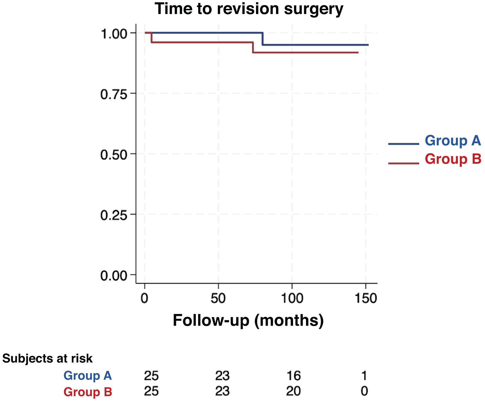
A total of 9 patients (11 THAs) died during follow-up from causes unrelated to their prostheses. As shown in the Kaplan–Meier curves (Fig. 1), the proportion of patients free from revision surgery did not differ significantly between groups, being 96% in Group A and 92% in Group B (p=.591).
Discussion
The switch from the Pinnacle®-Porocoat® cup to the Pinnacle®-Gription® cup resulted in an increase in both porosity and the coefficient of friction, which theoretically improves implant integration and durability.9 However, the translation of these benefits to primary total hip arthroplasty in humans remains a matter of controversy.
Our study, with a long-term follow-up, found no significant differences between the Porocoat® and Gription® coatings in terms of functional scores (HHS and MAP), radiographic outcomes (positioning, radiolucent lines, and osteolysis) or revision rates. These findings are consistent with studies focusing on patients undergoing prosthetic revision, in which highly porous coatings did not demonstrate clear superiority in terms of survival or clinical outcomes compared with conventional coatings.13 However, some authors have expressed concern regarding early radiographic findings in highly porous acetabular cups. Several studies on Tritanium® components (highly porous design) have shown an increase in the rates of radiolucent lines and poorer clinical scores in the short term, raising doubts about the universal benefit of increased porosity.10–12 In our cohort, radiolucent lines were observed exclusively in 2 patients, at 15 and 18 months, both in the Gription® group, although these were not clinically significant, as they were less than 1mm and non-progressive.
From a biomechanical perspective, the initial stability of the implant is enhanced by greater surface roughness and a high coefficient of friction. However, despite these theoretical advantages, cadaveric studies have shown only a minimal impact on the mechanical stability of the cup.20 Furthermore, the contribution of the implant's elasticity to long-term fixation remains debatable. Evidence suggests that increased friction plays a more significant role in primary stability than the modulus of elasticity.21 Few studies specifically evaluate the long-term differences between the various surface coatings of acetabular components.
Generally, available evidence suggests that greater porosity does not necessarily lead to better clinical outcomes, with only minimal, non-significant differences observed.10,22 Similarly, comparisons between StikTite® and Roughcoat® surfaces showed no significant differences in terms of implant migration or fixation after 10 years of follow-up.23 Furthermore, a long-term observational study compared the Plasmacup® component with porous-coated acetabular cups and found equivalent clinical outcomes and osseointegration rates after more than 10 years.24
These findings are consistent with our results, which suggest that both surface coatings are safe options for primary total hip arthroplasty in the long term.
All patients in this series received highly cross-linked polyethylene or ceramic inserts. These materials are known to reduce wear and the risk of osteolysis over time,25 and this factor, together with precise component positioning and consistent surgical technique, likely contributed to the absence of aseptic loosening.
Regarding intraoperative complications, only one acetabular fracture occurred, in a patient in the Porocoat® group. This was treated with mesh and a femoral head autograft, resulting in a favourable postoperative outcome. This complication did not differ significantly between the groups and is likely attributable to individual patient factors, such as bone quality or surgical technique.
Among the postoperative complications, one episode of post-traumatic dislocation was observed in a patient in the Gription® group, occurring 9 years after surgery, which was satisfactorily resolved by closed reduction, with no further episodes of instability. Furthermore, an acute THA infection was documented in a patient in the Porocoat® group, treated by debridement, antibiotic therapy, and implant retention (DAIR), with a favourable clinical outcome. During follow-up, two patients (one in each group) presented with Vancouver type B1 periprosthetic fractures, both of which were treated by open reduction and internal fixation.
No significant differences were observed between groups in the incidence or severity of intra- or post-operative complications, which reinforces the consistency of the results and the safety of both types of coating.
LimitationsThis study has some limitations. Firstly, the biases inherent in observational studies, although mitigated by the prospective collection of clinical data, cannot be completely ruled out, as the radiological analysis was performed retrospectively. However, this risk was minimised by a blinded procedure.
Secondly, the sample size was small (50 patients), which reduces the statistical power to detect any existing differences. Furthermore, all acetabular cups were implanted using supplementary fixation with screws in accordance with the current surgical protocol, which may have influenced initial stability. However, this procedure was applied uniformly in both groups.
Finally, this is a single-centre study, which limits the external validity and generalisability of the results.
ConclusionsUncemented acetabular cups with Porocoat® and Gription® coatings have demonstrated adequate osseointegration and stable fixation over time. There were excellent results in primary hip arthroplasty, both clinically and radiologically, with no superiority of one over the other observed in this long-term cohort.
Level of evidenceLevel of evidence III.
Ethical approvalThe ethics committee approved the study.
FundingNo funding was received to undertake this study.
Conflict of interestsThe authors have no conflicts of interests to declare.
We would like to thank Araceli Berjillos and Carlos Nicolás Pérez for their support, and our colleagues in the Orthopaedic Hip Unit for their assistance during the surgical procedures.


