



Medial meniscus injuries are the most prevalent knee injuries, frequently causing pain, instability, and orthopaedic surgeries. Although several clinical tests are used for meniscal pathology, most were developed for general meniscal lesions rather than specifically for medial meniscus posterior root tears (MMPRT). Evidence on root-specific physical examination signs remains limited. Recently, the Akmese manoeuvre has been introduced as a non-invasive diagnostic tool, assessing pain or tenderness during physical examination. This study aimed to determine the accuracy of the Akmese sign for the clinical diagnosis of MMPRT.
MethodsThis prospective cohort diagnostic study included patients aged 18–55 years scheduled for arthroscopic surgery following a clinical diagnosis of medial meniscus injury between January 2022 and July 2023. Diagnostic performance metrics, including sensitivity, specificity, positive and negative predictive values (PPV, NPV), and likelihood ratios (LR+, LR−), were calculated, using arthroscopy as the reference standard. The interobserver reliability of the Akmese manoeuvre was assessed using the kappa coefficient.
ResultsA total of 201 individuals met the eligibility criteria, with a mean age of 38.4±9.8 years. The Akmese sign was positive in 26 patients, and MMPRT was confirmed by arthroscopy in 28 patients. The Akmese sign demonstrated a sensitivity of 85.7%, specificity of 98.8%, PPV of 92.3%, NPV of 97.7%, LR+ of 74.1, and a LR− of 0.15.
ConclusionIn this selected surgical population, the Akmese manoeuvre showed promising diagnostic performance and very good interobserver reliability for identifying MMPRT. The sign may be useful as an adjunctive clinical tool to support preoperative suspicion, targeted MRI review, and surgical planning, but it should not be considered a replacement for MRI or arthroscopic assessment.
Las lesiones del menisco medial constituyen las lesiones de rodilla más frecuentes, y suelen ocasionar dolor, inestabilidad y requerir tratamiento quirúrgico ortopédico. Aunque varias pruebas clínicas se utilizan para la afección meniscal, la mayoría fueron desarrolladas para lesiones meniscales en general, y no específicamente para las roturas de la raíz posterior del menisco medial (RRPMM). La evidencia sobre los signos de exploración física específicos para la raíz meniscal sigue siendo limitada. Recientemente, la maniobra de Akmese ha sido propuesta como una herramienta diagnóstica no invasiva, basada en la evaluación del dolor o la sensibilidad durante la exploración física. El objetivo de este estudio fue determinar la exactitud del signo de Akmese para el diagnóstico clínico de las RRPMM.
MétodosSe realizó un estudio prospectivo de cohorte con diseño de prueba diagnóstica que incluyó pacientes de 18 a 55 años programados para artroscopia tras un diagnóstico clínico de lesión del menisco medial, entre enero de 2022 y julio de 2023. Se calcularon los parámetros de rendimiento diagnóstico, incluyendo sensibilidad, especificidad, valores predictivos positivo y negativo (VPP, VPN) y razones de verosimilitud (RV+, RV−), utilizando la artroscopia como estándar de referencia. La concordancia interobservador de la maniobra de Akmese se evaluó mediante el coeficiente kappa.
ResultadosUn total de 201 pacientes cumplieron los criterios de inclusión, con una edad media de 38,4±9,8 años. El signo de Akmese fue positivo en 26 pacientes, y se confirmaron RRPMM en 28 pacientes mediante artroscopia. El signo de Akmese mostró una sensibilidad del 85,7%, especificidad del 98,8%, VPP del 92,3%, VPN del 97,7%, una RV+ de 74,1 y una RV− de 0,15.
ConclusiónEn esta población quirúrgica seleccionada, la maniobra de Akmese mostró un rendimiento diagnóstico prometedor y muy buena concordancia interobservador para identificar RRPMM. El signo puede ser útil como herramienta clínica complementaria para apoyar la sospecha preoperatoria, la revisión dirigida de la resonancia magnética y la planificación quirúrgica, pero no debe considerarse un reemplazo de la MRI ni de la evaluación artroscópica.
Medial meniscal lesions are common knee injuries and may cause pain, mechanical symptoms, functional limitation, and the need for orthopaedic surgery.1–3 Among these lesions, medial meniscus posterior root tears (MMPRT) are particularly relevant because disruption of the posterior root compromises meniscal hoop-stress function and may lead to meniscal extrusion, increased tibiofemoral contact pressure, cartilage overload, and progressive joint degeneration.3–6
MMPRT can be clinically difficult to identify because symptoms may overlap with other medial meniscal lesions or degenerative knee conditions.3–6 Arthroscopic visualisation remains the reference standard for confirming meniscal root tears.3,5 Magnetic resonance imaging (MRI) is widely used in the preoperative diagnostic pathway; however, the identification of root-specific findings depends on image quality, MRI protocol, and radiological interpretation.7–11 Characteristic imaging features such as root discontinuity, radial tear signs, ghost sign, truncation signs, and meniscal extrusion may increase suspicion of MMPRT, but these findings are not always consistently recognised in routine practice.7,10,11
Traditional physical examination tests for meniscal pathology, including McMurray, Apley, Thessaly, and joint-line tenderness, were developed for general meniscal lesions rather than specifically for posterior root tears.12–15 Therefore, their diagnostic value for MMPRT is limited and should not be interpreted as directly equivalent to root-specific diagnostic assessment. In this context, a simple clinical manoeuvre capable of raising suspicion of MMPRT before surgery could be useful, especially when MRI access or specialist musculoskeletal radiology interpretation is limited.
The Akmese sign was first described as a clinical manoeuvre for diagnosing MMPRT.16 The test is based on the presence of marked medial joint-line tenderness in near extension that decreases or disappears during hyperflexion.16 This clinical pattern may reflect changes in the mechanical behaviour of the posterior root and adjacent meniscal tissue during knee flexion. Although promising, external evidence regarding the diagnostic accuracy of the Akmese sign remains limited.
This study aimed to evaluate the diagnostic accuracy of the Akmese sign for detecting MMPRT, using arthroscopic visualisation as the reference standard, among patients scheduled for arthroscopic surgery for suspected medial meniscal lesions in Peruvian healthcare facilities. We hypothesised that the Akmese sign would show high specificity and clinically useful sensitivity for identifying MMPRT in this selected surgical population.
MethodsDesign and contextThis prospective diagnostic test evaluation study was conducted at three healthcare facilities in Peru. The manuscript was prepared according to the Standards for Reporting Diagnostic Accuracy Studies (STARD) 2015 guideline.17
Population and sampleThe study population consisted of patients aged 18–55 years who were scheduled for arthroscopic surgery for suspected medial meniscal lesions between January 2022 and July 2023. Patients were eligible if they had clinical suspicion of a medial meniscal lesion, preoperative MRI available, and a planned arthroscopic procedure. Patients were excluded if they had concurrent lateral meniscal tears, ligament injuries, mucoid degeneration of the anterior cruciate ligament, and a history of synovectomy for pigmented villonodular synovitis of the knee.
A priori sample size calculation was performed using Epidat v4.2, referencing Akmese et al.16 With a 95% confidence level, expected sensitivity of 86.1%, and specificity of 99.1%, the required sample size was 198. An additional 10% was included to account for potential losses or rejections, yielding a target enrolment of 220 participants.
Diagnostic testThe diagnostic test evaluated was the Akmese sign, a manoeuvre performed during physical examination to detect pain or tenderness for the clinical diagnosis of MMPRT. With the patient in a supine position, the lower extremity was placed in a “figure-four”, inducing slight varus alignment to relax the medial collateral ligament and medial capsule. The medial joint line was palpated for pain or tenderness while knee flexion was reduced to 10° to 20° (near extension). The joint line was palpated again during hyperflexion (110°–130°). A positive Akmese sign was defined as severe tenderness near extension and minimal or no tenderness during hyperflexion.16 The Akmese sign was assessed after anamnesis and before review of MRI reports or images, and before arthroscopic confirmation of the diagnosis.
Reference standardThe reference standard for diagnosing MMPRT was intraoperative arthroscopic visualisation of the lesion.
Other variablesAdditional variables included sex, age, preoperative varus alignment (femorotibial angle >10°), body mass index (BMI), MRI findings, surgical procedure performed, and comorbidities.
Data collectionData were collected from the three healthcare facilities, involving patient enrolment, medical record review, and operative report analysis between January 2022 and July 2023. A structured data collection sheet was used to minimise errors and ensure consistency during data entry into a primary database. Inclusion and exclusion criteria were applied systematically.
ProceduresAll surgeries were performed by two orthopaedic surgeons with over five years of experience. Training sessions were conducted between December 2021 and February 2022, followed by quarterly refreshers to enhance interobserver concordance. During training, the surgeons were briefed on data collection protocols and instructed on performing the Akmese manoeuvre. A pilot study involving 20 patients was conducted to standardise the application of the Akmese sign. The Akmese sign was evaluated at the beginning of the clinical examination. The evaluators did not review MRI reports or images before performing the manoeuvre, and arthroscopic findings were not known at the time of index-test assessment. Inter-rater reliability was assessed using Cohen's kappa coefficient, with values above 0.80 considered optimal.18 This coefficient was chosen due to the involvement of two observers.
AnalysisThe data collection forms obtained from the hospitals were exported into a Microsoft Excel 2016 spreadsheet. A process of independent double coding was conducted, and any inconsistencies identified were resolved by reviewing the database and, when necessary, the corresponding medical records. Following quality control, the database was imported into Stata v16.0 (StataCorp, TX, USA) for analysis. To evaluate interobserver agreement between the two orthopaedic surgeons, the kappa coefficient (κ) was calculated. A pilot study involving 20 patients yielded a kappa coefficient of 0.89 (95% CI: 0.67–1.00), indicating very good agreement.
A bivariate analysis was conducted to compare the characteristics of the population with MMPRT (categorical dichotomous variable). For numerical variables with a normal distribution, the Student's t-test was used. For categorical variables, the Chi-square test or Fisher's exact test was applied, depending on whether expected cell counts were ≥5.
Based on the results of the Akmese sign and arthroscopic findings, sensitivity, specificity, predictive values, and likelihood ratios were calculated. All results were presented with 95% confidence intervals (95% CI).
EthicsThe research project was reviewed and approved by the Institutional Review Board of Universidad Peruana de Ciencias Aplicadas (FCS-SCEI/043-01-23). As this study was based on consecutive case reviews of medical records from the three healthcare facilities, access to these documents was requested from orthopaedic surgeons working in the research department. Study participants provided informed consent regarding the objectives, risks, and benefits of the study.
To ensure privacy and confidentiality, each medical record was assigned a numerical code, protecting patient identity. Only the research team had access to this information, which was not used for purposes unrelated to the study.
ResultsThe initial population consisted of 220 adults aged 18–55 years who received care at healthcare facilities in the Lima region, Peru, between January 2022 and July 2023. The application of eligibility criteria resulted in the exclusion of 19 participants, leaving a final study population of 201 individuals. Additionally, during the pilot test, an interobserver agreement kappa coefficient of 0.89 (95% CI: 0.67–1.00) was obtained, indicating very good concordance between the specialists performing the evaluation.
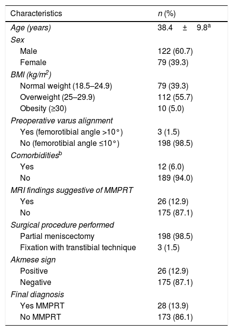
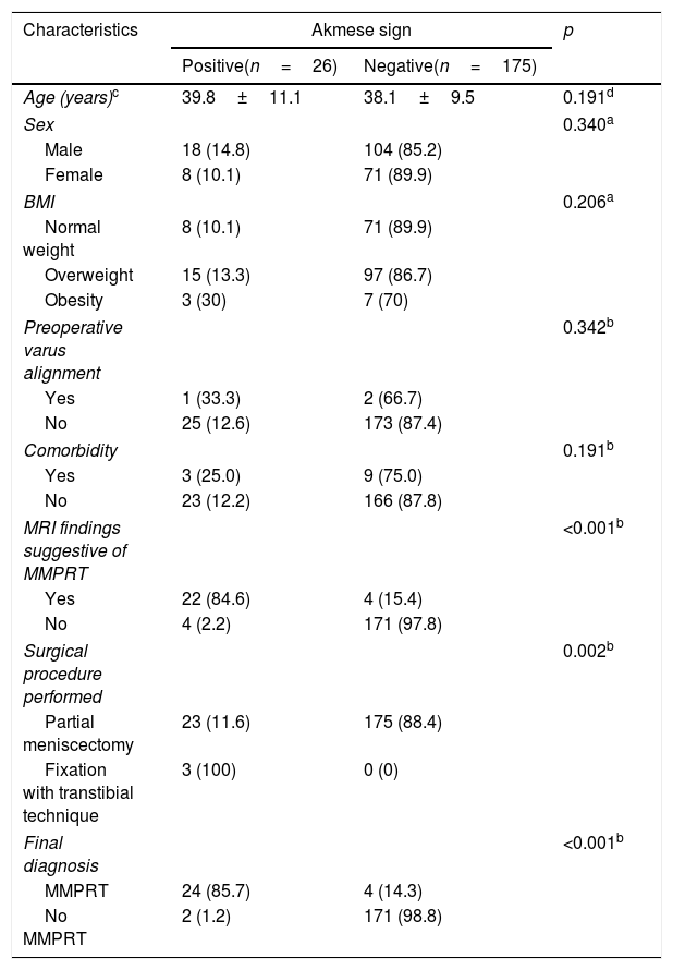
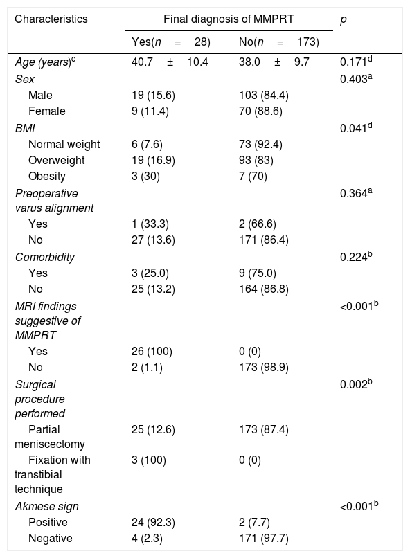
Characteristics of the study populationAmong the total participants, 60.7% were male, and the mean age was 38.4±9.8 years. Additionally, the majority (60.7%) had overweight or obesity, and 6% had at least one comorbidity. The Akmese sign was positive in 26 patients (12.9%), while MMPRT was confirmed arthroscopically in 28 patients (13.9%). MRI findings suggestive of MMPRT were present in 26 patients (12.9%). Regarding surgical procedures, 198 patients underwent partial meniscectomy and 3 underwent fixation with transtibial technique (Table 1). The Akmese sign was significantly more frequent among patients with MRI findings suggestive of MMPRT (p<0.001) and among those who underwent fixation with transtibial technique (p=0.002) (Table 2). Intraoperative MMPRT was significantly more frequent among individuals with higher BMI (p=0.041), MRI findings suggestive of MMPRT (p<0.001), fixation with transtibial technique (p=0.002), and a positive Akmese sign (p<0.001) (Table 3).
General characteristics of the study population (n=201).
| Characteristics | n (%) |
|---|---|
| Age (years) | 38.4±9.8a |
| Sex | |
| Male | 122 (60.7) |
| Female | 79 (39.3) |
| BMI (kg/m2) | |
| Normal weight (18.5–24.9) | 79 (39.3) |
| Overweight (25–29.9) | 112 (55.7) |
| Obesity (≥30) | 10 (5.0) |
| Preoperative varus alignment | |
| Yes (femorotibial angle >10°) | 3 (1.5) |
| No (femorotibial angle ≤10°) | 198 (98.5) |
| Comorbiditiesb | |
| Yes | 12 (6.0) |
| No | 189 (94.0) |
| MRI findings suggestive of MMPRT | |
| Yes | 26 (12.9) |
| No | 175 (87.1) |
| Surgical procedure performed | |
| Partial meniscectomy | 198 (98.5) |
| Fixation with transtibial technique | 3 (1.5) |
| Akmese sign | |
| Positive | 26 (12.9) |
| Negative | 175 (87.1) |
| Final diagnosis | |
| Yes MMPRT | 28 (13.9) |
| No MMPRT | 173 (86.1) |
BMI: body mass index; MRI: magnetic resonance imaging; MMPRT: medial meniscus posterior root tear.
Characteristics of the population according to the Akmese sign results.
| Characteristics | Akmese sign | p | |
|---|---|---|---|
| Positive(n=26) | Negative(n=175) | ||
| Age (years)c | 39.8±11.1 | 38.1±9.5 | 0.191d |
| Sex | 0.340a | ||
| Male | 18 (14.8) | 104 (85.2) | |
| Female | 8 (10.1) | 71 (89.9) | |
| BMI | 0.206a | ||
| Normal weight | 8 (10.1) | 71 (89.9) | |
| Overweight | 15 (13.3) | 97 (86.7) | |
| Obesity | 3 (30) | 7 (70) | |
| Preoperative varus alignment | 0.342b | ||
| Yes | 1 (33.3) | 2 (66.7) | |
| No | 25 (12.6) | 173 (87.4) | |
| Comorbidity | 0.191b | ||
| Yes | 3 (25.0) | 9 (75.0) | |
| No | 23 (12.2) | 166 (87.8) | |
| MRI findings suggestive of MMPRT | <0.001b | ||
| Yes | 22 (84.6) | 4 (15.4) | |
| No | 4 (2.2) | 171 (97.8) | |
| Surgical procedure performed | 0.002b | ||
| Partial meniscectomy | 23 (11.6) | 175 (88.4) | |
| Fixation with transtibial technique | 3 (100) | 0 (0) | |
| Final diagnosis | <0.001b | ||
| MMPRT | 24 (85.7) | 4 (14.3) | |
| No MMPRT | 2 (1.2) | 171 (98.8) | |
BMI: body mass index; MRI: magnetic resonance imaging; MMPRT: medial meniscus posterior root tear.
Characteristics of the population by intraoperative arthroscopic findings.
| Characteristics | Final diagnosis of MMPRT | p | |
|---|---|---|---|
| Yes(n=28) | No(n=173) | ||
| Age (years)c | 40.7±10.4 | 38.0±9.7 | 0.171d |
| Sex | 0.403a | ||
| Male | 19 (15.6) | 103 (84.4) | |
| Female | 9 (11.4) | 70 (88.6) | |
| BMI | 0.041d | ||
| Normal weight | 6 (7.6) | 73 (92.4) | |
| Overweight | 19 (16.9) | 93 (83) | |
| Obesity | 3 (30) | 7 (70) | |
| Preoperative varus alignment | 0.364a | ||
| Yes | 1 (33.3) | 2 (66.6) | |
| No | 27 (13.6) | 171 (86.4) | |
| Comorbidity | 0.224b | ||
| Yes | 3 (25.0) | 9 (75.0) | |
| No | 25 (13.2) | 164 (86.8) | |
| MRI findings suggestive of MMPRT | <0.001b | ||
| Yes | 26 (100) | 0 (0) | |
| No | 2 (1.1) | 173 (98.9) | |
| Surgical procedure performed | 0.002b | ||
| Partial meniscectomy | 25 (12.6) | 173 (87.4) | |
| Fixation with transtibial technique | 3 (100) | 0 (0) | |
| Akmese sign | <0.001b | ||
| Positive | 24 (92.3) | 2 (7.7) | |
| Negative | 4 (2.3) | 171 (97.7) | |
BMI: body mass index; MRI: magnetic resonance imaging; MMPRT: medial meniscus posterior root tear.
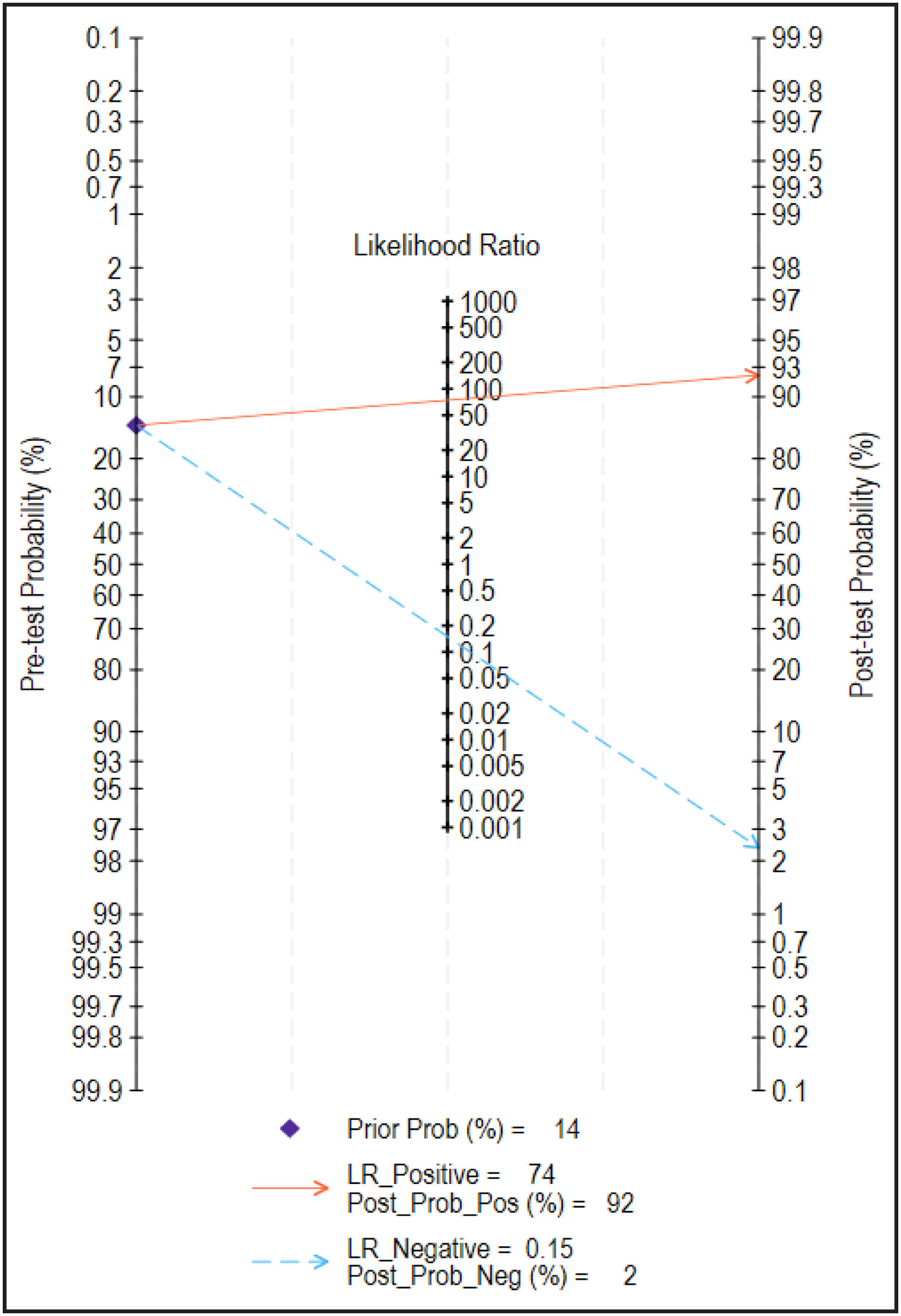
The diagnostic accuracy of the Akmese sign for MMPRT is presented in Table 4. Based on arthroscopic confirmation, the Akmese sign correctly identified 24 of 28 patients with MMPRT and was negative in 171 of 173 patients without MMPRT, corresponding to 24 true positives, 2 false positives, 171 true negatives, and 4 false negatives. Sensitivity and specificity were 85.7% and 98.8%, respectively, while the positive and negative predictive values were 92.3% and 97.7%, respectively. Fig. 1 shows the Fagan diagram, which, given a pre-test probability of 13.9%, reports positive and negative likelihood ratios of 74.1 and 0.15, respectively.
Accuracy of the Akmese sign for diagnosing MMPRT*
| Sensitivity | Specificity | PPV | NPV | LR+ | LR− | OR | |
|---|---|---|---|---|---|---|---|
| Akmese sign | 85.7%(67.3%–96.0%) | 98.8%(95.9%–99.9%) | 92.3%(74.9%–99.1%) | 97.7%(94.3%–99.4%) | 74.1(18.5–297) | 0.15(0.06–0.36) | 513(95.2–2674) |
PPV: positive predictive value; NPV: negative predictive value; LR: likelihood ratio; OR: diagnostic odds ratio.

The main finding of this study is that the Akmese sign showed good diagnostic performance for identifying medial meniscus posterior root tears (MMPRT) among patients scheduled for arthroscopic surgery because of suspected medial meniscal lesions. Using arthroscopic visualisation as the reference standard, the Akmese sign showed high specificity and clinically useful sensitivity, suggesting that it may be useful as an adjunctive clinical tool to raise preoperative suspicion of MMPRT in a selected surgical population.
The prevalence of arthroscopically confirmed MMPRT in this study was 13.9%, which is similar to that reported in the original study describing the Akmese sign.16 This supports the clinical relevance of evaluating root-specific diagnostic tools in patients with suspected medial meniscal pathology. However, the present results should be interpreted within the context of the study population. All included patients had already been selected for arthroscopic surgery and therefore had a higher pre-test probability of intra-articular pathology than patients evaluated in emergency care, sports medicine clinics, or non-surgical orthopaedic settings. Consequently, the predictive values and overall diagnostic performance observed here should not be directly extrapolated to broader clinical populations.
Clinical diagnostic frameworkThe Akmese sign should not be interpreted as a standalone diagnostic test. Clinical history remains central in the evaluation of suspected MMPRT, including the onset and mechanism of symptoms, posterior or medial knee pain, mechanical symptoms, functional limitation, and risk factors such as age, BMI, varus alignment, and degenerative joint changes.3,5,6 Similarly, physical examination should be interpreted as part of a broader diagnostic pathway rather than as an isolated finding. The value of the Akmese sign may lie in adding root-specific suspicion during the clinical assessment, particularly when other meniscal tests are non-specific.12–16
Traditional clinical tests such as McMurray, Apley, Thessaly, and joint-line tenderness were developed for general meniscal pathology and are not specific for posterior root tears.12–15 Therefore, direct comparison between these tests and the Akmese sign should be made cautiously. In contrast, the Akmese manoeuvre is conceptually directed at MMPRT and is based on the change in medial joint-line tenderness between near extension and hyperflexion.16 This may explain its potential usefulness in identifying posterior root involvement rather than general medial meniscal injury.
Role of MRI and combined assessmentMRI remains an important component of the preoperative evaluation of suspected MMPRT. However, MRI accuracy depends on technical quality, imaging planes, radiologist experience, and recognition of root-specific signs.10,11,19 Findings such as posterior root discontinuity, radial tear signs, ghost sign, truncation signs, and meniscal extrusion can support the diagnosis of MMPRT.1,10,11 The Akmese sign may therefore be most useful when it prompts a targeted MRI review or alerts the surgical team to the possibility of a root lesion before arthroscopy. This combined approach is likely more clinically appropriate than relying on either physical examination or MRI alone.3,5,19
Clinical relevanceThe clinical relevance of a positive Akmese sign should be understood within the diagnostic and preoperative decision-making pathway for MMPRT. A positive Akmese sign should not be considered a standalone diagnosis or a direct determinant of the surgical procedure. Rather, it may help clinicians identify patients in whom MMPRT should be specifically suspected, prompt a more focused review of the posterior root on MRI, support preoperative counselling, and alert the surgical team to the possibility that root repair may be required if the lesion is technically repairable. This distinction is important because surgical treatment was not standardised in this diagnostic accuracy study. In the present cohort, only 3 of the 28 patients with arthroscopically confirmed MMPRT underwent fixation with transtibial technique, whereas 25 underwent partial meniscectomy. Although meniscal root repair is generally preferred when MMPRT is technically repairable, the final surgical procedure depends on factors assessed before and during surgery, including tissue quality, chronicity of the lesion, degenerative changes, cartilage status, limb alignment, feasibility of repair, and patient-specific characteristics.2–5,20–23 Therefore, the surgical procedure variable should be interpreted descriptively and not as evidence comparing partial meniscectomy with meniscal root repair.
Nevertheless, preoperative suspicion of MMPRT remains clinically relevant because MMPRT is not simply a generic meniscal lesion. Posterior root tears have important biomechanical and prognostic implications and may be associated with meniscal extrusion, increased tibiofemoral contact pressure, cartilage overload, and progression of degenerative joint disease.1,3–6,9 Identifying a possible MMPRT before arthroscopy may help ensure careful intraoperative inspection of the posterior root, allow appropriate surgical preparation, and improve patient counselling about the possible intraoperative finding of a root tear and the available surgical options. However, whether use of the Akmese sign changes treatment selection, repair rates, or postoperative outcomes was beyond the scope of this diagnostic accuracy study and would require prospective management-impact studies with standardised criteria for repairability and treatment allocation.
LimitationsSome limitations must be acknowledged. First, the Akmese manoeuvre relies on patient-reported pain, introducing subjectivity that could be influenced by factors such as analgesic use or individual pain thresholds. However, most patients in this study sought care for persistent pain despite analgesic treatment, likely minimising this concern. Additionally, comorbidities such as neuropathies or metabolic disorders, which could affect pain perception, were rare in this cohort (fewer than 10% of participants). Second, only 28 patients had arthroscopically confirmed MMPRT. Although the total sample size was based on an a priori calculation, the limited number of target-condition cases may reduce the precision and stability of some diagnostic estimates, particularly the likelihood ratios and diagnostic odds ratio, as reflected by their wide confidence intervals. Third, the manoeuvre's accuracy depends on proper execution by trained clinicians and may be susceptible to assessment bias if evaluators are aware of imaging or other diagnostic information. To reduce this risk, the Akmese sign was assessed before arthroscopic confirmation, and evaluators did not review MRI reports or images before performing the manoeuvre. In addition, the manoeuvre was performed by experienced orthopaedic surgeons, and interobserver reliability was very good in the pilot assessment, with a kappa coefficient of 0.89. However, because the study was embedded in routine surgical care, complete blinding of the operating surgeon to all clinical information could not be guaranteed. Fourth, external validity is limited because the study was conducted in a selected surgical population from specific healthcare facilities in Peru. Therefore, the findings should not be directly extrapolated to emergency care, sports medicine, non-surgical orthopaedic settings, or populations with a lower pre-test probability of MMPRT.
ConclusionIn this selected population of patients scheduled for arthroscopic surgery for suspected medial meniscal lesions, the Akmese sign showed promising diagnostic performance for identifying MMPRT. The manoeuvre may be useful as an adjunctive clinical tool to support preoperative suspicion, targeted MRI review, and surgical planning when repair is feasible. However, it should not be considered a replacement for MRI or arthroscopic assessment, nor should it be interpreted as determining surgical treatment. These findings should not be extrapolated to general or non-surgical populations without further validation.
Level of evidenceLevel of evidence: III.
ORCID IDStefany M. Paucar-Vergara: 0000-0001-6311-4599
Joseph S. Urdanivia-Martos: 0000-0002-3515-4654
Authors’ contributionConceptualization: SMPV, JSUM; Data curation: SMPV, JSUM, CJTH; Formal analysis: All authors; Methodology: All authors; Writing – original draft: SMPV, JSUM; Writing – review & editing: CJTH. All authors have read and agreed to the published version of the manuscript.
Ethical approvalThe research project was reviewed and approved by the authors’ affiliated institutions.
Consent to participateStudy participants provided informed consent regarding the objectives, risks, and benefits of the study.
FundingSelf-funded.
Conflicts of interestsNone to declare.
We would like to express our gratitude to Dr. Diego Urdanivia Martos and Dr. Javier Urdanivia Martos for their assistance in data collection.


