


Rhinosporidiosis is caused by Rhinosporidium seeberi, a parasitic organism of the family Rhinosporideacea family, class Micomycetozoa. The disease is endemic in India; however, some cases were reported in Europe, Africa, North America, and South America. The aim of the present study is to report three cases of rhinosporidiosis in wild horses in different cities of Buenos Aires province, Argentina. We confirm the presence of R. seeberi in the analyzed samples using histopathological and PCR sequencing techniques.
La rinosporidiosis es una enfermedad causada por Rhinosporidium seeberi, un organismo parasitario clasificado en la familia Rhinosporideacea, clase Micomycetozoa. Es una enfermedad endémica de la India, pero se notificaron algunos casos en Europa, África, América del Norte y América del Sur. El objetivo del presente estudio fue describir tres casos de rinosporidiosis en caballos de vida libre en diferentes ciudades de la provincia de Buenos Ares, Argentina. Confirmamos la presencia de R. seeberi en las muestras analizadas utilizando técnicas histopatológicas, PCR y secuenciación.
Rhinosporidiosis is a disease caused by Rhinosporidium seeberi, a parasitic organism classified in the family Rhinosporideacea, class Micomycetozoa3,15. It is an endemic disease in India; however, some cases have been reported in Europe, Africa, North America, and South America4,9–11. Niño et al.12 described an endemic area of rhinosporidiosis in the province of Chaco, northern Argentina, and reported 33 cases of equine rhinosporidiosis in 1964. Rhinosporidium seeberi infects mammals including humans, cats, dogs, horses, and cattle2–4,7,8,13.
The natural reservoir of R. seeberi is lacustrine or stagnant water, in tropical and subtropical regions. In susceptible hosts, the lesions are frequently observed in nasal mucosae, partly because contaminated water is the source of infection7,11,14.
Infected animals present clinical signs characterized by nasal secretion, sneezing, epistaxis, breathing noise1, and occasionally, obstruction of the nasal cavity. Less frequently, the ocular conjunctiva can be affected causing epiphora. Taking into account that there is no drug treatment, surgical removal of the polypoid structures is the treatment of choice2; however, recurrence can occur4.
The microorganism does not grow in culture media, thus, the diagnosis is based on histolopathological findings in tissues. The method used to identify the agent is based on the amplification of the small subunit rRNA gene sequence from infected tissue5,11.
The aim of the present study is to describe three cases of rhinosporidiosis in wild horses in different cities of Buenos Aires province, Argentina identified by histopathology and PCR-sequencing trials.
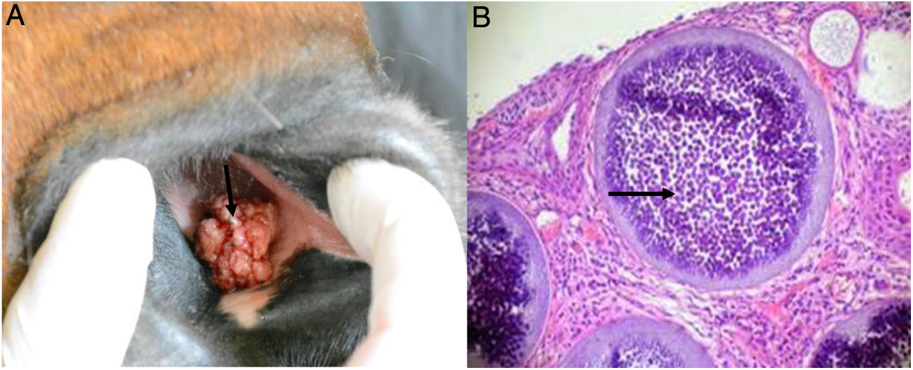
For confirmation of the causal agent, the pedunculated masses of three horses were removed by surgical excision (Fig. 1A). The fractionated tissues were fixed in 10% buffered formalin for the histopathological studies. The remaining tissue was used in PCR trials. The fixed tissues were stained with periodic acid-Schiff (PAS) and spherules (sporangia) of variable size (100–300μm) were observed. Sporangia had thin and weakly stained walls (Fig. 1B).
 Adult horse showing a multilobulated mass arising from the nasal mucosa (black arrow). (B) Thin section of nasal mucosa with mature sporangia (black arrow) and a myriad of endospores. Stained with PAS 100×.")
DNA was extracted from fresh biopsy samples (25mg) by using a commercially available kit (DNeasy Tissue Kit, QIAGEN) according to the manufacturer's protocol. R. seeberi specific primers Rhino 2F (5′-TTTGTGTAGGGGTTCCTCGC-3′) and Rhino 2R (5′-GCAAAACCGTTGCTCCAACT-3′) (7) were used in a PCR. The R. seeberi specific amplification product from the 3 samples were sequenced by Macrogen (www.macrogen.com). The PCR amplified three specific fragments of 377bp expected for R. seeberi (Suppl. Fig. 1). The amplification products were sequenced, showing 99% homology with Rhinosporidium spp. (ex Canis familiaris) (accession number AY372365.1). The program used to generate the phylogenetic tree was Geneious Version 2020.1 created by Biomatters (http://www.geneious.com) (Suppl. Fig. 2).
The three reported rhinosporidiosis cases in wild horses were from the following cities: Chascomús city located at geographical coordinates 35°34′30′S58°00′32′0, Ensenada city at 34°51′S 57°54′0 and La Plata city at 34°56′00′S57°57′00′0 (cities from Buenos Aires province, Argentina).
Two horses exhibited clinical signs of mild cough and rhinitis and the third one had epistaxis and nasal discharge. Therefore, the clinical examination was followed by a nasopharyngeal and laryngeal endoscopy, showing irregular pedunculated structures in shape and sizes (approximately 2.3×2.0×1.4cm each). These pendunculated structures revealed the following:
- -
-Isolate 1 (Chascomús) pedunculated masses in the larynx of a 15-year old mare.
- -
-Isolate 2 (Ensenada) pedunculated masses in the larynx of a 10-year-old male horse.
- -
-Isolate 3 (La Plata) pedunculated masses in the nose of a 4-year-old mare.
The polypoid structures were stained with PAS and microscopically observed. They consisted of a core of mature fibrovascular tissue superficially lined by a hyperplastic epithelium. Inside the granulated tissue and in the submucosa, there were conspicuous spherules (sporangia) of variable size (100–300mm in diameter) that showed a thin and weak wall.
The morphological features of the agent observed in all the cases presented here were characteristic, and could be presumptively identified as R. seeber based on histopathological features. Nevertheless, a differential diagnosis with polypoid or granulomatous rhinitis caused by a fungal infection such as Coccidioides immitis, Chrysosporium parvum, Cryptococcus neoformans, Conidiobolus coronatus, and other causes, such as equine nasal amyloidosis and neoplasia2,6,8 is needed. For this reason, the use of a specific PCR reaction and sequencing could allow us to reach a precise diagnosis in a shorter time.
In our study, the three analyzed samples amplified a specific fragment of 377bp expected for Rhinosporidium spp. Moreover, the sequence analysis confirmed these findings.
In Argentina, the occurrence of rhinosporidiosis is uncommon, and is rarely suspected by clinical veterinarians, thus it is underestimated o misdiagnosed. Furthermore, if the pedunculated structures are located in anatomical sites other than in the nasal cavity or conjunctiva, the disease can be even harder to diagnose by the practitioners.
It should be remarked that since there is no specific medical treatment for rhinosporidiosis, it is very important to make a routine clinical diagnosis in wild horses, taking into account that the infected animals not only share the stagnant water and lacustrine environment but also the feeders and drinkers with the healthy animals. These clinical surveys could allow us to delimit the geographical areas with a high prevalence of rhinosporidiosis so that there could be a chance to relocate the breeder farms of polo horses or racehorses. Considering that Argentina is an exporter of highly competitive horses, the early diagnosis of this disease will contribute to its control, preventing its spread to other places.
Conflict of interestThe authors declare that they have no conflicts of interest.
This research was supported by resources from the Course of Medical Mycology, School of Veterinary Medicine, National University of La Plata, Argentina.
The following are the supplementary data to this article:
