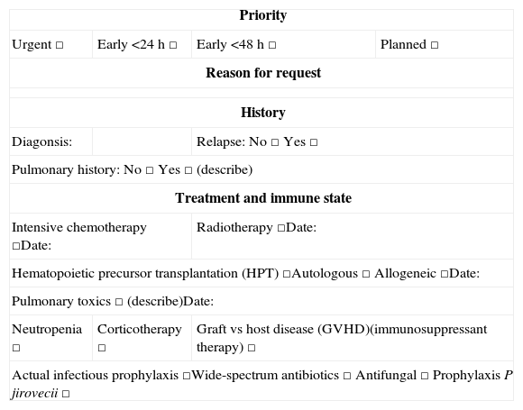
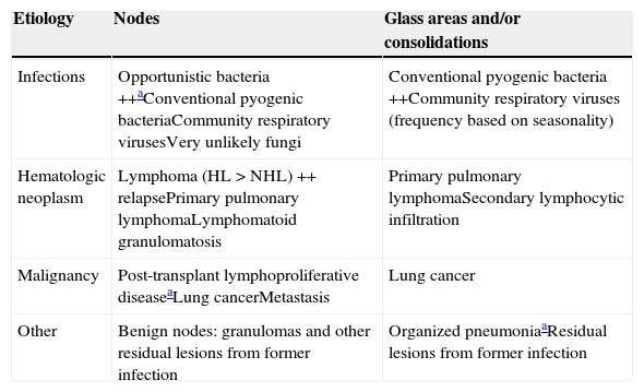
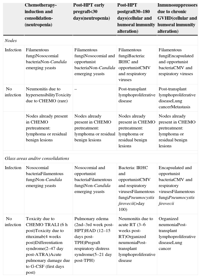
Lung disease is very common in patients with hematologic neoplasms and varies in function of the underlying disease and its treatment. Lung involvement is associated with high morbidity and mortality, so it requires early appropriate treatment. Chest computed tomography (CT) and the analysis of biologic specimens are the first line diagnostic tools in these patients, and sometimes invasive methods are necessary. Interpreting the images requires an analysis of the clinical context, which is often complex. Starting from the knowledge about the differential diagnosis of lung findings that radiologists acquire during training, this article aims to explain the key clinical and radiological aspects that make it possible to orient the diagnosis correctly and to understand the current role of CT in the treatment strategy for this group of patients.
La patología pulmonar en la historia de un paciente con neoplasia hematológica es muy frecuente y variable en función de la enfermedad de base y la terapia recibida. La morbimortalidad asociada es alta, por lo que requiere un tratamiento correcto y precoz. La tomografía computarizada (TC) torácica, junto con el análisis de muestras biológicas, son las herramientas de diagnóstico de primera línea empleadas en estos pacientes, y en determinados casos se requieren métodos invasivos. La interpretación de las imágenes exige el análisis de un contexto clínico en muchas ocasiones complejo. Partiendo del conocimiento que adquiere el radiólogo en su formación sobre el diagnóstico diferencial de los hallazgos pulmonares, el objetivo de este trabajo es explicar los aspectos clínicos y radiológicos claves que permiten orientar correctamente el diagnóstico y asimilar el papel actual de la TC en la estrategia terapéutica de este grupo de enfermos.
Artículo
Comprando el artículo el PDF del mismo podrá ser descargado
Precio 19,34 €
Comprar ahora


