


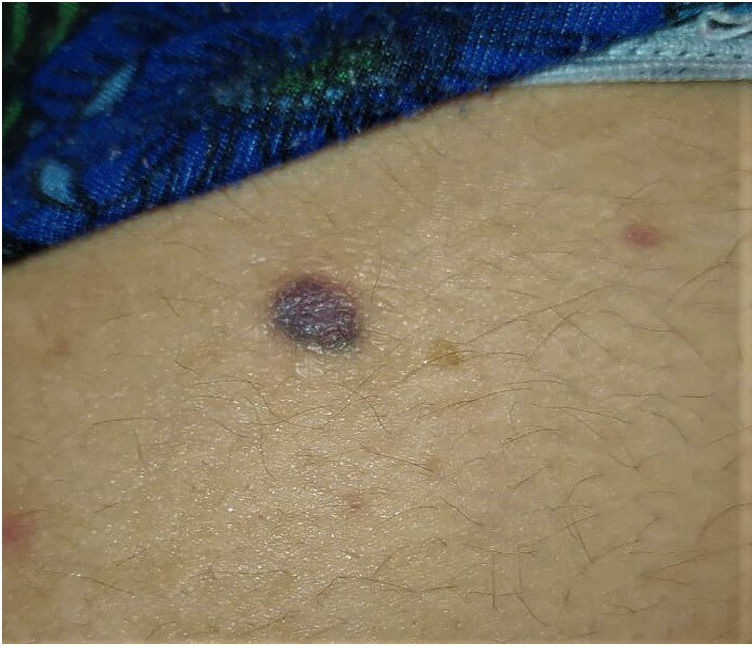

A 34-year-old woman with no remarkable past medical history presented with a slow-growing asymptomatic lesion on her right buttock that had been present for 20 years. Physical examination revealed a well-defined, round, bluish plaque with a diameter of 15mm (Fig. 1).

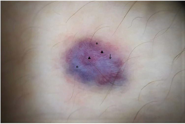
Polarized light dermoscopy showed global multichromatic pigmentation (blue, red, white), dotted and irregular linear vessels, bright white lines, and structureless bluish-white and reddish areas (Fig. 2).
, bright white lines (black arrow) and structureless bluish-white and reddish areas (*).")

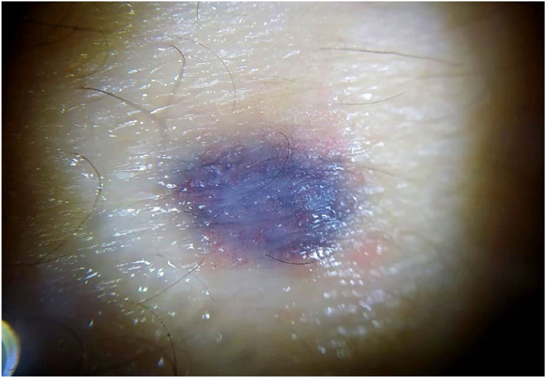
Nonpolarized dermoscopy showed a homogeneous blue pattern (Fig. 3).

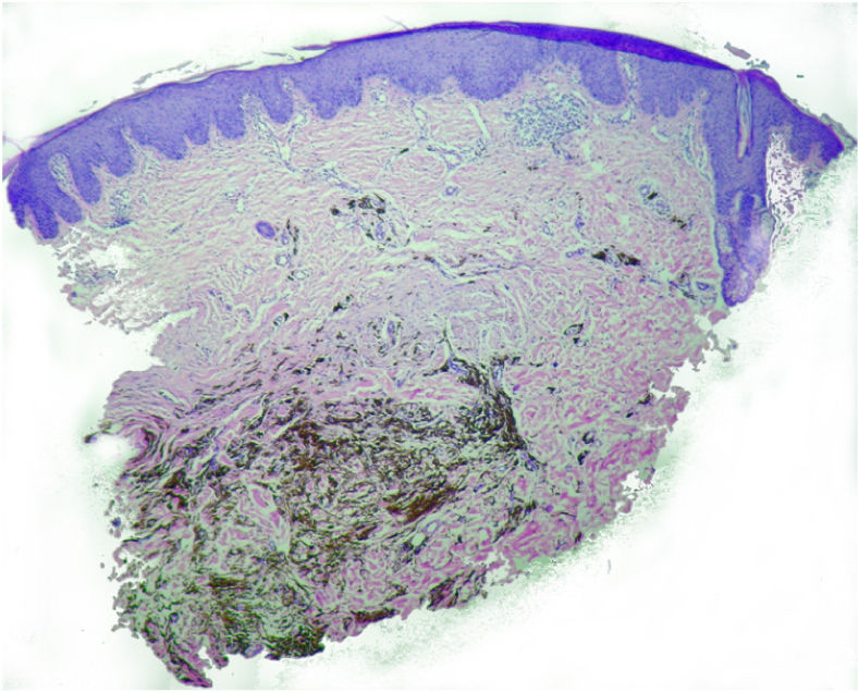
Histologic examination of a skin biopsy specimen showed a preserved epidermis with superficial parakeratosis and, in the deep reticular dermis, a dense proliferation of round dendritic melanocytes with darkly pigmented melanophages (Fig. 4).
.")
What Is Your Diagnosis?
DiagnosisSkin biopsy confirmed a diagnosis of cellular blue nevus.
CommentBlue nevi are benign lesions composed of a proliferation of dendritic melanocytes in the dermis. They can be congenital or acquired and present clinically as solitary or multiple papules, nodules, or plaques with a blue, gray-blue, or bluish-brown color.1–3 Onset is typically in childhood, adolescence, or early adulthood. Most lesions are located on the face, scalp, extremities, and buttocks.2,3
Blue nevi can mimic both melanocytic and nonmelanocytic lesions, including melanoma, cutaneous melanoma metastases, Spitz/Reed nevus, and basal cell carcinoma.1–3
The hallmark nonpolarized dermoscopic feature is a homogeneous blue pattern without a pigment network or any other distinctive structures.1–3 Less frequently, this global pattern may also have a diffuse blackish or brown color, or a combination of 2 colors (dichromatic pattern) such as blue-brown, blue-black, or blue-gray.1 Multichromatic pigmentation with a combination of blue, brown, white, black, red, and/or gray is uncommon and has been reported in just 15.8% of blue nevi.1
Vascular structures (polymorphous, dotted, comma, irregular linear, and arborizing vessels) are rare local findings. Some case series have shown these features to be significantly associated with the global multichromatic pattern.1–3
Presence of hypopigmented white areas, which histologically correspond to dermal fibrosis, is another uncommon local feature.1
Bright white lines, visualized only under polarized light, are not pathognomonic and have not been linked to blue nevus in any of the reports identified.
Observation of a solitary lesion present since childhood in a young patient with no personal or family history of melanoma combined with a homogeneous blue pattern seen on dermoscopy is strongly suggestive of a diagnosis of blue nevus.3 Melanoma, however, cannot be ruled out when dermoscopy shows a bluish lesion with multichromatic pigmentation and other local criteria, such as vessels. In such cases, surgical excision is mandatory.1
In conclusion, we have presented a case of cellular blue nevus in which polarized light dermoscopy showed the uncommon global multichromatic pattern and the bright white lines associated with this benign entity. Based on these findings, the differential diagnosis is broad.
Conflicts of InterestThe authors declare that they have no conflicts of interest.


