Buscar en
Revista de Senología y Patología Mamaria - Journal of Senology and Breast Disease
Toda la web
Inicio Revista de Senología y Patología Mamaria - Journal of Senology and Breast Dise... Soft fibroma of the nipple: Clinical and histopathological characteristics
Journal Information
Vol. 34. Issue 1.
Pages 60-61 (January - March 2021)
Share
Share
Download PDF
More article options
Vol. 34. Issue 1.
Pages 60-61 (January - March 2021)
IMAGES AND VIDEOS
Full text access
Soft fibroma of the nipple: Clinical and histopathological characteristics
Fibroma blando del pezón: características clínicas e histopatológicas
Visits
5141
Daniela Yucumáa,
Corresponding author
yucumad@javeriana.edu.co

Corresponding author.
, Diego F. Téllez Beltránb, Juanita Carolina Chaves Cortésb, Sergio Cervera Bonillab
a Clinical Epidemiology and biostatistics, Pontificia Universidad Javeriana, Bogotá, Colombia
b General Surgery Department, Hospital Universitario San Ignacio, Pontificia Universidad Javeriana, Bogotá, Colombia
Related content
This item has received
Article information
Full Text
Bibliography
Download PDF
Statistics
Figures (2)
Full Text

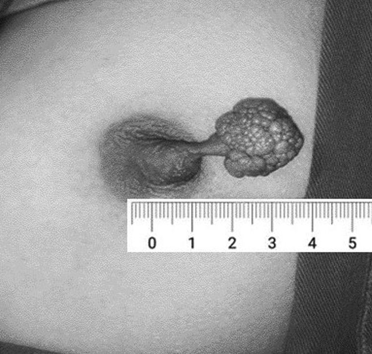
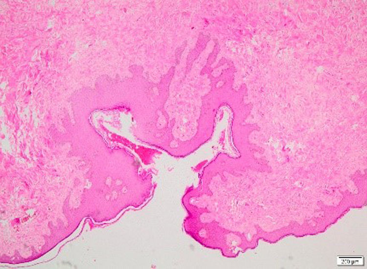
A 22-year-old woman presented with 5-years history of a pedunculated polyp originating from her left nipple, which appeared during her puerperium and had gradually enlarged over the past 10-months. No galactorrhea nor any other signs or symptoms were reported by the patient. The lesion measured 3.5×2.5×1.5cm and had a cauliflower-like surface (Fig. 1). No other findings were observed on the adjacent skin. Initially, the lesion was considered to be a papilloma and total surgical excision was considered and performed. Histopathological analysis showed a polypoidal lesion covered by squamous epithelium with fibro collagenous hypocellular stroma and small vessels in the stroma (Fig. 2). Neither necrosis nor mitosis were identified. Based on these findings, the lesion was finally classified as a soft fibroma as this lesion presents with proliferation of fibroblasts between collagen and scattered vessels, with a keratinizing squamous cell epithelium.1,2 Based on these findings, we diagnosed the lesion as a soft fibroma. At the ambulatory follow-up, patient exhibited a normal and uneventful recovery, without recurrences. Only a few cases of fibromas occurring on the nipple have been published in the literature. Here, we present a rare case of a cauliflower-like fibroma arising from the nipple that occurred in a female patient.

Figure 1.

Polypoid lesion originating from the left nipple.

(0.09MB).
Figure 2.

Histopathological findings: polypoidal lesion covered by squamous epithelium with fibrocollagenous hypocellular stroma and small vessels in the stroma (H&E).

(0.09MB).
Confidentiality of data

The authors declare that they have followed the protocols of their Center on the publication of patient data.

Conflict of interest

The authors declare that they have no conflict of interest.

References
[1]
B.F. Seo, S.N. Jung.
Fibroma of the nipple.
[2]
V.M. Doctor, M.V. Sirsat.
Florid papillomatosis (adenoma) and other benign tumours of the nipple and areola.
Br J Cancer, 25 (1971), pp. 1-9
Copyright © 2020. SESPM
Article options
Tools
es en pt

¿Es usted profesional sanitario apto para prescribir o dispensar medicamentos?

Are you a health professional able to prescribe or dispense drugs?

Você é um profissional de saúde habilitado a prescrever ou dispensar medicamentos