


To compare the effects of application mode of one self-etching adhesive on the shear bond strength of metallic orthodontic brackets.
MethodsSeventy-five healthy bovine incisors were divided into 5 groups (n=15). The self-etching primer (Transbond Plus, 3M Unitek) was applied on the enamel actively and passive for 0 (control), 5 and 10s, followed by air jet application and light cured for 10s (600mW/cm2). The metal brackets were bonded with adhesive (Transbond XT) and light cured for 20s each proximal surface (mesial and distal). The shear bond strength was determined after water storage at 37°C for 24h. The specimens were tested using a universal testing machine (Instron 3342). Once debonded, each specimen was examined to identify the failure mode. The bond strength data were subjected to One-way Anova and Tukey tests (α=0.05) and failure mode data were analyzed by Kruskal–Wallis test (α=0.05).
ResultsNo significant difference in bond strength was found between 5 groups. Increasing the application time and applying agitation of self-etching primers did not affect the shear bond strength (p=0.487). There were no differences between failure mode values in all tested groups (p=0.88) and score 1 was predominant.
ConclusionsThe shear bond strength of the self-etching adhesive is not influenced by the application mode.
Comparar os efeitos do modo de aplicação de um adesivo autocondicionante, na resistência ao cisalhamento de brackets metálicos em esmalte bovino.
MétodosSetenta e cinco dentes bovinos hígidos foram divididos em 5 grupos (n=15). O sistema adesivo autocondicionante (Transbond Plus, 3M Unitek) foi aplicado no esmalte de forma ativa e passiva por 0 (controle), 5 e 10 segundos, seguido de aplicação de jato de ar e fotoativação por 10s (600mW/cm2). Os brackets metálicos foram colados com resina fotopolimerizavel (Transbond XT, 3M Unitek) e fotoativado por 20s em cada face proximal (mesial e distal). A resistência ao cisalhamento foi determinada após armazenamento em água a 37°C por 24 horas. Os espécimes foram testados usando uma máquina de ensaio universal (Instron 3342). Uma vez descolados, cada espécime foi examinado para identificar o modo de fratura. Os dados da resistência ao cisalhamento foram submetidos aos testes One-way Anova e Tukey (α=0,05), enquanto que o modo de fratura foi examinado com o teste de Kruskal–Wallis (α=0,05).
ResultadosNão foram encontradas diferenças de resistência de união significantes entre os 5 grupos. O aumento do tempo de aplicação e agitação do primer auto-condicionante não afetou a resistência ao cisalhamento (p=0,487). Não foi observado diferença do modo de fratura nos grupos testados (p=0,88), o score 1 foi predominante em todos os grupos.
ConclusõesA resistência ao cisalhamento do adesivo auto-condicionante não é influenciada pelo modo de aplicação.
The adhesive systems used for orthodontic bracket bonding may be presented in different forms. The etch-and-rinse adhesive are those in which phosphoric acid is used to etch the substrate surface, and with the self-etching types acidic primers are used to demineralize the enamel.1–3 Orthodontic bracket bonding performed with adhesive systems with the use of phosphoric acid show high shear bond strength values. However, the innumerable clinical steps involve may prolong the time when the orthodontic appliance is being assembled, and cause iatrogenic damage to the enamel.4–7 Etching with phosphoric acid may demineralize approximately 10–30μm of enamel.8 Moreover, phosphoric acid may cause a reduction in the mechanical properties of the etched enamel, due to demineralization and thus lead to fracture of this substrate.9
The use of self-etching adhesive systems has the advantage of reducing the number of steps and minimizing risk of eventual errors occurring during the adhesive technique.10–12 These adhesive systems generally contain methacrylated phosphoric acid esters (derived from phosphoric acid) that demineralize the tooth surface by the removal of calcium ions.13
The SEP (self-etching) used in orthodontics have the advantage of simultaneously demineralizing and infiltrating into the tooth surface, and this mechanism is only possible due to the low pH of this material (pH<1)14 In addition to pH, there are innumerable other factors that may potentially contribute to the bond strength between enamel and the orthodontic bracket, including the type of enamel, adhesive composition, bracket base design, bracket material, oral medium, clinician's skills, acid concentration, and duration of etching time.15–17
The use of SEP is efficient in bracket bonding, but the bond strength results and clinical behavior are still below the standard obtained with etch-and-rinse adhesives. However, their behavior may change according to the application mode of these adhesive systems to enamel. Little is known about the application time and mode of application of these adhesive systems, or a combination of these factors on the bond strength of orthodontic brackets.
Therefore, the aim of this study was to evaluate the shear bond strength of a self-etching system applied in different modes (active and passive) and times (0, 5 and 10s). The null hypothesis was that the application mode could not interfere in the shear bond strength of self-adhesive.
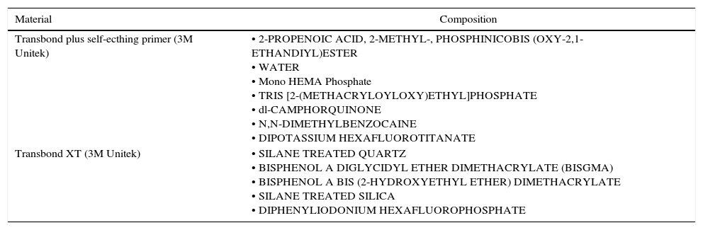
Material and methodsA total of 75 healthy bovine incisors were selected. The teeth were embedded in PVC tubes with acrylic resin, so that only the coronal portion remained visible. After this, the vestibular surfaces were treated with pumice stone and a rubber cup for 10s, then washed and dried. The teeth were divided into 5 groups (n=15) according to time and application mode of the adhesive system Transbond Plus Self Etching Primer (3M/Unitek, Monrovia, CA, USA) (Table 1). Light activation of adhesive system was performed with an Optilux 501 for 10s (600mW/cm2, Kerr, Orange, CA, USA):
- •
Group SEP0 (control): The adhesive was applied only on the surface, a light jet of air was applied for 1–2s, and then light activated;
- •
Group SEPNR5 (no rubbing): The adhesive was applied on the surface, waiting for 5s, a light jet of air applied for 1–2s, and then light activated;
- •
Group SEPR5 (active): The adhesive was applied on the surface with agitation for 5s, a light jet of air applied for 1–2s, and then light activated;
- •
Group SEPNR10 (no rubbing): In this group, the adhesive was applied on the surface, waiting for 10s, a light jet of air applied for 1–2s, and then light activated;
- •
Group SEPR10 (active): The adhesive was applied on the surface with agitation for 10s, a light jet of air applied for 1–2s and then light activated.
Composition of materials used in this research.a
| Material | Composition |
|---|---|
| Transbond plus self-ecthing primer (3M Unitek) | • 2-PROPENOIC ACID, 2-METHYL-, PHOSPHINICOBIS (OXY-2,1-ETHANDIYL)ESTER • WATER • Mono HEMA Phosphate • TRIS [2-(METHACRYLOYLOXY)ETHYL]PHOSPHATE • dl-CAMPHORQUINONE • N,N-DIMETHYLBENZOCAINE • DIPOTASSIUM HEXAFLUOROTITANATE |
| Transbond XT (3M Unitek) | • SILANE TREATED QUARTZ • BISPHENOL A DIGLYCIDYL ETHER DIMETHACRYLATE (BISGMA) • BISPHENOL A BIS (2-HYDROXYETHYL ETHER) DIMETHACRYLATE • SILANE TREATED SILICA • DIPHENYLIODONIUM HEXAFLUOROPHOSPHATE |
Seventy-five metal, Standard Edgewise (3M Unitek, Monrovia, CA, USA) superior central incisor brackets were used. The area of each bracket base was calculated (mean=15.84mm2) by using a digital pachymeter (Absolute Digimatic, Mitutoyo, Tokyo, Japan). Transbond XT resin (3M Unitek, Monrovia CA, USA) was applied at the base of the bracket, which was placed on the vestibular surface of the tooth by using orthodontic forceps and a tensiometer (Odeme Biotechnology, Joaçaba, SC, Brazil), with a force of 300g to ensure a uniform resin thickness. After that, light activation was performed for 20s on each bracket proximal surface (mesial and distal), so all brackets were light activated for 40s individually.
After bracket bonding, the test specimens were stored in distilled water at 37°C for 24h. The shear bond strength test was performed by universal test machine (Instron 3342, Canton, MA, USA) at a speed of 1.0mm/min., using a chisel (Odeme Biotechnology) applied on the bracket/enamel interface. The force required to debond the brackets was recorded in Newtons (N) and divided by the area of the brackets (mm2), thus the values are presented in MegaPascal (MPa).
Statistical analysis was performed by using SigmaPlot 12 software (SigmaPlot v. 12.3, Systat Software Inc., San Jose, USA). All the data were analyzed as regards normality of distribution by means of the Kolmogorov–Smirnov test (α=0.05). The shear bond strength data were submitted to the One-Way analysis of variance and Tukey tests (α=0.05).
After the bond strength test, all the specimens were analyzed under a microscope (Kozo Optical and Electronical Instrumental, Nanjing, China), at 10× magnification. It could evaluate the fracture patterns and adhesive remnant index (ARI): score 0, without remnant composite on the tooth; score 1, less than 50% remnant composite on the tooth; score 2, over 50% remnant composite on the tooth; and score 3, all the composite on the tooth, with a distinct impression of the bracket supporting screen. The nonparametric Kruskal–Wallis test was used to test for the significant differences in ARI scores among the groups. A p value <0.05 was considered significant.
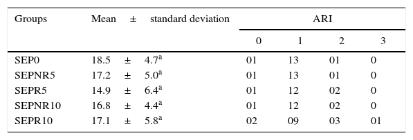
ResultsThe shear bond strength (MPa) and adhesive remnant index (ARI) for the different bonding protocols are shown in Table 2. The mean shear bond strength values in shear bond strength indicated there was no significant difference among all groups (P=0.487), by One-Way ANOVA. Table 2 shows the adhesive remnant index (ARI) scores for the adhesive.
Shear bond strength values (MPa) and adhesive remnant index (ARI) of experimental groups.
| Groups | Mean±standard deviation | ARI | |||
|---|---|---|---|---|---|
| 0 | 1 | 2 | 3 | ||
| SEP0 | 18.5±4.7a | 01 | 13 | 01 | 0 |
| SEPNR5 | 17.2±5.0a | 01 | 13 | 01 | 0 |
| SEPR5 | 14.9±6.4a | 01 | 12 | 02 | 0 |
| SEPNR10 | 16.8±4.4a | 01 | 12 | 02 | 0 |
| SEPR10 | 17.1±5.8a | 02 | 09 | 03 | 01 |
Similar letters means no difference in statistical analysis.
Kruskal–Wallis test was perfomed to ARI, the score 01 was predominant into all groups. According to statistical analysis all the groups exhibited similar bracket failure modes (p=0.88) (Table 2). Fig. 1(A–D) shows specimens with different ARI scores.
Discussion score 0: without remnant composite on the tooth, (B) score 1: less than 50% remnant composite on the tooth, (C) score 2: over 50% remnant composite on the tooth, (D) score 3: all the composite on the tooth, with a distinct impression of the bracket supporting screen.")
In the present study the application mode of self-etch adhesive did not show any significant effects in shear bond strength, leading us to accept the null hypothesis.
Some studies have suggested a long active application of SEPs may increase the enamel surface roughness, thus improve interlocking of the adhesive material with the enamel surface, and thereby increase the shear bond strength values.16,18 The increase in bond strength of SEPs to enamel would possibly indicate a better clinical behavior of this material, which would prevent premature bracket debonding, saving the patient from having to make several visits to the professional's dental office13 in order to perform removal of the resin remainders from the enamel surface, and new bracket bonding.
However, there was no statistical difference found between the groups tested in this study, active and increase the time of self-adhesive system on the enamel surface was not capable of increasing the shear bond strength values of metal brackets to the bovine tooth enamel surface. Another study,15 also found no differences in the bond strength values, by increasing the time from 3 to 5s, or to 15s. In order to try and understand these results, the authors evaluated the aspect of enamel after the different application methods, and observed a similar etching pattern on the enamel surface. Other study19 show no significant difference on the shear bond strength for different application times of 3, 10 and 30s. In this case, the authors observed a slight increase in etching efficacy, especially for an application time of 30s.
These results are believed to be due to the low degree of conversion values (DC%) of the adhesive system used.20 These low degree of conversion values of self-etching adhesives may lead to these materials continuing to demineralize the enamel surface,21–23 even after their polymerization, since these materials present a low pH (>1).14 This demineralization of the enamel surface would occur over the course of time with the action of the acidic monomers, and would only be interrupted by the buffer effect of enamel.24 Some self-etching systems present low DC values, which may be due to the large volume of solvent, which harms the polymerization reaction of the adhesive.25 Recently, a study demonstrated evaporation of the solvent was an important step in increasing the bond strength values of metal brackets bonded to bovine teeth.14
Another factor that may have contributed to no statistical difference among the groups tested was perhaps, the light activation appliance used. In this study, an halogen light polymerizing appliance was used, which may have contributed to a low degree of conversion of the material. A recent study26 suggested light emitting diode (LEDs) appliances must be considered, due to the high degree of conversion values obtained when these appliances are used on orthodontic adhesives.
The results of ARI scores showed that increasing the application time and agitation of self-etching primer did not produce significant increases in the amount of adhesive remaining on the tooth surfaces, it is clearly possible to observe a predominance of score 1 in all the groups. These results and those of another study suggest that increasing the application time and agitation should not increase the risk of enamel fracture and time for tooth clean-up after debonding, nevertheless, they were unable to improve the shear bond strength.19
These results suggests that, prolonging the time and performing an active application of the self-etching adhesive system did not provide any benefits to the shear bond strength of metallic orthodontic brackets on bovine enamel. Thus, a fast application of the adhesive system for bonding of orthodontic brackets will lead to reducing chair time while still maintaining sufficient bond strengths between the brackets and enamel.
ConclusionIn the present study, the application mode of a self-etching adhesive system used to bonding bracket showed no difference in the shear bond strength values to bovine enamel.
Ethical disclosuresProtection of human and animal subjectsThe authors declare that no experiments were performed on humans or animals for this study.
Confidentiality of dataThe authors declare that they have followed the protocols of their work center on the publication of patient data.
Right to privacy and informed consentThe authors declare that no patient data appear in this article.
Conflicts of interestThe authors have no conflicts of interest to declare.
This study was supported by a grant from the Foundation for the Support of Scientific and Technological Research of Maranhão (FAPEMA – BEPP 6527/2014).