


To investigate the applicability of Near-Infrared Spectroscopy (NIRS) for cortical hemodynamic assessment tool as an aid in the study of child development.
Data sourceSearch was conducted in the PubMed and Lilacs databases using the following keywords: “psychomotor performance/child development/growth and development/neurodevelopment/spectroscopy/near-infrared” and their equivalents in Portuguese and Spanish. The review was performed according to criteria established by Cochrane and search was limited to 2003 to 2013. English, Portuguese and Spanish were included in the search.
Data synthesisOf the 484 articles, 19 were selected: 17 cross-sectional and two longitudinal studies, published in non-Brazilian journals. The analyzed articles were grouped in functional and non-functional studies of child development. Functional studies addressed the object processing, social skills development, language and cognitive development. Non-functional studies discussed the relationship between cerebral oxygen saturation and neurological outcomes, and the comparison between the cortical hemodynamic response of preterm and term newborns.
ConclusionsNIRS has become an increasingly feasible alternative and a potentially useful technique for studying functional activity of the infant brain.
Investigar a aplicabilidade da espectroscopia de luz próxima ao infravermelho (NIRS) para avaliação da hemodinâmica cortical como ferramenta auxiliar no estudo do desenvolvimento infantil.
Fontes de dadosRevisão integrativa de literatura feita nas bases de dados PubMed e Lilacs, a partir da combinação das palavras-chave: “psychomotor performance/child development/growth and development/neurodevelopment/NIRS/spectroscopy/near-infrared” e seus correspondentes em português e espanhol. A pesquisa seguiu protocolo adaptado dos critérios estabelecidos pela Cochrane e teve como limite temporal de 2003 a 2013. Foram incluídas publicações nos idiomas inglês, português e espanhol.
Síntese dos dadosForam localizados 484 artigos, dos quais 19 foram selecionados, 17 transversais e dois longitudinais, todos publicados em periódicos estrangeiros. A análise dos artigos permitiu agrupá-los, quanto à sua abordagem, em estudos funcionais e estudos não funcionais do desenvolvimento infantil. Os estudos funcionais abordaram o processamento de objetos eo desenvolvimento de habilidades sociais, da linguagem e cognitivo. Os estudos não funcionais discutiram a relação entre a saturação de oxigênio cerebral e o desfecho neurológico e a comparação entre a resposta hemodinâmica cortical de recém-nascidos prematuros e a termo.
ConclusõesA NIRS se torna, cada vez mais, uma opção viável e uma técnica potencialmente útil para estudos de atividade funcional do cérebro infantil.
Near Infrared Spectroscopy (NIRS) represents a breakthrough in techniques used for brain function assessment. This tool has been considered promising for the evaluation of children's cerebral cortex function, contributing to the increase in knowledge related to neurodevelopment and cognition in children.1–4
The action mechanism of spectroscopy is based on the fact that neural activity is accompanied by changes in blood oxygenation, cerebral blood flow and volume. Thus, different wavelengths within the near infrared spectrum (780-2,500 nm) are used, capturing different characteristics of light absorption and dispersion in biological tissue. The light originates from a source, migrates through the tissue and is captured by a detector. Considering that tissue dispersion is a constant, the attenuation of the amount of light captured by the detector can be calculated, providing an indirect measure of activity in this tissue. That is, variations in the concentration of oxyhemoglobin (HbO2), deoxyhemoglobin (HHb) and total hemoglobin (HBT) are calculated, which allows a quantitative and qualitative assessment of hemodynamics and neuronal activation.5,6
Compared with other neuroimaging techniques, NIRS has the advantage of being a noninvasive, portable, quiet, relatively low-cost and safer method, less sensitive to motion artifacts, as it does not require a tracer or carrier substance to be injected into the blood stream and does not require irradiation.1 Additionally, it allows children to move on their caregiver's lap, where they remain more comfortable and, therefore, more likely to complete the examination (Fig. 1). Another advantage is that as newborns and infants tend to have fine hair and their skulls are thin and small, the ratio of loss of signal due to dispersion is less than that for participating adults.6
 on the mother")
Although the assessment of cerebral hemodynamics seems to be advantageous, it is important to identify how the methodology has been used and in what kind of research in child-related areas. The aim of this study was to carry out an integrative review of the literature published in indexed journals in the period of 2003-2013, on the use of NIRS to assess cerebral hemodynamics as an auxiliary tool in the study of normal childhood development.
MethodAn integrative review was carried out following an adaptation of the Cochrane criteria, which included: definition of the study databases, definition of target audience, time limit, definition of keywords, inclusion criteria for the selection of studies, study quality assessment, synthesis and interpretation of results.
The search was carried out in the PubMed and Lilacs databases, using a combination of the following keywords: “Psychomotor Performance”/“Child Development”/“Growth and Development”/Neurodevelopment, “NIRS”/“Spectroscopy, Near-Infrared” and their equivalents in Portuguese and Spanish.
The inclusion criteria for articles were: type of study (cohort, case-control, cross-sectional, randomized trials), target audience (children 0-7 years), language of publication (English, Portuguese and Spanish), available as full-text in digital media and temporal limits (June 2003 to June 2013).
The titles were selected by reading the abstracts in order to determine whether they addressed the subject of this research and if they met the inclusion criteria. The next step consisted in recovering the articles and read them in full. This step was performed in two stages: first, two researchers read and selected the articles independently and, second, the information was cross-checked and the articles were selected in agreement.
The next step was to identify the central ideas of each study, which were then grouped according to recurring themes, into thematic categories. These categories were analyzed, allowing the articulation among the assessed topics and the development of the knowledge synthesis.
ResultsBased on the combination of the previously mentioned descriptors and databases, 484 articles were located. After applying the inclusion criteria, 19 articles were selected, of which 17 were cross-sectional and two longitudinal studies.
The difference between the number of located publications and the number of selected publications is due to the fact that most of the identified studies consisted of review articles, with samples at age ranges above the one that was established for this study, studies carried out in animals, studies that were limited to exploring methodological aspects of the spectroscopy technique, studies published prior to the period established in the inclusion criteria, articles in other languages and not available in digital form.
All selected articles were published in 12 international journals, a heterogeneous distribution, highlighting the predominance of publications in the NeuroImage journal (26.3%). As for the distribution of articles by year of publication, there were four (21%) in 2012 and three (15.8%) each year in 2011, 2010, 2009 and 2007. The others were published in 2006 (5.3 %) and 2008 (10.5%).
Study analysis allowed us to group them concerning the use or not of stimulation paradigms to investigate cortical activation. Studies evaluating the neural activation during performance of stimulation paradigms were called functional studies. Functional studies addressed four topics related to child development, namely: object processing (Table 1), development of social skills and cognitive development (Table 2) and language development (Table 3). Non-functional studies were those that did not use specific stimulation paradigms, assessing only the spontaneous fluctuations of cortical hemodynamics (Table 4).
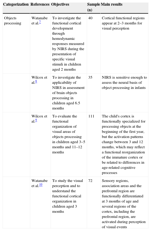
Characteristics of functional studies of cortical hemodynamics using NIRS to assess processing of objects.
| Categorization | References | Objectives | Sample (n) | Main results |
|---|---|---|---|---|
| Objects processing | Watanabe et al.7 | To investigate the functional cortical development through hemodynamic responses measured by NIRS during the presentation of specific visual stimuli in children aged 2 months | 40 | Cortical functional regions appear at 2–3 months for visual perception |
| Wilcox et al.8 | To investigate the applicability of NIRS in assessment of brain objects processing in children aged 6.5 months | 35 | NIRS is sensitive enough to assess the neural basis of object processing in infants | |
| Wilcox et al.9 | To evaluate the functional organization of visual areas of objects processing in children aged 3–5 months and 11–12 months | 111 | The child's cortex is functionally specialized for processing objects at the beginning of the first year, but the activation patterns change between 3 and 12 months, which may reflect a functional reorganization of the immature cortex or be related to differences in age-related cognitive processes | |
| Watanabe et al.10 | To study the visual perception and to understand the functional cortical organization in children aged 3 months | 72 | Sensory regions, association areas and the prefrontal region are functionally differentiated at 3 months of age and several regions of the cortex, including the prefrontal region, are activated during perception of visual events |
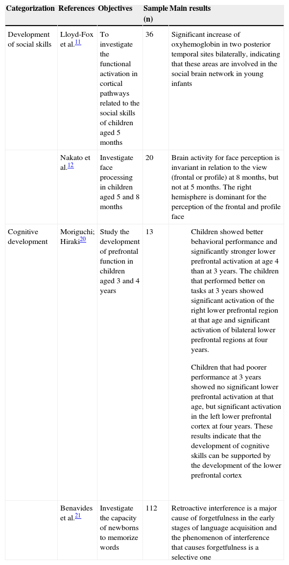
Characteristics of functional studies of cortical hemodynamics using NIRS to assess the development of social skills and cognitive development.
| Categorization | References | Objectives | Sample (n) | Main results |
|---|---|---|---|---|
| Development of social skills | Lloyd-Fox et al.11 | To investigate the functional activation in cortical pathways related to the social skills of children aged 5 months | 36 | Significant increase of oxyhemoglobin in two posterior temporal sites bilaterally, indicating that these areas are involved in the social brain network in young infants |
| Nakato et al.12 | Investigate face processing in children aged 5 and 8 months | 20 | Brain activity for face perception is invariant in relation to the view (frontal or profile) at 8 months, but not at 5 months. The right hemisphere is dominant for the perception of the frontal and profile face | |
| Cognitive development | Moriguchi; Hiraki20 | Study the development of prefrontal function in children aged 3 and 4 years | 13 |
|
| Benavides et al.21 | Investigate the capacity of newborns to memorize words | 112 | Retroactive interference is a major cause of forgetfulness in the early stages of language acquisition and the phenomenon of interference that causes forgetfulness is a selective one |
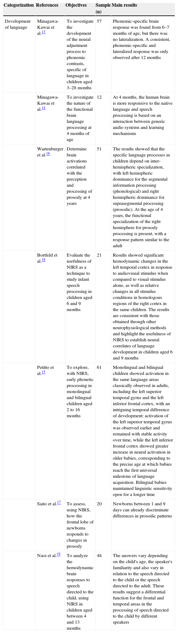
Characteristics of functional studies of cortical hemodynamics using NIRS to assess language development.
| Categorization | References | Objectives | Sample (n) | Main results |
|---|---|---|---|---|
| Development of language | Minagawa-Kawai et al.13 | To investigate the development of the neural adjustment process to phonemic contrasts, specific of language in children aged 3–28 months | 57 | Phonemic-specific brain response was found from 6–7 months of age, but there was no lateralization. A consistent, phonemic-specific and lateralized response was only observed after 12 months |
| Minagawa-Kawai et al.14 | To investigate the nature of the functional brain language processing at 4 months of age | 12 | At 4 months, the human brain is more responsive to the native language and speech processing is based on an interaction between generic audio systems and learning mechanisms | |
| Wartenburger et al.16 | Determine brain activations correlated with the perception and processing of prosody at 4 years | 51 | The results showed that the specific language processes in children depend on inter-hemispheric specialization, with left hemispheric dominance for the segmental information processing (phonological) and right hemispheric dominance for suprasegmental processing (prosodic). At the age of 4 years, the functional specialization of the right hemisphere for prosody processing is present, with a response pattern similar to the adult | |
| Bortfeld et al.18 | Evaluate the usefulness of NIRS as a technique to study infant speech processing in children aged 6 and 9 months | 21 | Results showed significant hemodynamic changes in the left temporal cortex in response to audiovisual stimulus when compared to visual stimulus alone, as well as relative changes in all stimulus conditions in homologous regions of the right cortex in the same children. The results are consistent with those obtained through other neurophysiological methods and highlight the usefulness of NIRS to establish neural correlates of language development in children aged 6 and 9 months | |
| Petitto et al.15 | To explore, with NIRS, early phonetic processing in monolingual and bilingual children aged 2 to 16 months | 61 | Monolingual and bilingual children showed activation in the same language areas classically observed in adults, including the left superior temporal gyrus and the left inferior frontal cortex, with an intriguing temporal difference of development: activation of the left superior temporal gyrus was observed earlier and remained with stable activity over time, while the left inferior frontal cortex showed greater increase in neural activation in older babies, corresponding to the precise age at which babies reach the first universal milestone of language acquisition. Bilingual babies maintained linguistic sensitivity open for a longer time | |
| Saito et al.17 | To assess, using NIRS, how the frontal lobe of newborns responds to changes in prosody | 20 | Newborns between 1 and 9 days can already discriminate differences in prosodic patterns | |
| Naoi et al.19 | To analyze the hemodynamic brain responses to speech directed to the child, using NIRS in children aged between 4 and 13 months | 48 | The answers vary depending on the child's age, the speaker's familiarity and also vary in relation to the speech directed to the child or the speech directed to the adult. These results suggest a differential function for the frontal and temporal areas in the processing of speech directed to the child by different speakers |
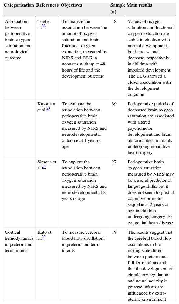
Characteristics of non-functional studies of cortical hemodynamics using NIRS.
| Categorization | References | Objectives | Sample (n) | Main results |
|---|---|---|---|---|
| Association between perioperative brain oxygen saturation and neurological outcome | Toet et al.22 | To analyze the association between the amount of oxygen saturation and brain fractional oxygen extraction, measured by NIRS and EEG in neonates with up to 48 hours of life and the development outcome | 18 | Values of oxygen saturation and fractional oxygen extraction are stable in children with normal development, but increase and decrease, respectively, in children with impaired development. The EEG showed a closer association with the development outcome |
| Kussman et al.23 | To evaluate the association between perioperative brain oxygen saturation measured by NIRS and neurodevelopmental outcome at 1 year of age | 89 | Perioperative periods of decreased brain oxygen saturation are associated with altered psychomotor development and brain abnormalities in infants undergoing reparative heart surgery | |
| Simons et al.24 | To explore the association between perioperative brain oxygen saturation measured by NIRS and neurodevelopment at 2 years of age | 27 | Perioperative brain oxygen saturation measured by NIRS may be a useful predictor of language skills, but it does not seem to predict cognitive or motor sequelae at 2 years of age in children undergoing surgery for congenital heart disease | |
| Cortical hemodynamics in preterm and term infants | Kato et al.25 | To measure cerebral blood flow oscillations in preterm and term infants | 19 | The results suggest that the cerebral blood flow oscillations in the resting state differ between preterm and full-term infants and that the development of circulatory regulation and neural activity in preterm infants are influenced by extra-uterine environment |
Regarding the methodology, all studies used multi-channel, continuous-wave NIR equipment with two wavelengths and most followed the standardization of the international 10-20 system of electroencephalography to locate optodes, with reported sample loss rate between 3% and 80%. The justifications for the losses, as specified by the authors, were: motion artifacts, obstruction by the hair, failure in the experimental protocol, crying and agitation, difficulty obtaining optical signal and intolerance to the equipment.
The studies on object processing were developed with children aged two to 12 months old and aimed to investigate cortical functional organization related to the visual perception of objects (color, shape and movement). The main results showed that, between two and three months old, it is already possible to identify functionally differentiated cortical regions for visual perception.7–10
About the development of social skills, the studies assessed functional activation in cortical pathways related to social skills of children between five and eight months of age, using images of the human face in different plans and different facial expressions. The results indicated that, at five months of age, there is already a specialized area of the temporal cortex activated by social stimuli.11,12 Furthermore, right hemisphere dominance was verified in the frontal plane perception and facial profile,12 whereas cortical activation occurs bilaterally for the perception of dynamic social stimuli.11
Studies on language development were carried out with children aged two months to four years, with the aim to analyze brain functional processing of language, mainly related to phonetic processing,13–15 prosody,16,17 the lateralization of speech13,14,16,18 and the influence of the speaker's familiarity in speech perception.19 The results were consistent with those obtained through other neurophysiological methods, emphasizing that: responses to specific language phonemic contrasts are present at six months of age; however, these become consistent and lateralized only after 12 months.13 Differences in prosodic patterns were discriminated by infants between one and nine days of life17 and functional specialization of the right hemisphere for prosody processing is present, with a response pattern similar to that of the adult, at four years of age.16
The approach of cognitive development involved the study of cognitive flexibility in children aged three and four years, and memory in newborns. The main findings indicated that the development of cognitive flexibility skill is related to the development of the inferior prefrontal cortex and suggest that children develop prefrontal activations between the ages of three and four years.20 About neonatal memory, a study investigated the capacity of newborns to memorize words, focusing on the causes of forgetfulness in early childhood, testing the capacity of neonates to recognize words after a period of familiarization. It was observed that newborns are already capable of memorizing words hours after birth.21
The non-functional studies discussed the association between cerebral oxygen saturation and neurological outcome;22–24 and the comparison between the cortical hemodynamic response of preterm and full-term infants.25 All used continuous wave equipment, ranging from two to 24 channels and only one used the international 10-20 system for positioning of the optodes with sample loss ranging from 14% to 56%. The reported causes for these losses were: neonatal death, diagnosed syndrome, follow-up losses and failure in the experimental protocol.
Children younger than 12 months were evaluated and the main results suggest that brain oxygen saturation values and oxygen extraction fraction may be related to neurological outcomes22–24 and that the oscillations in cerebral blood flow in the resting state differ between preterm and full-term infants.25
DiscussionBefore the advent of neuroimaging techniques, the association between brain regions and neurodevelopment was obtained primarily by clinical neuropsychological investigations of patients with brain injury and post-mortem examinations. With the advancement of this technology, it became possible to investigate not only the brain areas involved in a particular skill, but also the neural circuits involved in a particular function.26
Noninvasive methods have been explored to make inferences about neural correlates of processes linked to human development. Among these methods, some record magnetic (magnetoencephalography, MEG) or electrical fluctuations (electroencephalogram, EEG and amplitude-integrated electroencephalography, aEEG) that occur in neural activity; while others, such as functional magnetic resonance imaging (fMRI) and functional near-infrared spectroscopy (fNIR spectroscopy or fNIRS), measure local changes in cerebral hemodynamic activity, which can be used to make inferences about the underlying neural activity.1
Many of these techniques, which are well-established for use in adults, have restrictions for use in children. Among all brain imaging techniques, the fMRI is considered the “gold standard” for noninvasive functional mapping of the human brain.26 This technique stands out from the others due to its high spatial resolution and by displaying well-established routines of data acquisition and analysis, facilitating its use in research, among other reasons, because it is easier to make comparisons between results from different studies. However, as the MEG, the fMRI requires the participant to remain motionless, usually wrapped or restrained. There are a few published studies in children using these techniques; however, these works are generally limited to the study of children while sleeping, sedated or very young ones.
For many years, the first choice for neuroimaging studies in children while awake was EEG, a technique with high temporal resolution, but relatively low spatial resolution.6 In this field, continuous brain monitoring through aEEG has been used in neonates to assess real-time brain function and for long periods, allowing a better classification of the encephalopathy severity, early detection of subclinical seizures and the monitoring of treatment response.27 Abnormalities found in the aEEG early in life have strong predictive values of abnormal results at one year of age.28
Compared with the aforementioned techniques, NIRS offers a new direction for the study of child development, as it has the following advantages over these methods: better temporal resolution, better safety level, it is silent and less sensitive to motion artifacts, requiring less rigid stabilization of the head and body without the need for a labeled or carrier substance to be injected into the bloodstream.2
The most commonly used and simpler NIRS method involves measuring the intensity of diffusely reflected light with sources that emit light continuously. Instruments that acquire such measurements are referred to as continuous-wave systems.29 All studies discussed here resorted to this method, which, although it does not provide quantitative measurements of absolute concentrations of different types of hemoglobin, provides estimates of changes in their levels from a baseline value, thus reflecting variations in tissue oxygen use.5
The use of multiple channels with different combinations of sources and detectors has been described in the literature in recent years. Until the early 1990s, almost all NIRS systems employed one or two measurement channels, but over time, the number of channels in the available systems has increased, improving spatial resolution.29 Of all the reviewed studies, most of them used multichannel acquisition systems, allowing greater coverage of the region of interest.
The advantages of increasing the number of channels are clear. However, this results in the inevitable increase in weight and size of the device that maintains the optodes positioning in the scalp. This may explain the greater proportion of optical data loss due to excessive movement artifacts. The losses reported in the literature vary from 12.5 to 70%,6 similar to the losses found in this review: 3-80% in functional studies and 14-56% in non-functional studies. A possible explanation for the greater loss in functional studies would be the use of more complex data collection protocols, employing a greater numbers of channels.
Although NIRS has been used for more than 35 years, it was not applied to children up until the mid-1990s. Investigations in this field have expanded rapidly, providing evidence that NIRS can be used to collect information about hemodynamics correlated to the neural activity in children from an early age, using tasks that assess cognitive skills, language acquisition, visual perception, social cognition and other functional aspects of the brain during childhood.30 Moreover, non-functional NIRS studies have demonstrated its potential as a prognostic tool, based on the non-invasive monitoring of cerebral hemodynamics and oxygenation.22,31–33
Based on the publications analyzed, it was possible to recognize advances in the use of NIR spectroscopy in the study of child development; however, some methodological obstacles inherent in the use of this technology must be considered. Consistent with the results of other studies reviewing NIRS, it was observed that there is great variability in the methods used for data acquisition and analysis. The use of different wavelength combinations and different separations between sources and detectors could affect the captured response. In the 19 studies included in the review, we identified seven different wavelength combinations used. The separation between optrodes was more uniform, being two or three centimeters, which is adequate for the pediatric population.
Another question that should be addressed concerns the large differences in sample sizes of the studies (between 12 and 112 subjects) and significant losses due to the quality of the generated signal. While there is a good signal-to-noise ratio for optical imaging, the variation that is inherent in infant behavior requires that there be at least ten babies in each age group.34 The reviewed studies analyzed samples with 12 or more children. However, one study that proposed to evaluate the association between the amount of oxygen saturation, the fractional cerebral tissue oxygen extraction and the result of development,22 with an initial sample of 18 children, lost nine of them due to death, ending up with a final sample of nine children and found developmental change in only one. Despite the importance of studies like this to extend the current body of knowledge that emphasize the validity of NIRS to study infant brain development, the interpretation of results obtained with small sample groups should be very cautious and their projection to other contexts or populations becomes impaired.
Moreover, the difficulty of determining the locations for positioning of optodes using external markers, especially in children, should be considered. A current trend in NIRS studies, and which was identified in 13 of the 19 articles included in this review, is the use of the international 10-20 system for electroencephalography for the positioning of optodes.
It is also necessary to define the number of experiments required to obtain a significant response. Finding the balance between the number of repetitions required to capture a reliable response without making the test long and stressful, seems to be a delicate aspect of research using NIRS in children. This is due to the fact that inadequate signals and motion artifacts often make it necessary to repeat the tests. The studies reviewed here showed not only a large variability in the number of experiments, as well as in the duration of tests, reinforcing the premise that there is yet no consensus in the literature about this aspect. Some authors emphasize that the use of long experiments could lead to a diminished response over time, as the body adapts to the stimulus that is repeated many times. Additionally, the study designs in blocks, with long periods of stimulation and rest, has potential risk for false positive/negative changes in the signal due to any low frequency fluctuation at baseline or motion artifacts.35 On the other hand, some authors recommend that stimuli should be repeated at least 10 times, considering that spontaneous changes in cerebral blood volume are common, for instance, the so-called Mayer waves or slow vasomotion at 0.1 Hz. Such changes are about the same size as the functional activations and, therefore, may be misidentified. Repetition allows making averages of time series, reducing the influence of spontaneous changes, as they are not synchronized with the stimuli.36
It is worth noting that this review aimed to emphasize NIRS as an auxiliary tool in the study of normal childhood development. For this reason, no studies that addressed developmental disorders were included, due to high specificity of each of them. However, it is necessary to stress that the current literature on NIRS has also emphasized the adequacy of this technology for the study of child development disorders and there is a growing number of publications in this area. In this context, studies on Autism Spectrum Disorder,37,38 Attention Deficit Hyperactivity Disorder39 Cerebral Palsy40–42 and Down Syndrome43 have been highlighted.
In conclusion, NIRS is increasingly becoming a practical alternative and potentially useful technique for studies of functional activity of the infant brain. The development of equipment more adequate for use in children has increased, so that the results obtained when using NIRS technology are more reliable. It is worth mentioning that the spatial location of signals will never achieve the accuracy of fMRI, but in conjunction with other techniques, such as EEG, NIRS is emerging as an important non-invasive tool for the study of the developing brain.
FundingThis article is part of the research “Near Infrared Spectroscopy in the Prediction of Neurodevelopment of Preterm Infants at 4 and 8 months Corrected Age,” which received financial support from Fundação de Amparo à Pesquisa de Minas Gerais – FAPEMIG. Process number: APQ-01182-13.
Conflicts of interestThe authors declare no conflicts of interest.
