


Our study shows the case of a psychopath prisoner who has been in jail for three years accused of rape. The subject was evaluated through the Psychopathy Checklist-Revised (PCL-R) and Quantitative Electroencephalogram (QEEG). QEEG measures used in this study were absolute power and occipital Alpha medium frequency. Values were transformed into Z score and compared with the QEEG normative database. Results showed a Theta excess and Alpha decrease; moreover occipital Alpha medium frequency was below the norm for the subject's age. Findings suggest a cortical hypoactivation; some theories suggest that some psychopath's features can be explained by this low activation.
Nuestro estudio muestra el caso de un psicópata encarcelado desde hace 3 años por violación. Se evaluó al sujeto mediante la Escala de Psicopatía Revisada (PCL-R) y electroencefalograma cuantitativo (QEEG). Las medidas QEEG utilizadas fueron la potencia absoluta y la frecuencia media del Alpha occipital. Los valores fueron transformados a puntuación Z y comparados con las normas QEEG. Los resultados mostraron un exceso generalizado de Theta y una reducción de Alpha, también se encontró una frecuencia del Alpha occipital por debajo de lo esperado para la edad del sujeto. Los hallazgos sugieren una hipoactivación cortical, consistente con algunas teorías que explican algunos rasgos psicopáticos por esta baja activación.
Psychopathy has been the first personality disorder recognized by psychiatry.1 It is defined as a group of interpersonal, affective and behavioral traits such as: antisocial behavior, lack of empathy and regrets; also superficial charming, criminal versatility, flatten affection, irresponsibility, glibness, parasitic lifestyle, and a poor control of the impulses, among others. One of the most important aspects of psychopathy is the search of constant stimulation, expressed through impulsive conducts, leaving jobs and easy boredom.
The Psychopathy Checklist-Revised2 (PCL-R) is currently the main standard to evaluate psychopathy. It consists of a twenty-item semi-structured survey. Scoring goes from 0 to 40 where 30 is the suggested score to determine psychopathy. This scale has been validated in Mexican population.3 Te PCL-R items are grouped into two factors: interpersonal/affective (factor 1) and social deviation (factor 2). There are also two items that do not belong to any factor.
Many researchers have tried to find the possible causes and implications of psychopathy. A large number of theories lead to a different brain operation in psychopaths.4–6 Main findings show differences in the frontal lobes in charge of some functions as planning, social judgment, conduct inhibition and decision making.5,7
One way to study the brain is through the electroencephalogram (EEG), in which cortical electrical activity is recorded in electrodes placed on the scalp. Previous studies undertaken via visual inspection of the electroencephalogram have shown some anomalies in psychopaths, mainly, increased slow rhythms.8,9 However the EEG visual analysis is very subjective due to inter- and intrapersonal fluctuations in the interpretation of an EEG traced by specialists.10 So, in order to get a more objective result, we can use the Quantitative Electroencephalogram (QEEG), that digitalises the electrical brain signals to quantify and analyze them by computing.
There are a few studies about psychopathy and QEEG; for example, a researcher11 carried out a study in 31 psychopath prisoners compared with 27 non-psychopath prisoners. For the analysis the researcher took into consideration the Cuban QEEG norms,12 which defines the expected individual EEG standards according to age and gender. He found that psychopaths get farther from the average (an excess of activity) that non-psychopaths in Beta activity located in left parietal–temporal area and bioccipital zones. The study also showed a decrease in the Alpha activity in left center-temporal and center-parietal. The author attributed that to possible failures in the cortical development.
Medium frequency (MF) of the occipital electrical brain activity between 7.5 and 13Hz (Alpha) has been considered as a clinical indicator of brain maturity13 and autonomous activation state.14,15 It is also possible to get this measure via QEEG.
Our study shows the case of a psychopath prisoner evaluated through QEEG.
Clinical caseA 29-year-old male prisoner, and with about 16 year-schooling, was the clinical case. He has been in jail for three years accused of rape; he did not have any criminal record before this crime. In this study we will refer to him as “E2”. E2 was sexually abused during his childhood and in the same period he suffered a closed brain injury on the right forehead that caused him a consciousness loss. He was not found as drug consuming.
E2 was assessed using PCL-R and his criminal record was checked to validate PCL-R results. E2 got a 30 score; therefore he could be classified as a psychopath. Factor 1 score was 14 and factor 2 was 12. PCL-R non-specific factor questions were also recorded.
Electrical brain activity from E2 in resting state, with eyes closed was recorded. 32-NuAmps-channel amplifying and Neuroscan acquisition software were used. Standard 10–20 position system of electrodes collocations was used for the 19 system positions (Fp1, Fp2, F3, F4, C3, C4, P3, P4, O1, O2, F7, F8, T3, T4, T5, T6, FZ, CZ, PZ); linked ears were used as reference. About 3minute EEG closed eyes activity was recorded; signal was taken from 0.5 at 30Hz.
An EEG visual inspection was carried out that aimed to reject any kind of artifacts. In addition an automatic artifact rejection was made using software. Signal was filtered from 1.5 at 30Hz so as to ensure that artifact or noise activity (e.g. eye movement) was not included. This process was according to the Cuban QEEG norms12 which just studied a 1.56 at 19.14Hz range of activity. The EEG was divided in ninety-one 1020-ms epochs.
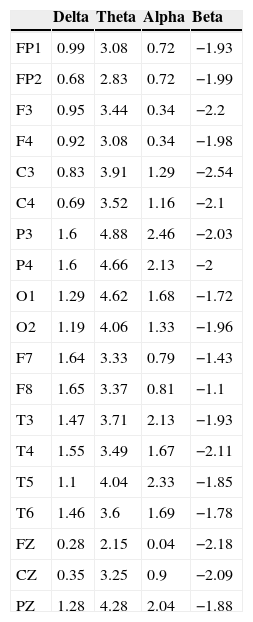
The software analysis used in this study was Neuroscan Edit (to filter and clean the signal) and Neuronic QEEGT (to transform the signal and compare it with the norms). Absolute power was computed through Fourier Fast Transform (FFT). Furthermore the occipital Alpha medium frequency was calculated. For each analysis EEG signal was divided into the four clinical bands recommended by QEEG norms12: Delta (1.56–3.52), Theta (3.91–7.42), Alpha (7.81–12.50) and Beta (12.89–19.14). The QEEG map shows E2's brain electrical activity (Fig. 1).

Obtained data were transformed into Z scoring in order to compare them with QEEG regulations. For a significant P<0.05 level extreme Z scores are required, it means >1.96 or <−1.96.
An excess of activity in Theta band was found, same as that was statistically significant in all electrodes. Major deviation focused on left side, mainly in parietal zones.
A significant Alpha increase was also found in biparietal zones, in left temporal zone and in center-parietal zone.
A significant Beta activity decrease was found in a bilateral way in frontal and parietooccipital zones, as much as in half frontal line and in the right temporal front zone.
No significant difference in Delta band was found.
The previous mentioned results can be consulted in Table 1.
Z scoring got by E2 according to QEEG normative basis.
| Delta | Theta | Alpha | Beta | |
|---|---|---|---|---|
| FP1 | 0.99 | 3.08 | 0.72 | −1.93 |
| FP2 | 0.68 | 2.83 | 0.72 | −1.99 |
| F3 | 0.95 | 3.44 | 0.34 | −2.2 |
| F4 | 0.92 | 3.08 | 0.34 | −1.98 |
| C3 | 0.83 | 3.91 | 1.29 | −2.54 |
| C4 | 0.69 | 3.52 | 1.16 | −2.1 |
| P3 | 1.6 | 4.88 | 2.46 | −2.03 |
| P4 | 1.6 | 4.66 | 2.13 | −2 |
| O1 | 1.29 | 4.62 | 1.68 | −1.72 |
| O2 | 1.19 | 4.06 | 1.33 | −1.96 |
| F7 | 1.64 | 3.33 | 0.79 | −1.43 |
| F8 | 1.65 | 3.37 | 0.81 | −1.1 |
| T3 | 1.47 | 3.71 | 2.13 | −1.93 |
| T4 | 1.55 | 3.49 | 1.67 | −2.11 |
| T5 | 1.1 | 4.04 | 2.33 | −1.85 |
| T6 | 1.46 | 3.6 | 1.69 | −1.78 |
| FZ | 0.28 | 2.15 | 0.04 | −2.18 |
| CZ | 0.35 | 3.25 | 0.9 | −2.09 |
| PZ | 1.28 | 4.28 | 2.04 | −1.88 |
Extreme values >1.96 (excess) and <−1.96 (decrease) are considered statistically significant (P<0.05).
The obtained occipital Alpha medium frequency was O1: 8.55Hz and O2: 8.57Hz, which is inferior to the expected one according to the subject's age and got a −3.58 and −3.43 Z score respectively, that is remarkably low.
DiscussionIn human beings slow waves (Delta, Theta) are expected to be displaced by faster ones (Beta, Alpha) when growing.13 The average adult is supposed to have more Beta and Alpha activity than Theta and Delta. For that reason an excess in slow waves is related to brain immaturity16 and a cortical hypoactivation, which could explain the constant need of stimulation in psychopaths, so as to compensate and improve activation levels.17
On the other hand, the occipital Alpha medium frequency has been related to autonomic activation processes, where a lower frequency would indicate a lower activation. This is part of the Vagotonia Theory,14 that suggests that violent criminals have an excessive vagal tone, meaning parasympathetic processes predominate over sympathetic, which leads to a cortical and cardiac hypoactivation, same as that is related to higher testosterone levels and a more aggressive behavior.4
Our results are congruent with that hypoactivation, not just because of the diffuse Theta excess, but also because of the reduced Alpha medium frequency.
Even though the reason of a parietal and temporal Alpha excess found in this research is not clear yet, in general terms Alpha activity is supposed to reflect a relaxation and repose state, although it has been said that it reflects inactive or idle brain zones10 (considering that it appears in stimulation absence, specially visual).
So that we can speculate that temporal Alpha excess is related to a lower cortical activation in the temporal area, as temporal lobes have been associated to emotional processing and also because some variations in these areas (such as epilepsy) can produce aggressive behavior,18 the results are meaningful.
In the left hemisphere more deviations than the average were found, which is congruent with some studies that suggest that there is a left lateralized brain disfunction in crime and violent behavior disorders.18,19
About E2's history, it is well known that sexually abused children are more prone to becoming sexual offender adults than non-abused children.20 There are some data pointing a Delta and Theta excess and Alpha decrease in sexual offenders.21 On the other hand a bigger PCL-R factor 1 score over factor 2 has been related with more violent behavior among sexual offenders.22 Concerning E2's childhood closed brain injury, it is said that frontal lobe damage could lead to an aggressive behavior.23 However, that evidence is not enough to explain this case.
Psychopathy is a more complex phenomena than a negative past, so we have compiled all the relevant facts, but further research is needed to establish the relation between psychopathy and brain, specially for QEEG measurements. QEEG is a useful tool that can provide information about the violent brain, and this case illustrates an example of use.
FundingThis work was partially supported by UNAM project PAPIIT IN305313: “Conducta violenta y sus bases biológicas: neuroimagen, neuropsicología y electrofisiología”.
Conflict of interestThe authors declare that they have no conflict of interests.
The authors thank PAPIIT for support.