


Renal cell carcinoma (RCC) accounts for 2–3% of adult tumors, with a peak incidence between 60 and 70 years of age. More than 70% are diagnosed incidentally. The most prevalent and aggressive histological subtype is clear cell RCC (ccRCC), representing 80% of cases, followed by papillary (10%) and chromophobe (5%), while tumors of the collecting ducts and miscellaneous forms are extremely rare. The typical clinical presentation (flank pain, hematuria, and palpable mass) occurs in less than 1% of cases, whereas 65% of RCCs are detected incidentally and only 35% produce symptoms.1,2
Renal masses can be solid or cystic, simple or complex, and their morphology and temporal evolution contribute to differential diagnosis. A major challenge is differentiating primary RCC from benign renal masses, as up to 15–30% of partial nephrectomies ultimately correspond to benign lesions. MRI has incorporated the Clear Cell Likelihood Score (ccLS), an algorithm that improves diagnostic performance compared to CT in cystic lesions and angiomyolipomas. Complementarily, Bosniak criteria allow standardized classification of cystic lesions using ultrasound, CT, and MRI. Conversely, PET with 18F-FDG has significant limitations due to urinary excretion and variable uptake in ccRCC.2,3
Carbonic anhydrase IX (CAIX) is a highly specific biomarker for ccRCC, with overexpression in more than 94% of cases and no uptake in normal renal tissue or cystic lesions. CAIX is a transmembrane protein induced by hypoxia and regulated by the von Hippel-Lindau (VHL) pathway; its overexpression is associated with acidified tumor microenvironments and mechanisms of dedifferentiation and cell migration.1 This specificity has enabled the development of targeted radiopharmaceuticals, with girentuximab-a chimeric IgG1 kappa monoclonal antibody against CAIX-being the most advanced.2,3 Labeling with 89Zr, which has a long half-life, allows delayed acquisition (4–7 days) with high sensitivity. The phase III ZIRCON trial demonstrated a sensitivity of 86% and specificity of 87%, with a favorable safety profile.3
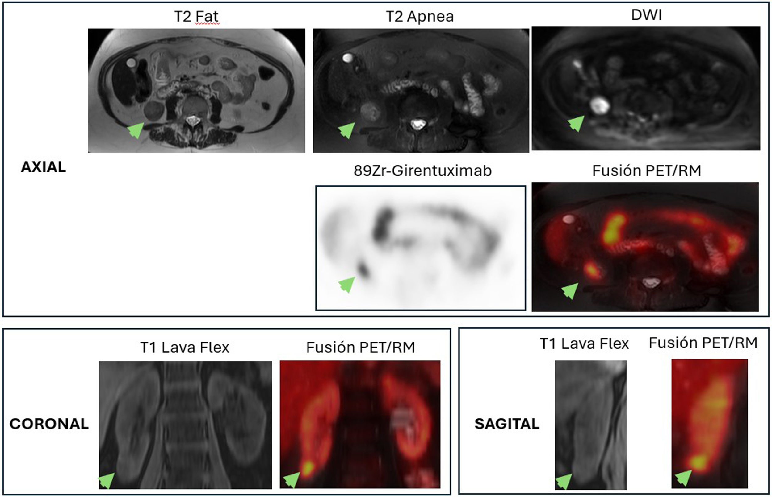
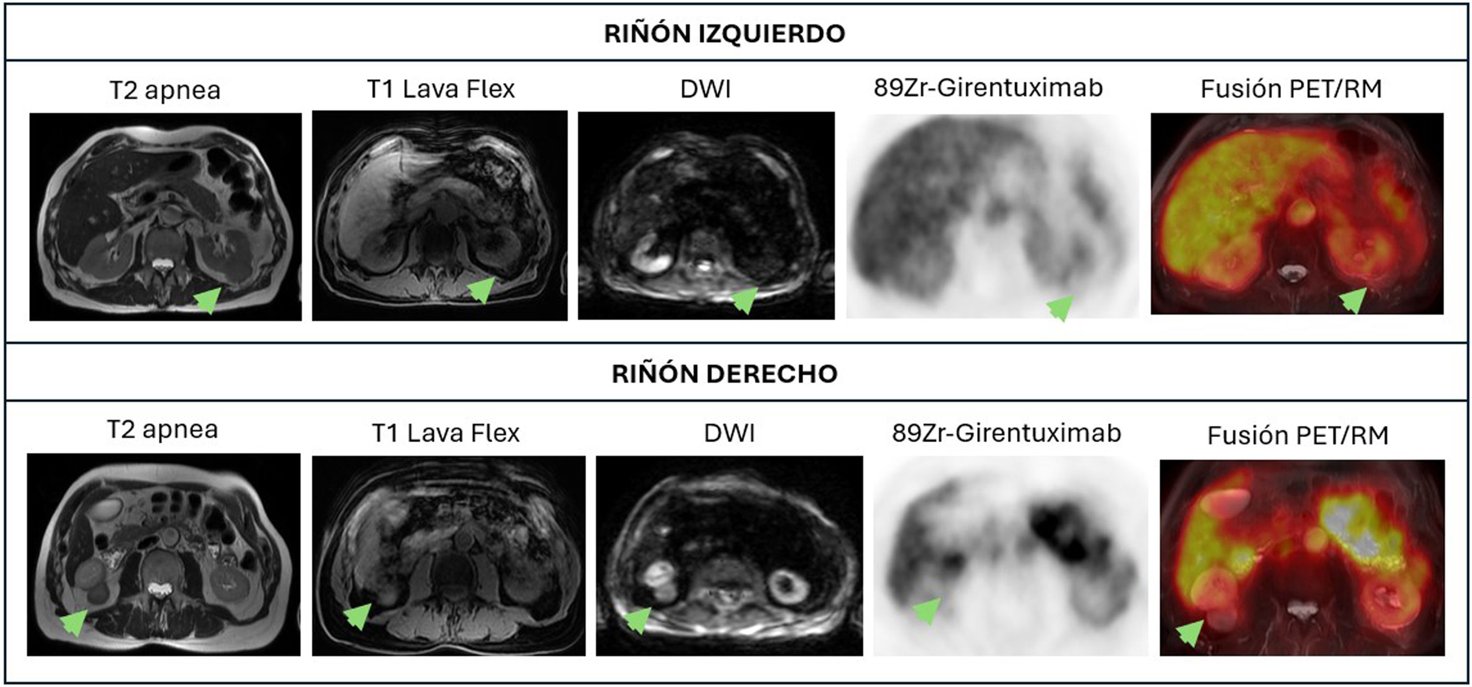
We present two clinical cases evaluated with PET/MRI using 89Zr-girentuximab within an operational protocol specifically developed for introducing the radiopharmaceutical at our center, after obtaining AEMPS authorization for its use. In the first case, a 72-year-old female patient presented with a cortical lesion in the right kidney showing significant tracer uptake, highly suggestive of ccRCC (Fig. 1). In contrast, the second case involved a 70-year-old male patient with a history of oncocytoma, showing two solid lesions without radiopharmaceutical uptake and morphology compatible with oncocytoma on MRI (Fig. 2).

72-year-old female; Clear Cell RCC.
Axial slices: T2 Fat, T2 Breath-hold, DWI, 89Zr-Girentuximab, PET/MRI Fusion
Coronal slices: T1 Lava Flex, PET/MRI Fusion
Sagittal slices: T1 Lava Flex, PET/MRI Fusion
A 72-year-old female patient with a solid cortical mass in the lower pole of the right kidney, under follow-up since 2024. PET/MRI performed 5 days after administration of 1.12 mCi of ⁸⁹Zr-girentuximab shows a partially exophytic 28 mm lesion, heterogeneous and with marked diffusion restriction (ADC < 1), presenting significant tracer uptake (SUVmax 10.5), highly suggestive of clear cell RCC.

70-year-old male; Bilateral Oncocytomas.
Left kidney, axial slices: T2 Breath-hold, T1 Lava Flex, DWI, 89Zr-Girentuximab, PET/MRI Fusion
Right kidney, axial slices: T2 Breath-hold, T1 Lava Flex, DWI, 89Zr-Girentuximab, PET/MRI Fusion
A 70-year-old male patient with a history of right partial nephrectomy for oncocytoma in 2022. PET/MRI performed 5 days after administration of 1.078 mCi of ⁸⁹Zr-girentuximab shows two solid renal lesions without tracer uptake: a posterior exophytic lesion in the lower pole of the right kidney (29 mm) and another cortical lesion in the mid-third of the left kidney with a central scar (33 mm), both without diffusion restriction and morphologically compatible with oncocytoma. Postsurgical changes show no radiopharmaceutical deposits.
Both studies demonstrated excellent correlation between CAIX expression and functional characterization using immunoPET. In the first case, partial nephrectomy confirmed the presence of ccRCC, while in the second case, follow-up was chosen.
These cases illustrate the potential of 89Zr-girentuximab to improve non-invasive characterization of indeterminate renal masses, reduce unnecessary biopsies, and optimize clinical management through targeted molecular evaluation.


