


A 66-year-old patient with a non-ulcerated skin lesion in the right twin region of one year of evolution. The result of the biopsy was an intradermal malignant melanoma with no connection to overlying epidermis, Clark level V, with more than 15 mm of growth in depth, without radial growth phase and with vertical growth phase of mixed type. It presented perivascular and lymphatic invasion with satellitosis and mitotic index 10 mitosis/mm2. Incomplete surgical excision regarding lateral and deep margins was performed. Lymph nodes were not removed. The result of the immunohistochemical techniques was: S-100 positive; Melan A: positive; HMB-45: positive; SOX-10: positive; P-53: negative; P-16: negative; Ki-67: moderate cell proliferation index and BRAF negative.
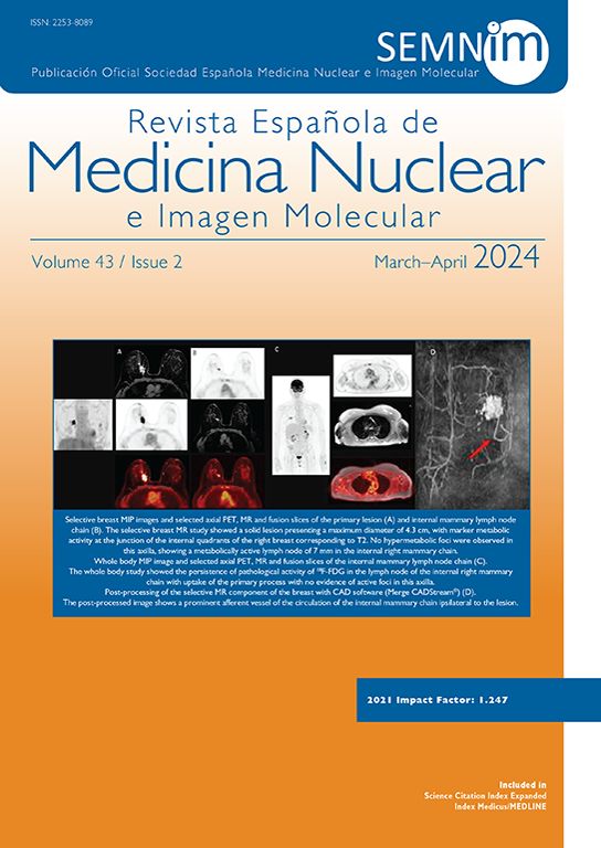
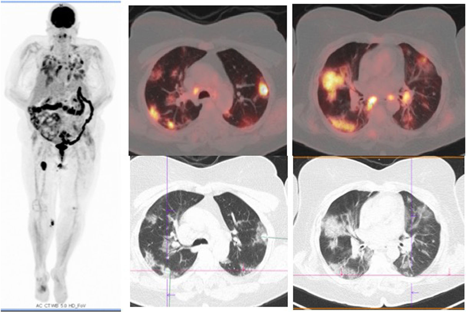
Having no relation to the overlying epidermis, positron emission tomography/computed tomography (PET/CT) was scheduled to identify the primary melanoma. PET/CT with a lung reconstruction algorithm, that is a fundamental tool for lung evaluation, identifies diffuse hypermetabolic parenchymal infiltrates, in addition to hypermetabolic nodular lesions in the right knee and ipsilateral inguinal region, in relation with lymphadenopathy secondary to its oncological process (Figs. 1 and 2).


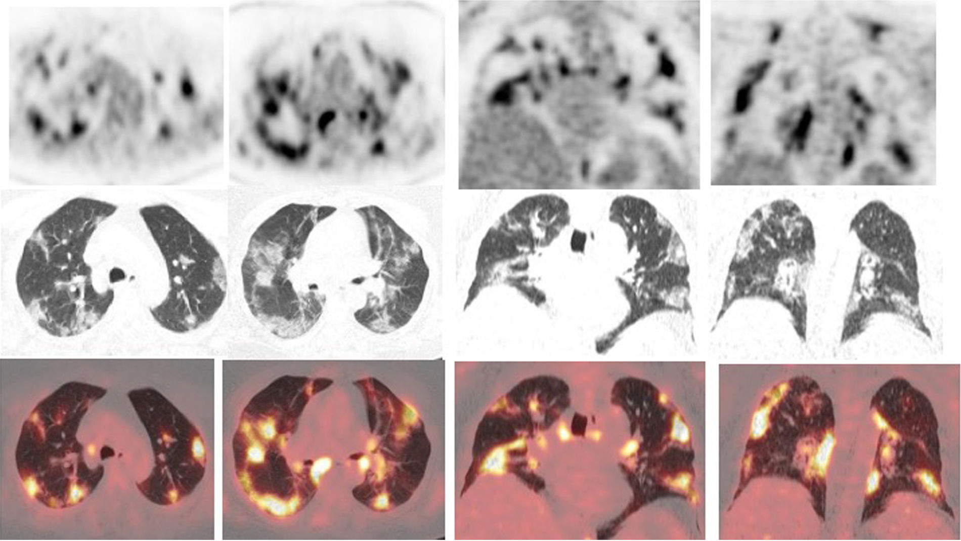
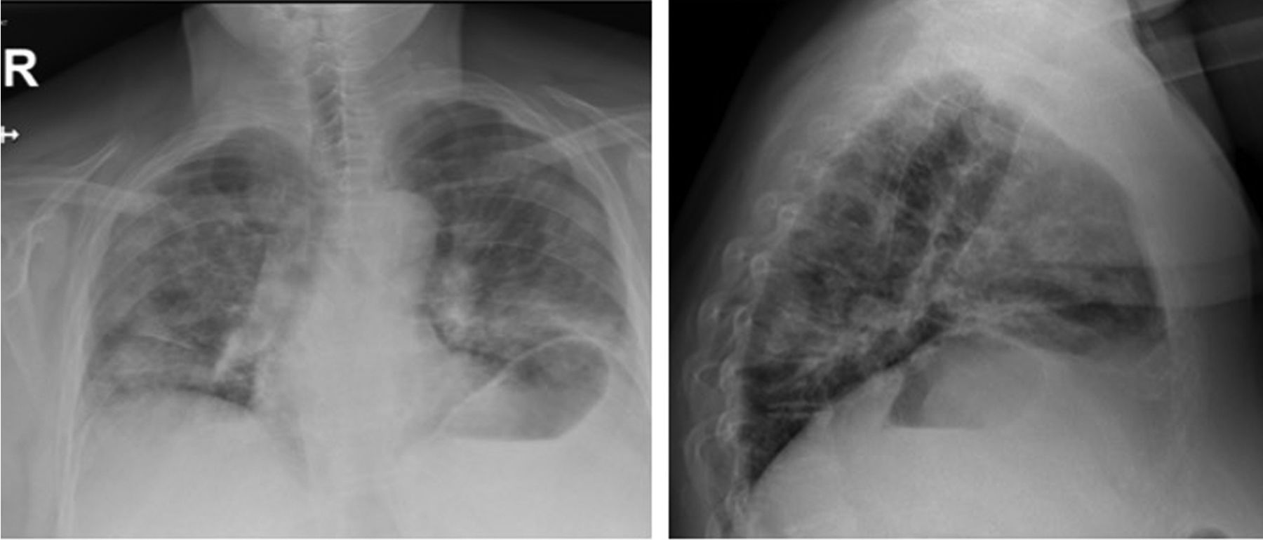
The patient was interviewed and reported fever, dyspnea, and polymyalgia of 8 days of evolution. Due to this symtoms and PET/CT image suggestive of viral pneumonie, the patient was referred to the emergency department where a chest x-ray (Fig. 3), for subsequent radiological follow-up, supported the diagnosis of suspected bilateral viral pneumonia by COVID-19. Infection is confirmed by COVIC-19 after collecting a nasal mucosa sample. The patient was deceased 4 days later.

Please cite this article as: Gómez-Caminero-López F, García-Talavera-San-Miguel P, Lucas Velázquez B, García Arroyo J, Díaz González LG, Gómez Grande A. Neumonía vírica bilateral por COVID-19 como hallazgo causal en 18F-FDG-PET-TC de estadificación de paciente con melanoma gemelar derecho. Rev Esp Med Nucl Imagen Mol. 2020;39:316–317.
