


We report the case of a 6-year-old patient who started with abdominal pain and left sciatica, which did not improve after applying symptomatic treatment. A complete analytical and imaging study was performed, which showed a lesion in left S1 corresponding to Garrè’s chronic diffuse sclerosing osteomyelitis. The diagnosis was confirmed by biopsy of the lesion. Treatment was established with corticosteroids and anti-inflammatory drugs, obtaining a clinical improvement, although in the follow-up imaging tests 2 years after the onset of the symptoms, the lesion persists but with a significant reduction in its size.
Presentamos el caso de un paciente de 6 años, que comenzó con dolor abdominal y ciatalgia izquierda sin mejoría después de aplicar un tratamiento sintomático. El estudio analítico completo y de imagen realizado mostró una lesión en S1 izquierda correspondiente a osteomielitis esclerosante crónica de Garrè, confirmando el diagnóstico mediante biopsia de la lesión. Se instauró tratamiento con corticoides y antiinflamatorio, que consiguen una remisión parcial de los síntomas, y en las pruebas de imagen realizadas 2 años después del inicio de los síntomas persiste la lesión, aunque con una disminución significativa de su tamaño.
We present the case of a 6-year-old patient who reported with occasional fever, no circadian rhythm, and severe and diffuse abdominal pain following an accidental fall. The symptoms persisted despite treatment with paracetamol and a soft diet for about 10 days. After this period, we decided to complete the study with an abdominal ultrasound and blood tests, which were normal. After approximately 15 days from the onset of symptoms the patient suffered severe, left, lumbosciatic pain and the abdominal symptoms and fever disappeared. We began treatment with non-steroidal anti-inflammatory drugs (NSAIDs) (oral ibuprofen 70mg/8h) and analgesic drugs (rectal metamizole 380mg/8h), while continuing with imaging studies. We obtained 2 plain radiographic projections of the lumbosacral region, which were normal. The study was completed with a magnetic resonance imaging (MRI) scan of the lumbosacral region which showed a bone lesion accompanied by bone oedema in the left half of the S1 vertebral body and the ipsilateral sacral wing. These could correspond to sequelae of the trauma, stress fractures or infectious processes, without ruling out other diseases.
Since the lumbosciatic symptoms persisted despite treatment with NSAIDs and analgesics, we decided to start treatment with corticosteroids (methylprednisolone 1mg/kg/day with gradual withdrawal), which temporarily managed to control the symptoms. Laboratory tests including blood count, biochemistry, coagulation, liver function, C-reactive protein, erythrocyte sedimentation rate, complement components C3 and C4, basal ACTH, bone alkaline phosphatase, serum markers for Epstein–Barr virus, cytomegalovirus, parvovirus, antistreptolysin O antibodies, antinuclear antibodies, anti-Ro and anti-neutrophil cytoplasmic antibodies, B27 human leukocyte antigen, urine amino acids, Mantoux and Rose Bengal staining were all normal or negative.
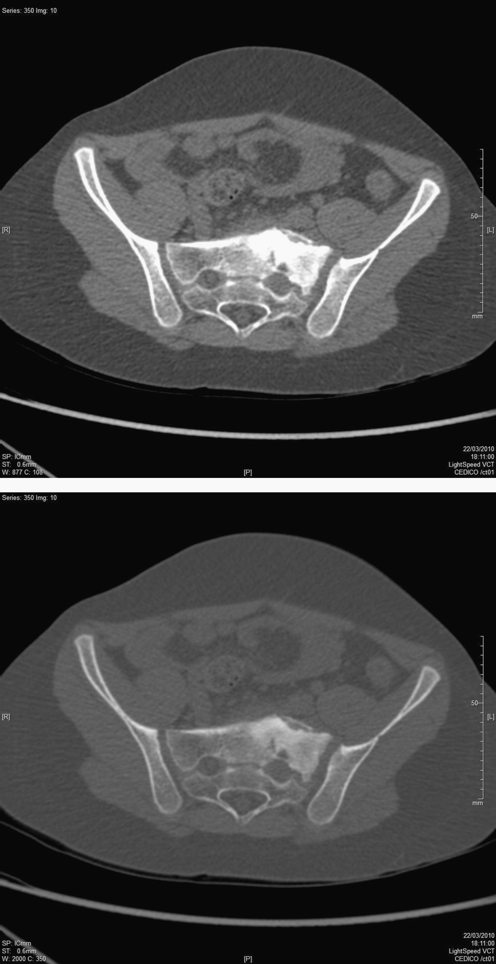
After 3 months of corticosteroid treatment we repeated the MRI scan, which still showed a small, focal area of residual bone oedema, which had become significantly reduced in size as compared to the previous study. This lesion still appeared in MRI controls performed 1 year later, so we obtained a computed tomography (CT) scan of the lumbosacral region, which showed a sclerotic lesion with periosteal reaction in the left wing of the sacrum. This suggested Garrè’s chronic diffuse sclerosing osteomyelitis (CDSO) (Fig. 1).

To confirm the suspected diagnosis we obtained a bone scintigraphy scan and biopsy of the lesion. The bone scintigraphy with technetium-99 bisphosphonate in 3 stages showed late uptake in the left sacroiliac region, thus suggesting a chronic inflammatory process. The biopsy revealed fibrofatty tissue and fragments of bone marrow with fibrous areas, but free of specific bone activity and without neoplastic growth. These histological features in conjunction with the radiographic characteristics led to the diagnosis of CDSO.
Approximately 2 years passed since the appearance of the first symptoms until the definitive diagnosis. During this period the patient suffered recurrent, left sciatic pain, with a variable recurrence ranging from 2 to 6 months and the symptoms lasting for 3–5 weeks in each episode.
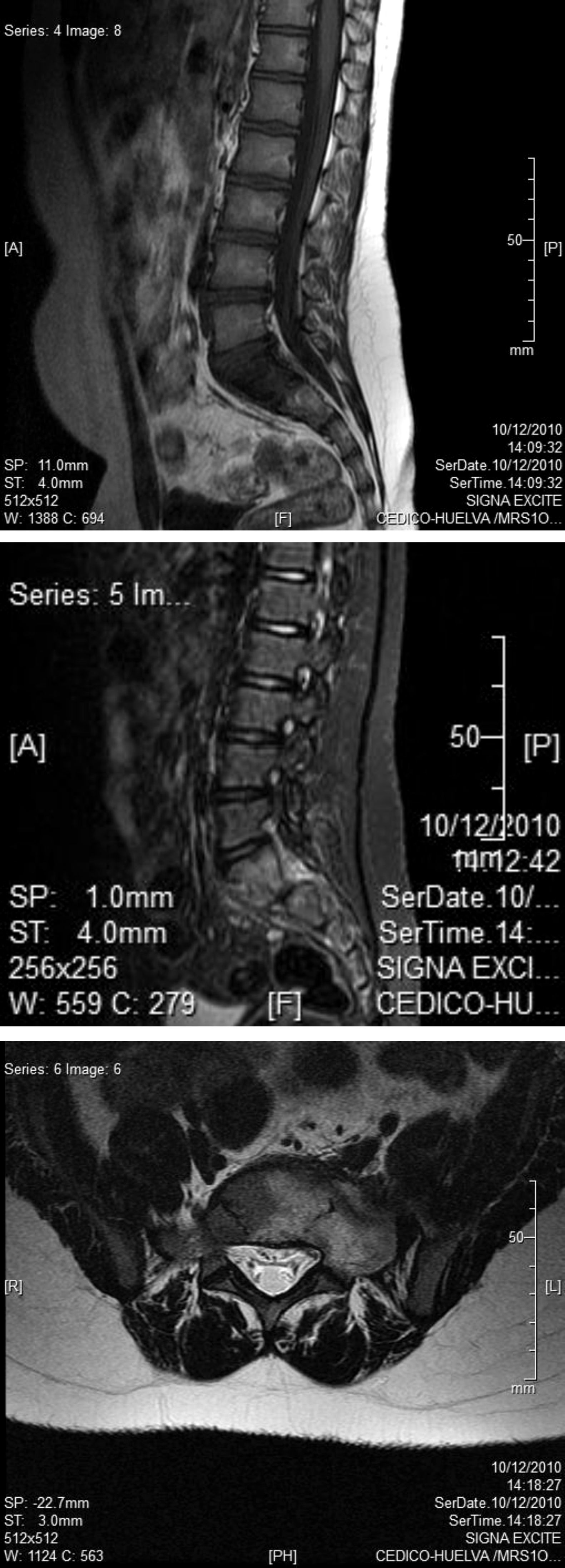
The patient continues to exhibit the previously described lesion in periodic MRI controls, without significant changes in its dimensions (Fig. 2).
Discussion
CDSO is a type of chronic recurrent multifocal osteomyelitis (CRMO).
It is suspected that CRMO may be an autoimmune disorder. It has been reported in association with several, chronic, autoimmune diseases, including inflammatory bowel disease, Wegener's granulomatosis, psoriasis and Takayasu vasculitis. It is possible that CRMO represents a juvenile form of seronegative spondyloarthropathy1–6 or a paediatric variant of SAPHO syndrome (synovitis, acne, pustulosis, hyperostosis and osteitis).7,8This disease usually appears before the age of 25 years, when the activity of osteoblasts in the periosteum is at its peak.12 The most common location is the mandible, but it can occur in any bone.12–15 Most of the patients with CRMO display elevated C-reactive protein or erythrocyte sedimentation rates during exacerbations, with normal white blood cell counts,5,6 but there are also cases in which analytical studies show strictly normal results.12 Rheumatoid factor, antinuclear antibodies and other serological findings are generally negative. B27 human leukocyte antigen does not appear to play a role in CRMO.1,6–11
Histological examination usually reports non-specific, subacute inflammation or chronic, non-suppurative inflammation with supracortical and subperiosteal foci of reactive bone with a marked osteoblastic activity and intertrabecular spaces filled with fibrovascular material containing inflammatory cells, primarily lymphocytes, as well as plasma cells, macrophages, histiocytes, multinucleated giant cells and some polymorphonuclear leukocytes.1,5,12,14 The clinical course of CRMO is characterised by an insidious onset with local pain and swelling in the region of the affected bone. The lesions go through exacerbation and remission periods, which always occur in the same place and may appear in other bones with each crisis.1,9,16 Some of the patients remain asymptomatic between crises. Most of the cases are prolonged in time, but are self-limiting. The symptoms tend to disappear and the lesions become resolved over time without severe sequelae.1,6,17
Radiographic examinations are useful before starting with more invasive diagnostic procedures and should, therefore, be the initial imaging tests.1 MRI is very important for the evaluation of symptomatic lesions since this is the most sensitive test to show local extension and activity.1,18–21 It has a better sensitivity than radiography and bone scintigraphy for the evaluation of bones, joints and soft tissues adjacent to the lesions and can show the ideal location for a guided biopsy, thus allowing a definitive diagnosis. It also has the advantage for patients of not emitting radiation, making it into the imaging method of choice for monitoring chronic lesions occurring in childhood and adolescence.1 Bone scintigraphy scans are useful to evaluate the extent of lesions, since some of them can be asymptomatic.1,5,9,10,18,22
The following protocol seems to be the most appropriate when conducting imaging tests in cases of CRMO: (1) radiography of symptomatic regions, supplemented by MRI if necessary; (2) bone scintigraphy scan, and (3) MRI scan for the evaluation of lesions detected by bone scintigraphy, but hidden in simple radiography.1
The differential diagnosis of sacral lesions should include metastasis, as well as all major benign and malignant neoplasms.23 Due to its clinical and radiographic features, this form of osteomyelitis can be misdiagnosed as osteosarcoma.12 In the present case, the main differential diagnosis was plasmacytoma, but we also considered other malignant tumours such as chordoma, myeloma, chondrosarcoma, osteosarcoma and Ewing tumours.12,24,25
The diagnosis of CDSO is usually based on the clinical course and conventional radiographic findings, complemented by bone scintigraphy and/or MRI scans and, if necessary, histopathological and microbiological tests to exclude tumours and infectious diseases.
The treatment of this disease is complex and there are multiple forms of treatment.26 Classically, it begins with medical treatment based on anti-inflammatory drugs, followed by corticosteroid therapy if this is not sufficient. Surgical treatment is reserved for cases that are refractory to medical treatment and involves debridement and curettage of the bone lesion, as well as intralesional corticosteroid therapy.
However, during recent years there have been reports of good responses to bisphosphonates, both in oral and intravenous forms (10mg of alendronate in a single, intravenous dose), for cases refractory to standard treatment.27–30 There are also good experiences with the use of calcitonin in these refractory cases.31
There have also been reports of new techniques in cases resorting to surgery such as the use of autologous bone grafts to fill the bone defect following curettage or the use of autologous bone grafts of vascularised fibula, with good bone integration and healing of the lesion.32,33
ConclusionCDSO is a rare disease and even more so among children and in a sacral location. Therefore, it is essential to achieve definitive diagnosis which rules out tumours as cause of the symptoms. This requires the use of imaging techniques and, in some cases, even invasive techniques such as biopsy, which lead to a definitive diagnosis. In the case of children, the most appropriate technique for monitoring the evolution of symptoms is serial magnetic resonance imaging scans. Regarding treatment, in this case we decided to use corticosteroids and anti-inflammatory drugs as primary treatment, with good results in the progression of the lesion. After 2 years of follow-up the lesion remains stable, although total remission of symptoms has not been achieved.
Level of evidenceLevel of evidence V.
Ethical responsibilitiesProtection of people and animalsThe authors declare that this investigation did not require experiments on humans or animals.
Confidentiality of dataThe authors declare that they have followed the protocols of their workplace on the publication of patient data and that all patients included in the study received sufficient information and gave their written informed consent to participate in the study.
Right to privacy and informed consentThe authors declare having obtained written informed consent from patients and/or subjects referred to in the work. This document is held by the corresponding author.
Conflict of interestsThe authors have no conflict of interests to declare.
Please cite this article as: Franco-Jiménez S, et al. Osteomielitis esclerosante crónica de Garrè en un ni¿no con afectación sacra. Rev Esp Cir Ortop Traumatol. 2013;57:145-9.
