


The vascular anatomy of the hand has already been widely described macroscopically. However, there are very few papers that study the pattern of normality of in vivo vascularisation that describe and analyse the main arteries of the hand. The aim of this paper was to carry out a study to serve as a reference for the normal values of size and flow of the radial and ulnar artery at the level of the wrist, and the digital radial and ulnar arteries at the level of the fingers.
Material and methodA descriptive observational cross-sectional study on 200 hands in 100 healthy volunteers aged between 20 and 30 years. Doppler-colour ultrasound was performed on the ulnar and radial arteries in the wrist, as well as on the radial and ulnar digital arteries in each finger. Once the measurements had been taken, a general comparative analysis was performed also taking laterality, dominance and gender into account.
ResultsIt was observed that the radial artery is larger in size than the ulnar at wrist level, however, it was the ulnar artery that showed flow dominance at this level. At finger level, the arteries are greater in size and flow in the areas of the fingers more protected from injury (digital ulnar artery in the first three and radial artery in the fourth and fifth digits).
La anatomía vascular de la mano ya ha sido ampliamente descrita a nivel macroscópico. Sin embargo, existen muy pocos trabajos que estudien el patrón de normalidad de la vascularización in vivo y describan y analicen las arterias principales de la mano. El objetivo de este trabajo es realizar un estudio que sirva de referencia para los valores normales de tamaño y flujo de la arteria radial y ulnar a nivel de la muñeca y de las arterias digitales radiales y ulnares a nivel de los dedos.
Material y métodoEstudio descriptivo observacional de corte trasversal sobre 200 manos en 100 voluntarios sanos entre 20-30años. Se realizó ecografía Doppler-color de las arterias ulnar y radial en la muñeca, así como de las arterias digitales radial y ulnar de cada dedo. Una vez tomadas las medidas se llevó a cabo un análisis comparativo de forma general y también teniendo en cuenta la lateralidad, la dominancia y el género.
ResultadosSe observó que existe un mayor tamaño de la arteria radial sobre la ulnar a nivel de la muñeca; sin embargo, es la arteria ulnar la que presentó dominancia de flujo a este nivel. A nivel de los dedos, en los tres primeros fue la arteria digital ulnar la que presentó un mayor tamaño y mayor flujo. No obstante, en el cuarto y quinto dedos fue la arteria digital radial la que presentó un mayor tamaño y dominancia de flujo.
ConclusiónHa quedado confirmado que la dominancia de flujo, pero no de tamaño, a nivel de la muñeca es de la arteria ulnar. A nivel de los dedos, existe un mayor tamaño y flujo de las arterias en las zonas de los dedos más protegidas de las lesiones (arteria digital ulnar en los tres primeros y radial en el cuarto y quinto).
The vascular anatomy of the hand has already been widely described macroscopically. The main arteries providing blood supply to the hand are the radial artery (RA) and the ulnar artery (UA); once they reach the hand they give rise to the superficial palmar arch, which in turn emits the common and own palmar digital arteries.1 There are many anatomical variations,2 but the presence is constant of the radial and ulnar artery at the wrist and the ulnar and radial digital arteries at the level of each finger.
In recent years, dynamic in vivo vascular study has been added to macroscopic vascular anatomical study thanks to ultrasound and Doppler ultrasound.3–5 This technique has generally been used to study vascularisation in impaired situations – e.g. in smokers – or to assess vascular changes when performing the Allen test.6–8 Despite all the studies carried out in impaired situations, there are very few papers that study the pattern of normality of vascularisation of the hand or vascular dominance between different arteries in a healthy population to serve as a reference.3,5,9
This study aims to describe, in a healthy Spanish population, the morphological pattern and dynamic flow of the radial and ulnar arteries at the level of the wrist and of each of the digital arteries. We will also analyse the dominance of one artery (radial or ulnar) over the other in the wrist and in each of the fingers and we will analyse the influence of the laterality of the hand (right or left), dominance (right or left) and gender. In this way we expect to achieve normal values that can serve as a reference for the study of different conditions.
Material and methodThe study is cross-sectional, descriptive, and observational in design. To assess the variables studied by ultrasound in a healthy Spanish population, a population of 100 volunteer subjects was selected. The participants were randomly selected in different locations, mainly in the areas near the Hospital Universitario Infanta Leonor; this includes facilities such as sports centres, university centres, public offices, shops and the health facilities themselves.
For the selection of the healthy population to serve as a reference and in whom a normal pattern could be estimated, people between the ages of 20 and 30 were taken as the inclusion criteria with no exclusion criteria.
The exclusion criteria were the following: patients under 20 years of age or over 30, a history of high blood pressure and/or diabetes, smoking habits in the last 5 years, vascular pathology, connective tissue disease, history of inflammatory disease, systemic metabolic diseases, Raynaud's disease and a history of trauma in the forearm or hand, including from the elbow joint to the proximal interphalangeal joints.
The chosen age range of the sample was between 20 and 30, since at this age growth is complete and there are usually no vascular conditions (such as arteriosclerosis) that could alter normal arterial flow. Of the 100 patients selected, 50 were men and 50 were women. The dominant hand was the right in 90 participants (45 women and 45 men) and the left in 10 (5 women and 5 men). The patients were first interviewed briefly, when they were informed of the study objectives and the reason for the questions about their medical history. After confirming that both inclusion and exclusion criteria had been met, all the participants were given an informed consent form to sign, as well as a patient information sheet referring to the research objectives and the subsequent handling of the data obtained. The study was approved by the Clinical Research Ethics Committee of the Hospital General Universitario Gregorio Marañón in Madrid (protocol code: HUIL-181025).
The variables studied were the following:
Epidemiological and general anthropometric variables: age, gender, height in cm, weight in kg, body mass index, dominant hand, considering the hand that the patient used to write as dominant.
Ultrasound study of the wrist and hand: the variables collected in each artery were the cross-sectional diameter of the artery in cm and the flow velocity in cm/s. These parameters were measured in the wrist in the radial artery and the ulnar artery, as well as in all the radial and ulnar digital arteries specific to each finger. The GE Logiq ultrasound system from GE Health Care® was used to take the measurements, with a GE i12L-RS probe, at a frequency range of 4.0–10.0MHz, designed for the examination of vascular structures, among others. The patients’ arms were first placed in a supination and elbow joint flexion position, with the wrist placed on a support surface to keep it relaxed.
All the patients underwent an ultrasound scan of first the right upper limb and then the left upper limb.
Once the hand was in position, the ultrasound settings were adjusted; mainly by activating the colour function to be able to locate the vessels and differentiate between vein and artery, and modifying the angle of the Doppler function, which should always be kept between 55° and 60°, never exceeding 60°.
First, the ultrasound study was performed on the right wrist, starting with the radial artery. The proximal palmar fold was taken as a superficial anatomical reference. The probe was placed on this fold perpendicular to the forearm, with the notch of the probe facing medially at all times (Fig. 1A). In this plane, the ultrasound scan showed the radial artery accompanied by the 2 veins and the styloid process of the radius, which was the anatomical reference at ultrasound level (Fig. 1B). Once the artery had been located, minimum pressure was exerted on the wrist to avoid reducing the diameter of the vessel and the transverse diameter was measured in cm. Once the diameter had been measured, in the same plane, the probe was rotated by 90°, the notch being orientated distally (Fig. 1C). Once the rotation had been made, the longitudinal section of the artery was observed, and the flow velocity was measured. First the colour function was activated, then the Steer function, at all times placing the angle in favour of the vessel (from 55° to 60°) and placing the centre of the vessel in the longitudinal section as a reference. Once all this had been done, the Doppler function was activated and the arterial flow velocity, in both systole and diastole, was observed on the screen. By freezing the screen, if the recorded pulse was regular, the ultrasound machine marked the arterial flow velocity at the time of the systolic peak, and this value was recorded for the study (Fig. 1D). If the recorded pulse was not regular, the manoeuvre was repeated.
 Procedure for measuring the diameter of the radial artery of the right wrist. Measured at the level of the proximal palmar fold. The probe is placed transversely to locate the artery (arrow) and the measurement is taken using the styloid of the radius as a reference (asterisk). (B) Procedure to measure the flow velocity of the radial artery of the right wrist. Measured at the level of the proximal palmar fold. The probe is placed longitudinally to locate the artery (white arrow) and the peak systolic value is taken if the pulse was regular (yellow arrow).")
(A) Procedure for measuring the diameter of the radial artery of the right wrist. Measured at the level of the proximal palmar fold. The probe is placed transversely to locate the artery (arrow) and the measurement is taken using the styloid of the radius as a reference (asterisk). (B) Procedure to measure the flow velocity of the radial artery of the right wrist. Measured at the level of the proximal palmar fold. The probe is placed longitudinally to locate the artery (white arrow) and the peak systolic value is taken if the pulse was regular (yellow arrow).
After study of the radial artery, we continued with the ulnar artery; the superficial anatomical reference was the same as for the radial artery. The probe was placed over the fold, this time in the medial region of the wrist, perpendicular to the forearm, with the notch facing medially. However, at ultrasound level the reference was the distal epiphysis of the ulna. After taking the diameter of the artery in the transverse plane, the probe was rotated 90° and the notch was oriented distally to measure the flow velocity of the ulnar artery in the longitudinal section.
Once the data collection in the arteries of the wrist was completed, the ultrasound of the finger arteries was performed. We started with the first finger at all times and in each finger, we first collected the variables of the digital radial artery and then the ulnar.
To measure the arterial diameter of both the radial and the ulnar digital arteries, the proximal interphalangeal joint was taken as the anatomical reference, placing the probe proximal to it and in a perpendicular position to the proximal phalanx (Fig. 2A). In the radial digital artery (Rx, x being the finger) the probe was placed in the lateral half of the finger, while in the ulnar digital artery (Ux, x being the finger) the probe was placed in the medial half. In this plane, to facilitate locating the arteries, the colour function of the ultrasound machine was activated. Once located, the diameter of the artery was taken (Fig. 2B). For the flow velocity, the measurement was taken in the same position, proximal to the IFP joint, with the probe in a longitudinal direction (Fig. 2C). As with the arteries in the wrist, we took the flow velocity value at the systolic peak, a value indicated by the ultrasound machine (Fig. 2D).
 Procedure for measuring the diameter of the ulnar digital artery of the third finger of the right hand. Measured proximal to the IFP joint. The probe is placed transversely to locate the artery adjacent to the flexor tendons (asterisk). (B) Procedure for measuring the flow velocity of the ulnar digital artery of the third finger of the right hand. Measured proximal to the IFP joint. The probe is placed longitudinally to locate the artery and the systolic peak value is taken if the pulse was regular (yellow arrow).")
(A) Procedure for measuring the diameter of the ulnar digital artery of the third finger of the right hand. Measured proximal to the IFP joint. The probe is placed transversely to locate the artery adjacent to the flexor tendons (asterisk). (B) Procedure for measuring the flow velocity of the ulnar digital artery of the third finger of the right hand. Measured proximal to the IFP joint. The probe is placed longitudinally to locate the artery and the systolic peak value is taken if the pulse was regular (yellow arrow).
After completing the measurements in the right hand, we proceeded to take samples in the left hand following the same method.
Once the abovementioned variables had been measured, the following comparative analysis was carried out:
- •
Comparative study of the size (transverse diameter) of the arteries in the wrist. The analysis was performed in 4 scenarios: in general; in relation to laterality; in relation to dominance, and in relation to gender.
- •
Comparative study of the flow velocity (systolic peak) of the arteries of the wrist. Conducted in the same 4 scenarios: generally, in relation to laterality, in relation to dominance, and in relation to gender.
- •
Comparative study of the size (transverse diameter) of the digital radial artery versus the digital ulnar artery in each of the five fingers generally; in each of the five fingers in relation to laterality; in each of the five fingers in relation to dominance, and in each of the five fingers in relation to gender.
- •
Comparative study of the flow velocity (systolic peak) of the radial digital artery versus the ulnar digital artery in each of the five fingers generally; in each of the five fingers in relation to laterality; in each of the five fingers in relation to dominance, and in each of the five fingers in relation to gender.
R for Mac software (version 3.5.1; R, Vienna, Austria) was used for the statistical analysis. The descriptive values for the study of the arteries are always presented in terms of median and interquartile range (Q1–Q3). Given the large sample size available for the study and the fact that the main variables of analysis, diameter and arterial flow, are continuous quantitative, the Student's t-test for independent samples was used to carry out comparisons between paired groups of individuals (by gender). Similarly, when making comparisons in the same individual (in terms of side, in terms of dominance) the Student's t-test for paired samples was used. In all cases the level of statistical significance was set at p<.05.
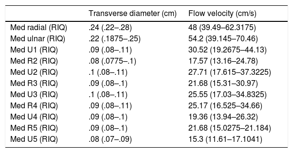
ResultsThe results of the descriptive study of the transverse diameter and of the arterial flow velocity at the time of the systolic peak in each of the 12 arteries studied can be seen in Table 1.
Results of the diameter (in centimetres) and flow velocity (in centimetres/second) of the radial artery (radial) and ulnar artery (ulnar) in the wrist and of the radial and ulnar digital arteries of the five fingers.
| Transverse diameter (cm) | Flow velocity (cm/s) | |
|---|---|---|
| Med radial (RIQ) | .24 (.22–.28) | 48 (39.49–62.3175) |
| Med ulnar (RIQ) | .22 (.1875–.25) | 54.2 (39.145–70.46) |
| Med U1 (RIQ) | .09 (.08–.11) | 30.52 (19.2675–44.13) |
| Med R2 (RIQ) | .08 (.0775–.1) | 17.57 (13.16–24.78) |
| Med U2 (RIQ) | .1 (.08–.11) | 27.71 (17.615–37.3225) |
| Med R3 (RIQ) | .09 (.08–.1) | 21.68 (15.31–30.97) |
| Med U3 (RIQ) | .1 (.08–.11) | 25.55 (17.03–34.8325) |
| Med R4 (RIQ) | .09 (.08–.11) | 25.17 (16.525–34.66) |
| Med U4 (RIQ) | .09 (.08–.1) | 19.36 (13.94–26.32) |
| Med R5 (RIQ) | .09 (.08–.1) | 21.68 (15.0275–21.184) |
| Med U5 (RIQ) | .08 (.07–.09) | 15.3 (11.61–17.1041) |
Rx: radial digital artery of finger x; Ux: ulnar digital artery of finger x.
Results of the 200 hands studied, shown as median (Med) and interquartile range (IQR).
The results of the same descriptive study broken down by right/left hand can be found in Table 2.
Results of the diameter (in centimetres) and flow velocity (in centimetres/second) of the 100 right hands and 100 left hands.
| Right | Left | |||
|---|---|---|---|---|
| Transverse diameter (cm) | Flow velocity (cm/s) | Transverse diameter (cm) | Flow velocity (cm/s) | |
| Med radial (RIQ) | .24 (.22–.28) | 49.92 (40.0675–65.4275) | .25 (.2175–.27) | 47.23 (38.71–61.0575) |
| Med ulnar (RIQ) | .22 (.1875–.25) | 54.47 (39.49–70.5475) | .22 (.1875–.24) | 53.88 (37.16–69.7175) |
| Med R1 (RIQ) | .08 (.08–.1) | 19.91 (13.94–28.8425) | .08 (.07–.09) | 19.38 (13.0675–26.42) |
| Med U1 (RIQ) | .09 (.08–.11) | 31.74 (18.58–44.13) | .09 (.08–.1125) | 29.81 (19.36–44.3125) |
| Med R2 (RIQ) | .09 (.08–.1) | 17.81 (13.4975–24.78) | .08 (.07–.1) | 16.72 (13.16–24.785) |
| Med U2 (RIQ) | .1 (.08–.11) | 28.87 (18.3875–37.94) | .1 (.08–.1125) | 27.1 (17.03–37.16) |
| Med R3 (RIQ) | .09 (.08–.11) | 22.45 (16.26–31.0625) | .09 (.08–.1) | 20.13 (14.71–29.705) |
| Med U3 (RIQ) | .1 (.09–.11) | 27.99 (17.465–35.9625) | .1 (.08–.12) | 24.78 (17.03–33.4975) |
| Med R4 (RIQ) | .09 (.08–.1) | 24.39 (16.5–33.46) | .1 (.08–.11) | 25.67 (16.8625–34-84) |
| Med U4 (RIQ) | .09 (.08–.1) | 20.58 (14.71–26.5325) | .09 (.08–.1) | 17.61 (13.7775–25.7425) |
| Med R5 (RIQ) | .09 (.08–.1) | 21.68 (15.49–30.7) | .09 (.08–.1) | 21.29 (14.5175–33.29) |
| Med U5 (RIQ) | .08 (.07–.09) | 15.49 (12.39–21.095) | .08 (.07–.09) | 14.59 (11.61–19.5425) |
The following results were obtained for the comparative analysis of the variables studied:
- 1.
In the comparative study of the size (transverse diameter) of the arteries of the wrist.
- •
In general, that is, without considering laterality, dominance or gender, a larger (statistically significant) size of the radial artery is observed with respect to the ulnar artery (Fig. 3A).
 of the radial and ulnar arteries at wrist level shown in cm. (A) General comparison (with no discrimination). (B) Comparison in terms of side (right wrist/left wrist). (C) Comparison in terms of dominance (dominant hand/non-dominant hand). (D) Comparison in terms of gender (male/female). The asterisk indicates statistically significant differences at p<.05.") Figure 3.
Figure 3.Comparative study of the size (transverse diameter) of the radial and ulnar arteries at wrist level shown in cm. (A) General comparison (with no discrimination). (B) Comparison in terms of side (right wrist/left wrist). (C) Comparison in terms of dominance (dominant hand/non-dominant hand). (D) Comparison in terms of gender (male/female). The asterisk indicates statistically significant differences at p<.05.
- •
In terms of laterality, the tendency is the same as described above, with a larger (statistically significant) size of the radial artery in both the right and left wrist (Fig. 3B).
- •
In terms of dominance, there is no statistically significant difference between the size of the radial ulnar arteries of the dominant hand and the size of the same arteries in the non-dominant wrist (Fig. 3C).
- •
In terms of gender, the two arteries in the wrist were found to be statistically significantly larger in the men than in the women (fig. 3D).
- •
- 2.
In the comparative study of the flow velocity (systolic peak) of the arteries of the wrist.
- •
In general. A higher (statistically significant) flow was observed of the ulnar artery with respect to the radial artery (Fig. 4A).
 of the arteries of the wrist shown in cm/s. (A) General comparison (with no discrimination). (B) Comparison in terms of side (right wrist/left wrist). (C) Comparison in terms of dominance (dominant hand/non-dominant hand). (D) Comparison in terms of gender (male/female). The asterisk indicates statistically significant differences at p<.05.") Figure 4.
Figure 4.Comparative study of flow velocity (systolic peak) of the arteries of the wrist shown in cm/s. (A) General comparison (with no discrimination). (B) Comparison in terms of side (right wrist/left wrist). (C) Comparison in terms of dominance (dominant hand/non-dominant hand). (D) Comparison in terms of gender (male/female). The asterisk indicates statistically significant differences at p<.05.
- •
In terms of laterality, the trend was the same, with greater flow of the ulnar artery (statistically significant) in both the right and the left wrist (Fig. 4B).
- •
In terms of dominance, no statistically significant differences were observed between the radial and ulnar artery flow of the dominant hand with respect to the same arteries in the non-dominant hand (Fig. 4C).
- •
In terms of gender, it was observed that in both arteries the flow is statistically significantly higher in the men than in the women (Fig. 4D).
- •
- 3.
In the comparative study of the size (transverse diameter) of the digital arteries in the fingers:
- •
In general, it was observed that in the first, second and third fingers the digital ulnar artery was larger (statistically significant) than the digital radial artery. In the fourth and fifth fingers, the digital radial artery was larger (statistically significant) than the digital ulnar artery (Fig. 5A).
 of the digital arteries in the fingers shown in cm. (A) General comparison (with no discrimination). (B) Comparison in terms of side (right hand/left hand). (C) Comparison in terms of dominance (dominant hand/non-dominant hand). (D) Comparison in terms of gender (male/female). The asterisk indicates statistically significant differences at p<.05.") Figure 5.
Figure 5.Comparative study of the size (transverse diameter) of the digital arteries in the fingers shown in cm. (A) General comparison (with no discrimination). (B) Comparison in terms of side (right hand/left hand). (C) Comparison in terms of dominance (dominant hand/non-dominant hand). (D) Comparison in terms of gender (male/female). The asterisk indicates statistically significant differences at p<.05.
- •
In terms of laterality, the trend was the same. Larger diameter of the digital ulnar artery in the first, second and third fingers, and larger diameter of the digital radial artery in the fourth and fifth fingers, both in the right and left hand (Fig. 5B).
- •
In terms of dominance, no statistically significant differences were observed between the digital radial and ulnar arteries of each finger of the dominant hand compared to the digital arteries of the same fingers in the non-dominant hand (Fig. 5C).
- •
In terms of gender, both the digital radial and digital ulnar arteries of the 5 fingers were found to be larger in the men than in the women (Fig. 5D).
- •
- 4.
In the comparative study of the flow velocity (systolic peak) of the digital arteries in the fingers:
- •
In general, it was observed that in the first, second and third fingers the flow of the digital ulnar artery was greater compared to the digital radial artery. However, in the fourth and fifth fingers it was observed that the flow of the digital radial artery was greater in comparison to the digital ulnar artery, all of these results being statistically significant (Fig. 6A).
 of the digital arteries in the fingers shown in cm/s. (A) General comparison (with no discrimination). (B) Comparison in terms of side (right hand/left hand). (C) Comparison in terms of dominance (dominant hand/non-dominant hand). (D) Comparison in terms of gender (male/female). The asterisk indicates statistically significant differences at p<.05.") Figure 6.
Figure 6.Comparative study of the flow velocity (systolic peak) of the digital arteries in the fingers shown in cm/s. (A) General comparison (with no discrimination). (B) Comparison in terms of side (right hand/left hand). (C) Comparison in terms of dominance (dominant hand/non-dominant hand). (D) Comparison in terms of gender (male/female). The asterisk indicates statistically significant differences at p<.05.
- •
In terms of laterality, the trend was the same. With greater flow of the digital ulnar artery in the first, second and third fingers and greater flow of the digital radial artery in the fourth and fifth fingers, in both the right and left hand (Fig. 6B).
- •
In terms of dominance, no statistically significant differences were observed between the digital radial and ulnar arteries of each finger of the dominant hand with respect to the digital arteries of the same fingers of the non-dominant hand (Fig. 6C).
- •
In terms of gender, it was observed that the flow of the arteries, both digital radial and digital ulnar of the 5 fingers, was greater in the men than in the women (Fig. 6D).
- •
 of the radial and ulnar arteries at wrist level shown in cm. (A) General comparison (with no discrimination). (B) Comparison in terms of side (right wrist/left wrist). (C) Comparison in terms of dominance (dominant hand/non-dominant hand). (D) Comparison in terms of gender (male/female). The asterisk indicates statistically significant differences at p<.05.")
 of the arteries of the wrist shown in cm/s. (A) General comparison (with no discrimination). (B) Comparison in terms of side (right wrist/left wrist). (C) Comparison in terms of dominance (dominant hand/non-dominant hand). (D) Comparison in terms of gender (male/female). The asterisk indicates statistically significant differences at p<.05.")
 of the digital arteries in the fingers shown in cm. (A) General comparison (with no discrimination). (B) Comparison in terms of side (right hand/left hand). (C) Comparison in terms of dominance (dominant hand/non-dominant hand). (D) Comparison in terms of gender (male/female). The asterisk indicates statistically significant differences at p<.05.")
 of the digital arteries in the fingers shown in cm/s. (A) General comparison (with no discrimination). (B) Comparison in terms of side (right hand/left hand). (C) Comparison in terms of dominance (dominant hand/non-dominant hand). (D) Comparison in terms of gender (male/female). The asterisk indicates statistically significant differences at p<.05.")
In this study describe the normal flow pattern of the radial and ulnar arteries of the wrist and the radial and ulnar digital arteries of each finger in a healthy Spanish population. In general terms, we observed that, regardless of the variables studied, at the level of the wrist the radial artery is of a greater size. However, it is the ulnar artery that presented flow dominance at this level. At finger level, we observed that in the first, second and third fingers it is the ulnar digital artery that presents a greater size, as well as flow dominance, over the radial digital artery. However, in the fourth and fifth fingers the digital radial artery was shown to be of greater size and flow dominance compared to the digital ulnar artery.
Regarding the ultrasound study of the size of the arteries of the wrist, in the current literature there are several studies that focus on the comparative analysis between the radial and ulnar arteries, with different conclusions. Ashraf et al.9 establish that the ulnar artery is larger in size than the radial. However, other studies conclude that the radial artery is larger at the wrist,3–5 or that they find no significant differences in sizes.10 Some of these studies also compared the size of these arteries according to laterality, and found no differences between the arteries of the right and left wrist,3 or differences between the dominant and non-dominant hand,4,10 but found a larger size of the radial artery in men compared to women.4,10 In our study we observed that, regardless of the discriminated variable, the size of the radial artery is always larger than the ulnar artery, in contrast to some of the abovementioned studies. In terms of laterality, we also found no differences in arterial size when comparing right and left hands. Similarly, we found no differences in terms of dominance. Finally, on analysing size in terms of gender, we did find statistically significant differences in the size of both the radial and ulnar arteries, which were larger in the men. To explain the difference in the results, we should mention that our sample was obtained from completely healthy patients, with demanding exclusion criteria, while many of these studies included a populations with diseases that could impair vascularisation.
The arterial flow in the arteries of the wrist has also been studied in several papers. There are studies describing a flow pattern in which the ulnar artery is the dominant artery in the wrist4,11; however, we also found studies describing a pattern of radial dominance.3,12 It seems that radial dominance is more likely in patients who smoke, since in smokers the ulnar artery is most affected.12 The harmful effect of smoking on the circulation of the hand is known,13,14 and finding radial dominance in patients who smoke does not go against our study, in which smoking was a criterion for exclusion. In our study it was observed that, in the wrist, the ulnar artery is dominant over the radial artery; our findings coincide with the first studies mentioned. If we take laterality into account, there are no marked differences between the flow of the left and right hand, the ulnar artery being the dominant artery in both hands. No previous studies of arterial flow in the wrist have been described that consider dominance, and in our study no statistically significant differences were observed between the flow from the dominant and the non-dominant hand. Finally, when considering gender, blood flow at the wrist was found to be higher in the men than in the women. Probably one of the most curious data of this study is the discrepancy between the size and flow of the radial and ulnar arteries, since, in spite of the larger size of the radial artery, the flow is less.
Regarding the size of the radial and ulnar digital arteries of each finger, the only article we found that studies the size of the digital vessels ultrasonically is the one by Trager et al.11 on 20 patients. They describe that the ulnar artery of the first, second and third fingers is larger than the radial, while the radial artery of the fourth and fifth fingers is the largest. The same results were obtained in our study.
Finally, our last comparative analysis refers to the flow of the digital arteries in the fingers. Similarly, the only article we found that studies the flow velocity of the digital arteries is that of Trager et al.11 They describe a greater flow in the arteries with a larger diameter, i.e. the ulnar of the first to the third fingers and the radial of the fourth and fifth fingers. In our study of 200 hands and 1000 fingers we found the same results. We were able to make a heat map in which the flow in each of the arteries of the fingers can be seen graphically (Fig. 7).

As a limitation of the study, we should point out that the measurements were made by a single scanner (always the same one), but the measurements were not compared between two different scanners for interobserver validation.
As a curiosity, it is worth noting that the size and flow is less in areas of the fingers “less protected from injury” and where greater support is provided, such as the ulnar area of the fourth and fifth fingers and the radial area of the first, second and third fingers, while the arteries in the more protected areas provide greater vascular support. This has a completely logical evolutionary explanation in which the vascular contribution of the finger is protected, giving greater importance to the arteries that have less risk of injury.
ConclusionThe pattern of normal flow of the radial and ulnar arteries of the wrist and the radial and ulnar digital arteries of each finger in a healthy Spanish population has been described. In general terms, we observed that, regardless of the variables studied, at wrist level the radial arteries are greater in size. However, it is the ulnar artery that presented flow dominance at this level. At finger level, we observed that in the first, second and third fingers it is the ulnar digital artery that was larger in size, as well as flow dominance, than the radial digital artery of these fingers. However, in the fourth and fifth fingers we found that it is the digital radial artery that is larger in size and flow dominance compared to the digital ulnar artery of the same fingers. It can be concluded that there is a greater size and flow of the arteries in the areas of the fingers most protected from injury (ulnar area of the first, second and third fingers and radial area of the fourth and fifth) and a smaller size and flow in the areas of greater support and probability of injury (radial area of the first, second and third fingers and ulnar area of the fourth and fifth).
Level of evidenceLevel of evidence III.
Conflict of interestsThe authors have no conflict of interests to declare.
Please cite this article as: Coderech Carretero J, Corella Montoya F, Grande Barez M, Corella Montoya MÁ, Ocampos Hernández M, Larrainzar-Garijo R. Descripción y análisis del patrón de normalidad de flujo dinámico y morfológico de las arterias principales de la muñeca y mano en población sana española. Rev Esp Cir Ortop Traumatol. 2020;64:167–176.
