Buscar en
Revista Colombiana de Reumatología (English Edition)
Toda la web
Inicio Revista Colombiana de Reumatología (English Edition) Systemic sclerosis and gastrointestinal involvement
Journal Information
Vol. 27. Issue S1.
Systemic Sclerosis
Pages 44-54 (April 2020)
Visits
5830
Vol. 27. Issue S1.
Systemic Sclerosis
Pages 44-54 (April 2020)
Review Article - Meta-analysis
Full text access
Systemic sclerosis and gastrointestinal involvement
Visits
5830
Jose Luis Tandaipana, Ivan Castellvíb,
Corresponding author
icastellvi@santpau.cat

Corresponding author at: Department of Rheumatology Hospital de la Santa Creu i Sant Pau Carrer de Mas Casanovas 90 08041, Barcelona, Spain.
a Department of Rheumatology, Hospital Universitari Mútua de Terrassa, Terrassa. Spain
b Autoimmune Diseases Unit, Department of Rheumatology, Hospital Universitari de la Santa Creu i Sant Pau, Barcelona. Spain
This item has received
Article information
Abstract
Full Text
Bibliography
Download PDF
Statistics
Figures (1)
Tables (1)
Table 1. Complications of gastrointestinal involvement in SSc.
Abstract

Systemic sclerosis (SSc) is a systemic autoimmune disease in which gastrointestinal manifestations are a frequent complication. Gastrointestinal involvement is present in up to 90 % of patients. The most affected areas are the esophagus and the anorectal tract. Reflux, heartburn and dysmotility are the leading causes of gastrointestinal discomfort. Disordered anorectal function can occur early in the course of SSc and is an important factor in the development of fecal incontinence. Current recommendations to treat gastrointestinal disorders in SSc include the use of proton pump inhibitors, prokinetics and rotating antibiotics. This review discusses the proposed pathophysiological mechanisms, the clinical presentation, the different diagnostic techniques and the current management of the involvement of each section of the gastrointestinal tract in SSc.

Keywords:
Systemic sclerosis
Dysmotility
Chronic intestinal pseudo-obstruction
Dysphagia
Fecal incontinence
Gastroesophageal reflux disease
Gastroparesis
Small intestinal bacterial overgrowth.
Full Text
Introduction

Gastrointestinal tract (GIT) involvement is common in patients with Systemic sclerosis (SSc), affecting up to 90 % of patients,1–4 and being an important cause of morbidity and mortality in the disease.5

A dysfunction of the microcirculation, the immune system and the fibrosis control mechanisms is present in the GIT in SSc like other complications of the disease. The esophagus is the most affected GIT area in SSc and dysphagia and gastro-esophageal reflux disease are the main manifestations.6,7 Gastric involvement is less frequent, although it may be responsible for delayed gastric emptying and gastric antral vascular ectasia.8–11 Intestinal involvement, although usually asymptomatic, can be life-threatening. The small intestine can suffer from intestinal stasis and predispose to bacterial overgrowth that causes severe diarrhea, abdominal pain and weight loss. Colonic involvement can cause severe constipation.12,13 The anorectal area can be affected in more than 50 % of patients and cause great impact in quality of life.14 The presence of primary biliary cholangitis (PBC) occurs relatively frequently in patients with SSc (7–17 %), especially in limited cutaneous Systemic Sclerosis (lcSSc).15,16

The University of California, Los Angeles Scleroderma Clinical Trial Consortium Gastrointestinal Tract 2.0 (UCLA GIT 2.0) instrument can be used to assess the presence and severity of gastrointestinal symptoms in patients with SSc as well as their impact on the quality of life. It has been validated and the patients themselves report their symptoms, through 34 questions, grouped into 7 subgroups: reflux, bloating, diarrhea, fecal dirt, constipation, emotional well-being and social functioning, scored according to severity.17

There are no specific randomized clinical trials to assess different therapies for the management of complications of GIT in patients with SSc, and most treatment options are based on evidence in other diseases or on expert recommendations.

In this manuscript we review the recent knowledge of the pathogenesis, clinical manifestations, tests and treatments used to manage the different gastrointestinal manifestations in SSc.

MethodsLiterature search methods

A bibliographic research was carried out until June 2019, to find studies that met the criteria for inclusion in the MEDLINE, EMBASE and Cochrane Library databases.

When conducting the research in Medline, this was done through Pubmed using the MeSH terms: systemic, orofacial sclerosis, microstomia, xerostomia, periodontal disease, dysmotility, gastroesophageal reflux disease, esophageal dysphagia, Barret esophagus, esophageal adenocarcinoma, gastroparesis, watermelon stomach, intestinal dysmotility, constipation, diarrhea, intestinal bacterial overgrowth, intestinal pseudo-obstruction, intestinal cystoid pneumatosis, diverticulosis, primary biliary cholangitis, autoimmune hepatitis, pancreatic insufficiency, fecal incontinence, rectal prolapse.

Only articles published in English language were included.

Article selection and information extraction

Selected articles were saved in a database; initially, those articles that had the keywords included in the abstract or in the title were taken into account. Subsequently, those articles that did not meet the inclusion criteria were ruled out, and a committee was held among the authors to unify the database and choose those articles that were relevant to this review.

Inclusion criteria

For the selection of studies, it was taken into account that they met the following inclusion criteria in terms of type of study, population, and intervention.

  • Type of studies: Meta-analyses, systematic reviews (SR) of randomized clinical trials (RCTs), RCTs, prospective and retrospective cohort studies, and institutional protocols have been considered for inclusion. It has not been used as a criterion that the studies had a minimum follow-up time or a minimum sample size.

  • Type of population: patients over 18 years old with SSc and gastrointestinal involvement.

  • Intervention: Studies describing the gastrointestinal condition, explaining the pathophysiology, clinic, diagnostic techniques, prognosis, treatment and complications.

Exclusion criteria

  • Articles without access to full text

  • Duplicate articles

  • Studies that were not conducted in humans

Results

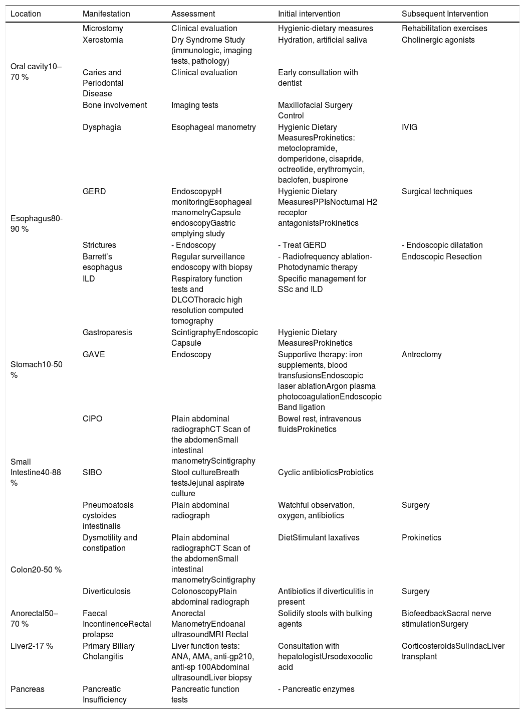
At the end of the research, a total of 2101 articles were analyzed by the researchers. After excluding duplicate articles, articles without access to full text or non-human studies, we obtained a total of 148 articles. Table 1 summarizes GIT complications in SSc and their approach that we found on literature.

Table 1.

Complications of gastrointestinal involvement in SSc.

Location  Manifestation  Assessment  Initial intervention  Subsequent Intervention 
Oral cavity10–70 %Microstomy  Clinical evaluation  Hygienic-dietary measures  Rehabilitation exercises 
Xerostomia  Dry Syndrome Study (immunologic, imaging tests, pathology)  Hydration, artificial saliva  Cholinergic agonists 
Caries and Periodontal Disease  Clinical evaluation  Early consultation with dentist   
Bone involvement  Imaging tests  Maxillofacial Surgery Control   
Esophagus80-90 %Dysphagia  Esophageal manometry  Hygienic Dietary MeasuresProkinetics: metoclopramide, domperidone, cisapride, octreotide, erythromycin, baclofen, buspirone  IVIG 
GERD  EndoscopypH monitoringEsophageal manometryCapsule endoscopyGastric emptying study  Hygienic Dietary MeasuresPPIsNocturnal H2 receptor antagonistsProkinetics  Surgical techniques 
Strictures  - Endoscopy  - Treat GERD  - Endoscopic dilatation 
Barrett’s esophagus  Regular surveillance endoscopy with biopsy  - Radiofrequency ablation- Photodynamic therapy  Endoscopic Resection 
ILD  Respiratory function tests and DLCOThoracic high resolution computed tomography  Specific management for SSc and ILD   
Stomach10-50 %Gastroparesis  ScintigraphyEndoscopic Capsule  Hygienic Dietary MeasuresProkinetics   
GAVE  Endoscopy  Supportive therapy: iron supplements, blood transfusionsEndoscopic laser ablationArgon plasma photocoagulationEndoscopic Band ligation  Antrectomy 
Small Intestine40-88 %CIPO  Plain abdominal radiographCT Scan of the abdomenSmall intestinal manometryScintigraphy  Bowel rest, intravenous fluidsProkinetics   
SIBO  Stool cultureBreath testsJejunal aspirate culture  Cyclic antibioticsProbiotics   
Pneumoatosis cystoides intestinalis  Plain abdominal radiograph  Watchful observation, oxygen, antibiotics  Surgery 
Colon20-50 %Dysmotility and constipation  Plain abdominal radiographCT Scan of the abdomenSmall intestinal manometryScintigraphy  DietStimulant laxatives  Prokinetics 
Diverticulosis  ColonoscopyPlain abdominal radiograph  Antibiotics if diverticulitis in present  Surgery 
Anorectal50–70 %  Faecal IncontinenceRectal prolapse  Anorectal ManometryEndoanal ultrasoundMRI Rectal  Solidify stools with bulking agents  BiofeedbackSacral nerve stimulationSurgery 
Liver2-17 %  Primary Biliary Cholangitis  Liver function tests: ANA, AMA, anti-gp210, anti-sp 100Abdominal ultrasoundLiver biopsy  Consultation with hepatologistUrsodexocolic acid  CorticosteroidsSulindacLiver transplant 
Pancreas  Pancreatic Insufficiency  Pancreatic function tests  - Pancreatic enzymes   

SSc=Systemic Sclerosis, IVIG=intravenous immunoglobulins, GERD=Gastroesophageal reflux disease, ILD=interstitial lung disease, DLCO=diffusing capacity for carbon monoxide, GAVE=gastric antral vascular ectasia, CIPO=Chronic intestinal pseudo-obstruction, SIBO=small intestinal bacterial overgrowth, ANA=antinuclear antibody, AMA=antimitochondrial antibodies.

DiscussionOral cavity

The oral cavity is affected in between 10–70 % of patients with SSc.18 The most common complications are external to the oral cavity such as microstomy and microcheilia, both present in 50–80 % of cases.19–21

Microstomy is mainly caused by fibrosis of the perioral tissues and produces a reduction in oral opening22; Occasionally it can affect speech or chewing and predispose to periodontal diseases.21 When it is observed in early stages, mouth stretching exercises, massages, Kabat technique (exercises on facial muscle stimulation) and kinesitherapy are recommended23 regularly to maintain the beneficial effect.24

Xerostomia occurs in 30–68 % of patients19,20 and overlap disease with Sjögren syndrome can appear in up to 23 % of cases.25 Oral dryness favors tooth decay, taste disturbance, atrophy and mouth infections.21,26 To reduce symptoms, the intake of small amounts of water and the use of artificial saliva mouthwashes are recommended.20,21 In periodontal disease microstomy, the increase in interincisal distance27,28 and a defective oral hygiene are involved. It is recommendable to perform periodic examinations by dentists with experience in SSc.28 Other less frequent complications are mandibular bone involvement or the presence of temporomandibular arthropathy.30 An increased risk of squamous cell carcinoma in the tongue has been found in patients with SSc.31

Esophagus

Esophageal involvement is the most frequent GIT affectation, present in up to 90 % of cases,6,7,32,33 that has been described more commonly in lcSSc.4 The pathological mechanisms proposed are: vascular damage with hypoperfusion and ischemia,22 a neurological damage from the microvascular changes of the vasa vasorum and a nervous compromise due to inflammatory and/or fibrotic infiltrate.24,35 These changes cause dysfunction of esophageal motility, mainly in the lower part.36 An autoimmune neurological component in gastrointestinal manifestations has also been described,27,28 the involvement of the acetylcholine receptor 3 antimuscarinic antibodies has been postulated37–39 by inhibiting the contractibility of smooth muscles.34 A decrease in the amplitude of the contractibility and a low resting pressure of the lower esophageal sphincter have been found,40,41 which are translated into symptoms, such as dysphagia, cough, heartburn, regurgitation or dyspepsia.34,42

Esophageal dysphagia is present in 4.3 % of patients with SSc,43 affecting swallowing. If it exists an exclusive dysphagia for solids, it is advisable to rule out an obstructive cause such as esophageal stricture.44 The presence of esophageal stenosis, esophageal candidiasis, immunosuppressive treatment and chronic acid suppression due to proton pump inhibitors (PPIs) has been associated with the presence of dysphagia in SSc.45

Gastroesophageal reflux disease (GERD) is present in up to 70 % of patients1,46–51 and it is a frequent clinical manifestation at the esophageal level.52,53 Due to the decrease in the pressure of the LES, the number of episodes of reflux increases and together with the lower motility capacity of the esophagus, gastric acid remains longer in the esophagus and can move to the trachea and pharynx.54 The neutralization of gastric acid by saliva is decreased and incomplete, worsened by dry oropharyngeal syndrome.55

Complications that have been associated with GERD are esophageal stenosis (30 %), Barrett esophagus (37 %), cough, bronchospasm or laryngospasm.48,56–60

In addition, esophageal involvement may contribute to interstitial lung disease (ILD).61,62 A retrospective review with more than 400 patients with SSc showed that a larger esophageal diameter was correlated with reductions in pulmonary functional values.44,62 Another study identified similar levels of pepsin in bronchial and gastric fluids in patients with SSc.63 pH monitoring has been suggested as a prognostic factor in patients with SSc with ILD.64

It is recommendable to perform esophageal evaluation of all patients with SSc, being the most useful procedures manometry, pH monitoring and endoscopy.42 Esophageal manometry is essential for the diagnosis of esophageal dysmotility,65 especially in the beginning stages. The high resolution manometry allows a better evaluation of the entire esophagus,66–69 however, this technique is not yet validated65; the esophageal pH monitoring, with or without impedance, allows the detection of gastroesophageal reflux. Abnormal pH monitoring has been observed in up to 85 % of patients with SSc46,70,71 but in clinical practice it is only used in patients with symptoms of resistant reflux.42 Endoscopy is the best tool to assess dysphagia or identify the presence of GERD and their complications. Barrett’s esophagus is caused by chronic GERD, requiring periodic endoscopic controls due to the presence of a lesion of the normal esophageal mucosa and its replacement by metaplastic mucosa.29 The presence of metaplastic mucosa is a relevant risk factor for adenocarcinoma in the esophagus.

Other less useful diagnostic tools are scintigraphy; magnetic resonance imaging; the esophagogram with barium or the endoscopic capsule.65,72

First measures for treatment of esophageal involvement in SSc are based on the hygienic-dietary recommendations like eating soft foods in small quantities; abundant fluid intake; avoid certain substances such as alcohol, caffeine, spices, among others; raise the head of the bed or eat about 2−3hours before bedtime.32,73 There are no randomized clinical trials that have found evidence of the different pharmacological options to treat esophageal complications in patients with SSc. The use of PPIs to treat GERD is recommended74 and may be useful for ulcers and esophageal stricture prevention.44,75 A decrease in esophagus adenocarcinoma has been seen in patients undergoing treatment with PPIs.76 The H2 receptor antagonist (anti-H2) would be indicated in cases refractory to PPIs.77

The use of prokinetics can relieve different motility disorders (dysphagia, GERD, abdominal distension, pseudo-obstruction, early satiety).74 Cisapride (5-HT4 agonist and 5-HT3 receptor antagonist) may improve gastric and esophageal motility, but also causes decreased tone of the lower esophageal sphincter and small intestine motility.78–81 Domperidone (D2 receptor antagonist) in association with PPIs has been shown to be useful to decrease the severity of motility symptoms.82 Metoclopramide has shown a tonal increase in the lower esophageal sphincter in patients with SSc and also improves motility.83–85 Prucalopride (5-HT4 agonist) showed an improvement in motility in a series of patients with SSc.86 Other alternatives are buspirone and baclofen that can improve GERD by increasing the lower sphincter tone and enhancing the esophageal contractions.72 According to the latest European Scleroderma Trials and Research Group (EUSTAR) recommendations the use of prokinetics in patients with SSc is suggested for symptomatic motility disorders.74 Regarding the use of immunomodulatory therapies, there is an observational study with the use of intravenous immunoglobulins in patients with SSc and GIT condition, evidencing a decrease in the frequency and intensity of GERD symptoms.87

Some patients with GERD refractory to medical treatment require invasive techniques such as esophagectomy, fundoplication, or Roux-en-Y bypass.29 Patients with esophageal stenosis may benefit from endoscopic dilations.29

Stomach

Around 50 % of patients with SSc have stomach involvement.13,60 The most common manifestations are delayed gastric emptying and gastric antral vascular ectasia (GAVE or watermelon stomach; Fig. 1).8,10,88 Although the specific mechanism of gastric disease is unknown, microvascular damage, a compromise of the peripheral nervous system and myogenic dysfunction have been proposed as possible causes.41 Gastroparesis can affect 10–80 % of patients.3,8,14,48,61,89,90

Fig. 1.

Gastric antral and vascular ectasia in a systemic sclerosis patient.

(0.15MB).

The clinical manifestations present are abdominal pain/distension, bloating, and more commonly, early satiety and postprandial nausea.91 The severity of the symptoms does not correlate with the degree of involvement.92,93

Scintigraphy is the recommended test to assess motility at this level91 and endoscopic pill can be useful in patients who do not tolerate scintigraphy.72 In SSc, the delayed emptying of liquids correlates with early satiety and anorexia.72 Other diagnostic tools are: breath testing, esophago-gastrointestinal manometry, single photon emission computed tomography (SPECT) or gastric magnetic resonance imaging.94–96

Treatment with hygienic-dietary measures and avoiding drugs that affect intestinal transit such as opioids or neuroleptics72 are the first measures to suggest. Metoclopramide (D2 receptor antagonist and 5-HT4 agonist) is the first-line drug for gastroparesis.91 Metoclopramide has been shown to improve nausea and post-prandial distention in patients with SSc.83,84 Cisapride and domperidone may be considered in some cases.72 In refractory patients, macrolides such as erythromycin have been shown to improve solid gastric emptying97,98 but they can reduce the transit of the small intestine and can change the QT interval.99 Buspirone (agonist of 5-HT1a receptor) can cause relief in functional dyspepsia but also causes a decrease in gastric emptying of fluids.72 Levosulpride (D2 receptor antagonist) seems to decrease gastric filling time. In studies without SSc patients, levosulpride has demonstrated superiority over metroclopramide or domperidone.72 Prucolopride (5-HT4 receptor agonist) improves duodenal motility and decreases the effects of gastroparesis.86 Ghrelin (neurohormone secreted by the stomach and small intestine) has been shown to improve gastric emptying in patients with gastroparesis with or without SSc.72,100 The use of antiemetic therapies (ondansetron, promethazine, meclizine or cannabinoids) to improve gastroparesis is not rare. More invasive procedures like botox injections and gastric stimulator implants, can be considered in severe cases.72

GAVE is found in up to 22.3 % of patients.88 GAVE probably is produced due to an alteration of the microvascular component of the SSc.8,101 A recent study found a greater presence of GAVE in patients with early diagnosis of SSc and patients with the diffuse subset (dcSSc).10 A negative association with anti-topoisomerase antibodies (ATA)102 and with anti-U1-ribonucleoprotein88 were also published. The association of GAVE with other autoantibodies is controversial.103 Main clinical manifestations include iron deficiency anemia or upper gastrointestinal bleeding.103 Is noteworthy the recommendation to rule out the presence of SSc in patients with GAVE.88,101 The diagnosis is endoscopic, showing multiple confluent small vascular ectasias with longitudinal orientation from the folds of the antrum to the pylorus.104 The initial treatment is supportive therapy, with iron supplements and/or red blood cell transfusions. If conservative therapy fails, endoscopic therapy would be required with laser ablation, argon plasma photocoagulation or endoscopic band ligation, especially for patients with GAVE-related bleeding.44 The antrectomy is only reserved for severe cases.105

Small intestine

Complications of the small intestine in SSc are frequent and the duodenum is the most affected (40–88 %).106 It is believed that small intestine alterations are the result of progressive histological lesions similar to those of other organs in SSc. Sjögren proposed a damage progression based on vascular involvement (grade 0), neurogenic deterioration (grade 1) and myogenic dysfunction (grade 2) with the replacement of normal smooth muscle with collagen fibrosis and atrophy.41 Dysmotility of the small intestine is due to neuropathic and myopathic changes, by vascular ischemia and causes nerve damage, smooth muscle atrophy and finally fibrosis.106 This contributes to small intestinal bacterial overgrowth (SIBO).107

Chronic intestinal pseudo-obstruction (CIPO) is characterized by a malfunction of the gastrointestinal motility with symptoms and signs of acute or chronic intestinal obstruction in the absence of mechanical occlusion.108 CIPO is usually secondary to atony, dilation and delayed transit within the small intestine, perforation may also occur due to serous fibrosis with loss of adhesion of the wall in the muscular layer.101 Jejunal diverticulum may develop due to protrusion of the intestinal wall.101 X-Ray can suggest its presence and baritated contrast shows dilated intestine loops with dilation and food accumulation.55 Alternative diagnosis methods are scintigraphy, capsule endoscopy or enterography.107

To avoid small intestine manifestations is very important that SSc patients maintain adequate fluid intake and avoid laxatives and high fiber foods because they can worse symptoms.29 In case of small intestine dysmotility and CIPO, the use of prokinetics can also be useful. Octeotride and cisapride help duodenal motility.81,109 Octreotide in combination with erythromycin improves abdominal pain and nausea109 but its prolonged use may favor cholelithiasis and intestinal perforation.110

The presence of SIBO occurs in between 43 and 60 % of patients with SSc and can cause nausea, vomiting, diarrhea, bloating, swelling and signs related to malabsorption (weight loss, steatorrhea, vitamin deficits).115 SIBO severity can be correlated with symptomatology.107,111,112 The gold standard for diagnosis is jejunal aspirate culture113 but in clinical practice other tests like hydrogen or methane breath test are used.114 The use of intermittent or rotary antibiotics can be useful.111,112

Pneumatosis cystoides intestinalis is characterized by multiple cysts with air content in the intestinal wall due to the elevation of intraluminal pressures caused by bacterial overgrowth.116 It is a rare radiographic finding with radiolucent cystic images due to the presence of air in the submucosa or sub-serosa, conservative management being recommended. Also, it can produce a pneumoperitoneum in case of rupture.13,117,118

Colon

Large bowel involvement (20–50 %) is usually asymptomatic119 but the presence of cardiac, pulmonary, renal or cutaneous involvement is related to the presence of colonic symptoms.13

Large bowel symptoms are probably caused by inflammation in the intestinal wall causing muscular atrophy, fibrosis and dysmotility. Colonic hypomotility can cause a delay in intestinal transit time and constipation with an increased bacterial overgrowth that can produce malabsortive diarrhea.120,121 Diverticulosis presents a risk of ulceration and infection due to fecal retention, and during the evolution of the disease the intestinal wall becomes stiff, doing a false resolution of the diverticulum.122

Large bowel chronic intestinal pseudo-obstruction is a rare complication that is expressed in the form of nausea, vomiting, bloating and changes in depositional rhythm, recommending conservative management, and electrical stimulation or surgery should be used as a last therapeutic resource for CIPO.13,121,122 In case of recent onset of constipation in patients with SSc, endoscopic and/or imaging studies are necessary to exclude malignancy, stenosis, diverticulosis and other colon diseases.121 The management is symptomatic29,121 using stimulant laxatives,77,123 in addition to other agents that help constipation such as metoclopramide, domperidone and prucalopride.99,123,124

Anorectal

Anorectal symptoms occur in 50–70 % of patients and affect the internal anal sphincter (IAS) producing fecal incontinence, constipation and rectal prolapse.125 IAS compromise may be due to a primary pathology of the muscles or neurons or secondary to ischemia/inflammation of the anorectal wall.7,126 The decrease in the recto-anal inhibitory reflex is additional evidence of peripheral nerve deterioration in SSc.127 Constipation and rectal prolapse occur as the disease progresses causing altered relaxation and restricted distension.128 Manometric studies show an inhibition of anorectal reflex and normal compression pressures,129 endoanal ultrasound or rectal magnetic resonance imaging can also assess the integrity and structural abnormalities.130,131 Diet recommendations can improve intestinal motility and the integrity of IAS, including fecal incontinence; antidiarrheals may be useful, but they should be used carefully as they can exacerbate constipation and induce rectal prolapse.13 Sacral nerve stimulation improves symptoms, but there is no long-term evidence.123

Liver

Liver disease in SSc is rare (1.5 % of patients) and PBC is the most frequent involvement.44 In fact, SSc is the systemic autoimmune disease most associated with PBC (7–17 %).132 lcSSc is more associated,133–136 as well as the presence of anti-centromere antibodies (ACA).137,138 Anti-mitochondrial antibodies (AMA) are present in up to 94 % of patients with PBC associated with SSc.32,139,140 The presence of AMA has been observed in up to 13 % and up to 3 % of patients with lcSSc and dcSSc, respectively, without presenting a clear relationship with underlying PBC.29

Patients usually are asymptomatic and the diagnosis is made by unexplained elevation of hepatic alkaline phosphatase and presence of AMA.141 Anti-gp210 and anti-sp100 antibodies increase sensitivity up to 100 %.29 Liver biopsy is only necessary for diagnosis when there are no antibodies specific for PBC.29

Ursodeoxycholic acid (UDCA) remains the treatment of choice for PBC, and liver transplantation is recommended in patients with late stage disease. Prednisone and budesonide have been shown to improve histology at an early stage, but there are no long-term studies. Sulindac and benzofibrate improve liver function in limited groups of patients with an incomplete response to UDCA.141 Patients with PBC and SSc have shown a slower progression in comparison with patients with isolated PBC.142 In patients with SSc and PBC is recommended monitoring every 1–2 years with liver function test.142 Autoimmune hepatitis is rare and usually associated with a lcSSc.143,144 Other liver diseases related toSSc are: Regenerative nodular hyperplasia, idiopathic portal hypertension, spontaneous liver rupture, massive hepatic infarction and hepatic duct obstruction related to vasculitis.145

Pancreas

Pancreatic disease in SSc is rare and sometimes can be confused with SIBO.32,146 In a case of suspected SIBO, pancreatic insufficiency should be ruled out if there is no response after starting antibiotics.147 Cases of pancreatic necrosis, acute hemorrhagic pancreatitis and chronic pancreatitis have also been reported.148

Conclusions

Gastrointestinal involvement is common in SSc and can affect the entire digestive tract. The esophagus is the most compromised area. Diverse clinical manifestations can appear and they have a great impact on the quality of life of the patients. The evaluation of the esophagus by manometry is recommended in all patients with SSc and the use of other tools to determine the gastrointestinal compromise depends on the different clinical manifestations presented by the patients. The management is based on hygienic-dietary measures as well as on the treatment of the different symptoms. It is important to identify the gastrointestinal condition to prevent long-term complications, as well as to exclude the causes of symptoms not related to SSc.

References
[1]
M. Abu-Shakra, F. Guillemin, P. Lee.
Gastrointestinal manifestations of systemic sclerosis.
Semin Arthritis Rheum., 24 (1994), pp. 29-39
[2]
M.A. Young, S. Rose, J.C. Reynolds.
Gastrointestinal manifestations of scleroderma.
Rheum Dis Clin North Am., 22 (1996), pp. 797-823
[3]
G. Gyger, M. Baron.
Gastrointestinal manifestations of scleroderma: recent progress in evaluation, pathogenesis, and management.
Curr Rheumatol Rep., 14 (2012), pp. 22-29
[4]
T. Schmeiser, P. Saar, D. Jin, M. Noethe, A. Muller, N. Soydan, et al.
Profile of gastrointestinal involvement in patients with systemic sclerosis.
Rheumatol Int., 32 (2012), pp. 2471-2478
[5]
V.D. Steen, T.A. Medsger Jr.
Severe organ involvement in systemic sclerosis with diffuse scleroderma.
Arthritis Rheum., 43 (2000), pp. 2437-2444
[6]
A. Akesson, F.A. Wollheim.
Organ manifestations in 100 patients with progressive systemic sclerosis: a comparison between the CREST syndrome and diffuse scleroderma.
Br J Rheumatol., 28 (1989), pp. 281-286
[7]
C.G. Roberts, L.K. Hummers, W.J. Ravich, F.M. Wigley, G.M. Hutchins.
A case-control study of the pathology of oesophageal disease in systemic sclerosis (scleroderma).
Gut., 55 (2006), pp. 1697-1703
[8]
I. Marie, P. Ducrotte, M. Antonietti, S. Herve, H. Levesque.
Watermelon stomach in systemic sclerosis: its incidence and management.
Aliment Pharmacol Ther., 28 (2008), pp. 412-421
[9]
T.A. McNearney, H.S. Sallam, S.E. Hunnicutt, D. Doshi, D.E. Wollaston, M.D. Mayes, et al.
Gastric slow waves, gastrointestinal symptoms and peptides in systemic sclerosis patients.
Neurogastroenterol Motil., 21 (2009), pp. 1269-e120
[10]
K.M. Ingraham, M.S. O’Brien, M. Shenin, C.T. Derk, V.D. Steen.
Gastric antral vascular ectasia in systemic sclerosis: demographics and disease predictors.
J Rheumatol., 37 (2010), pp. 603-607
[11]
I. Zuber-Jerger, A. Muller, F. Kullmann, C.M. Gelbmann, E. Endlicher, U. Muller-Ladner, et al.
Gastrointestinal manifestation of systemic sclerosis--thickening of the upper gastrointestinal wall detected by endoscopic ultrasound is a valid sign.
Rheumatology (Oxford)., 49 (2010), pp. 368-372
[12]
T. Ponge, S. Bruley des Varannes.
[Digestive involvement of scleroderma].
Rev Prat., 52 (2002), pp. 1896-1900
[13]
H. Sallam, T.A. McNearney, J.D. Chen.
Systematic review: pathophysiology and management of gastrointestinal dysmotility in systemic sclerosis (scleroderma).
Aliment Pharmacol Ther., 23 (2006), pp. 691-712
[14]
K. Franck-Larsson, W. Graf, A. Ronnblom.
Lower gastrointestinal symptoms and quality of life in patients with systemic sclerosis: a population-based study.
Eur J Gastroenterol Hepatol., 21 (2009), pp. 176-182
[15]
B. Marasini, M. Gagetta, V. Rossi, P. Ferrari.
Rheumatic disorders and primary biliary cirrhosis: an appraisal of 170 Italian patients.
Ann Rheum Dis., 60 (2001), pp. 1046-1049
[16]
S. Assassi, M.J. Fritzler, F.C. Arnett, G.L. Norman, K.R. Shah, P. Gourh, et al.
Primary biliary cirrhosis (PBC), PBC autoantibodies, and hepatic parameter abnormalities in a large population of systemic sclerosis patients.
J Rheumatol., 36 (2009), pp. 2250-2256
[17]
D. Khanna, R.D. Hays, P. Maranian, J.R. Seibold, A. Impens, M.D. Mayes, et al.
Reliability and validity of the University of California, Los Angeles Scleroderma Clinical Trial Consortium Gastrointestinal Tract Instrument.
Arthritis Rheum., 61 (2009), pp. 1257-1263
[18]
J. Raja, C.T. Ng, I. Sujau, K.F. Chin, S. Sockalingam.
High-resolution oesophageal manometry and 24-hour impedance-pH study in systemic sclerosis patients: association with clinical features, symptoms and severity.
Clin Exp Rheumatol., 34 (2016), pp. 115-121
[19]
I.H. Bajraktari, A. Kryeziu, F. Sherifi, H. Bajraktari, A. Lahu, G. Bajraktari.
Oral manifestations of Systemic Sclerosis and Correlation with anti-Topoisomerase I Antibodies (SCL-70).
Med Arch., 69 (2015), pp. 153-156
[20]
V. Crincoli, L. Fatone, M. Fanelli, R.P. Rotolo, A. Chiala, G. Favia, et al.
Orofacial Manifestations and Temporomandibular Disorders of Systemic Scleroderma: An Observational Study.
Int J Mol Sci., 17 (2016),
[21]
S. Jung, T. Martin, M. Schmittbuhl, O. Huck.
The spectrum of orofacial manifestations in systemic sclerosis: a challenging management.
Oral Dis., 23 (2017), pp. 424-439
[22]
G. Nagy, J. Kovacs, M. Zeher, L. Czirjak.
Analysis of the oral manifestations of systemic sclerosis.
Oral Surg Oral Med Oral Pathol., 77 (1994), pp. 141-146
[23]
A. Auluck.
Widening of periodontal ligament space and mandibular resorption in patients with systemic sclerosis.
Dentomaxillofac Radiol., 36 (2007), pp. 441-442
[24]
G. Pizzo, G.A. Scardina, P. Messina.
Effects of a nonsurgical exercise program on the decreased mouth opening in patients with systemic scleroderma.
Clin Oral Investig., 7 (2003), pp. 175-178
[25]
J. Avouac, C. Sordet, C. Depinay, M. Ardizonne, M.C. Vacher-Lavenu, J. Sibilia, et al.
Systemic sclerosis-associated Sjogren’s syndrome and relationship to the limited cutaneous subtype: results of a prospective study of sicca syndrome in 133 consecutive patients.
Arthritis Rheum., 54 (2006), pp. 2243-2249
[26]
C.H. Chu, C.M. Yeung, I.A. Lai, W.K. Leung, M.Y. Mok.
Oral health of Chinese people with systemic sclerosis.
Clin Oral Investig., 15 (2011), pp. 931-939
[27]
R.E. Wood, P. Lee.
Analysis of the oral manifestations of systemic sclerosis (scleroderma).
Oral Surg Oral Med Oral Pathol., 65 (1988), pp. 172-178
[28]
M. Baron, M. Hudson, S. Tatibouet, R. Steele, E. Lo, S. Gravel, et al.
The Canadian systemic sclerosis oral health study: orofacial manifestations and oral health-related quality of life in systemic sclerosis compared with the general population.
Rheumatology (Oxford)., 53 (2014), pp. 1386-1394
[29]
I.M. McFarlane, M.S. Bhamra, A. Kreps, S. Iqbal, F. Al-Ani, C. Saladini-Aponte, et al.
Gastrointestinal Manifestations of Systemic Sclerosis.
Rheumatology (Sunnyvale)., 8 (2018),
[30]
M. Dagenais, D. MacDonald, M. Baron, M. Hudson, S. Tatibouet, R. Steele, et al.
The Canadian Systemic Sclerosis Oral Health Study IV: oral radiographic manifestations in systemic sclerosis compared with the general population.
Oral Surg Oral Med Oral Pathol Oral Radiol., 120 (2015), pp. 104-111
[31]
C.T. Derk, M. Rasheed, J.R. Spiegel, S.A. Jimenez.
Increased incidence of carcinoma of the tongue in patients with systemic sclerosis.
J Rheumatol., 32 (2005), pp. 637-641
[32]
D.F. Kirby, S. Chatterjee.
Evaluation and management of gastrointestinal manifestations in scleroderma.
Curr Opin Rheumatol., 26 (2014), pp. 621-629
[33]
J. Wipff, Y. Allanore, F. Soussi, B. Terris, V. Abitbol, J. Raymond, et al.
Prevalence of Barrett’s esophagus in systemic sclerosis.
Arthritis Rheum., 52 (2005), pp. 2882-2888
[34]
R. Thonhofer, C. Siegel, M. Trummer, W. Graninger.
Early endoscopy in systemic sclerosis without gastrointestinal symptoms.
Rheumatol Int., 32 (2012), pp. 165-168
[35]
W.P. Naylor, C.W. Douglass, E. Mix.
The nonsurgical treatment of microstomia in scleroderma: a pilot study.
Oral Surg Oral Med Oral Pathol., 57 (1984), pp. 508-511
[36]
I. Bennani, R. Lopez, D. Bonnet, G. Prevot, A. Constantin, D. Chauveau, et al.
Improvement of Microstomia in Scleroderma after Carbon Dioxide Laser Treatment.
Case Rep Dermatol., 8 (2016), pp. 142-150
[37]
A. Montesi, A. Pesaresi, M.L. Cavalli, G. Ripa, M. Candela, A. Gabrielli.
Oropharyngeal and esophageal function in scleroderma.
Dysphagia., 6 (1991), pp. 219-223
[38]
M. Lahcene, N. Oumnia, N. Matougui, M. Boudjella, A. Tebaibia, B. Touchene.
Esophageal dysmotility in scleroderma: a prospective study of 183 cases.
Gastroenterol Clin Biol., 33 (2009), pp. 466-469
[39]
M. Matucci-Cerinic, L. Czirjak.
Immune-endothelial-nerve interaction: an explanation for the failure of the gastrointestinal system in systemic sclerosis?.
Ann Rheum Dis., 68 (2009), pp. 609-610
[40]
J. Hamel-Roy, G. Devroede, P. Arhan, L. Tetreault, A. Duranceau, H.A. Menard.
Comparative esophageal and anorectal motility in scleroderma.
Gastroenterology., 88 (1985), pp. 1-7
[41]
R.W. Sjogren.
Gastrointestinal motility disorders in scleroderma.
Arthritis Rheum., 37 (1994), pp. 1265-1282
[42]
K. Denaxas, S.D. Ladas, G.P. Karamanolis.
Evaluation and management of esophageal manifestations in systemic sclerosis.
Ann Gastroenterol., 31 (2018), pp. 165-170
[43]
K.E. Sigterman, B. van Pinxteren, P.A. Bonis, J. Lau, M.E. Numans.
Short-term treatment with proton pump inhibitors, H2-receptor antagonists and prokinetics for gastro-oesophageal reflux disease-like symptoms and endoscopy negative reflux disease.
Cochrane Database Syst Rev, (2013),
[44]
E. Savarino, M. Furnari, N. de Bortoli, I. Martinucci, G. Bodini, M. Ghio, et al.
Gastrointestinal involvement in systemic sclerosis.
Presse Med., 43 (2014), pp. e279-91
[45]
W.L. Campbell, J.C. Schultz.
Specificity and sensitivity of esophageal motor abnormality in systemic sclerosis (scleroderma) and related diseases: a cineradiographic study.
Gastrointest Radiol., 11 (1986), pp. 218-222
[46]
P. Stentoft, L. Hendel, S. Aggestrup.
Esophageal manometry and pH-probe monitoring in the evaluation of gastroesophageal reflux in patients with progressive systemic sclerosis.
Scand J Gastroenterol., 22 (1987), pp. 499-504
[47]
S.A. Kaye, Q.H. Siraj, J. Agnew, A. Hilson, C.M. Black.
Detection of early asymptomatic esophageal dysfunction in systemic sclerosis using a new scintigraphic grading method.
J Rheumatol., 23 (1996), pp. 297-301
[48]
S. Weston, M. Thumshirn, J. Wiste, M. Camilleri.
Clinical and upper gastrointestinal motility features in systemic sclerosis and related disorders.
Am J Gastroenterol., 93 (1998), pp. 1085-1089
[49]
R. Vardar, E. Vardar, S. Bor.
Is the prevalence of intestinal metaplasia at the squamocolumnar junction different in patients with progressive systemic sclerosis?.
Turk J Gastroenterol., 21 (2010), pp. 251-256
[50]
M. Lahcene, N. Oumnia, N. Matougui, M. Boudjella, A. Tebaibia, B. Touchene.
Esophageal involvement in scleroderma: clinical, endoscopic, and manometric features.
ISRN Rheumatol., 2011 (2011),
[51]
X. Liu, M. Li, D. Xu, Y. Hou, Q. Wang, Z. Tian, et al.
Prevalence and clinical importance of gastroesophageal reflux in Chinese patients with systemic sclerosis.
Clin Exp Rheumatol., 30 (2012), pp. S60-6
[52]
D.A. Johnson, W.E. Drane, J. Curran, E.L. Cattau Jr, C. Ciarleglio, A. Khan, et al.
Pulmonary disease in progressive systemic sclerosis. A complication of gastroesophageal reflux and occult aspiration?.
Arch Intern Med., 149 (1989), pp. 589-593
[53]
X.J. Zhang, A. Bonner, M. Hudson, M. Baron, J. Pope.
Association of gastroesophageal factors and worsening of forced vital capacity in systemic sclerosis.
J Rheumatol., 40 (2013), pp. 850-858
[54]
E.C. Ebert.
Esophageal disease in scleroderma.
J Clin Gastroenterol., 40 (2006), pp. 769-775
[55]
E.C. Ebert.
Gastric and enteric involvement in progressive systemic sclerosis.
J Clin Gastroenterol., 42 (2008), pp. 5-12
[56]
M.B. Orringer, L. Dabich, C.J. Zarafonetis, H. Sloan.
Gastroesophageal reflux in esophageal scleroderma: diagnosis and implications.
Ann Thorac Surg., 22 (1976), pp. 120-130
[57]
P.J. Clements, B. Kadell, A. Ippoliti, M. Ross.
Esophageal motility in progressive systemic sclerosis (PSS). Comparison of cine-radiographic and manometric evaluation.
Dig Dis Sci., 24 (1979), pp. 639-644
[58]
B.J. Zamost, J. Hirschberg, A.F. Ippoliti, D.E. Furst, P.J. Clements, W.M. Weinstein.
Esophagitis in scleroderma. Prevalence and risk factors.
Gastroenterology., 92 (1987), pp. 421-428
[59]
G. Lock, M. Pfeifer, R.H. Straub, M. Zeuner, B. Lang, J. Scholmerich, et al.
Association of esophageal dysfunction and pulmonary function impairment in systemic sclerosis.
Am J Gastroenterol., 93 (1998), pp. 341-345
[60]
P.J. Clements, R. Becvar, A.A. Drosos, L. Ghattas, A. Gabrielli.
Assessment of gastrointestinal involvement.
Clin Exp Rheumatol., 21 (2003), pp. S15-8
[61]
I. Marie, S. Dominique, H. Levesque, P. Ducrotte, P. Denis, M.F. Hellot, et al.
Esophageal involvement and pulmonary manifestations in systemic sclerosis.
[62]
C.B. Richardson, J.P. Singer.
Lung Transplantation for Scleroderma-related Lung Disease.
Curr Respir Care Rep., 3 (2014), pp. 79-87
[63]
R.B. Christmann, A.U. Wells, V.L. Capelozzi, R.M. Silver.
Gastroesophageal reflux incites interstitial lung disease in systemic sclerosis: clinical, radiologic, histopathologic, and treatment evidence.
Semin Arthritis Rheum., 40 (2010), pp. 241-249
[64]
P.M. Fisichella, N.P. Reder, J. Gagermeier, E.J. Kovacs.
Usefulness of pH monitoring in predicting the survival status of patients with scleroderma awaiting lung transplantation.
J Surg Res., 189 (2014), pp. 232-237
[65]
D.E. Furst, Y. Braun-Moscovic, D. Khanna.
Points to consider for clinical trials of the gastrointestinal tract in systemic sclerosis.
Rheumatology (Oxford)., 56 (2017),
[66]
J.N. Kimmel, D.A. Carlson, M. Hinchcliff, M.A. Carns, K.A. Aren, J. Lee, et al.
The association between systemic sclerosis disease manifestations and esophageal high-resolution manometry parameters.
Neurogastroenterol Motil., 28 (2016), pp. 1157-1165
[67]
L. Luciano, B. Granel, E. Bernit, J.R. Harle, K. Baumstarck, J.C. Grimaud, et al.
Esophageal and anorectal involvement in systemic sclerosis: a systematic assessment with high resolution manometry.
Clin Exp Rheumatol., 34 (2016), pp. 63-69
[68]
H.S.M. Abozaid, H.M.K. Imam, M.M. Abdelaziz, D.H. El-Hammady, N.A. Fathi, D.E. Furst.
High-resolution manometry compared with the University of California, Los Angeles Scleroderma Clinical Trials Consortium GIT 2.0 in Systemic Sclerosis.
Semin Arthritis Rheum, 47 (2017), pp. 403-408
[69]
N. Aggarwal, R. Lopez, S. Gabbard, N. Wadhwa, P. Devaki, P.N. Thota.
Spectrum of esophageal dysmotility in systemic sclerosis on high-resolution esophageal manometry as defined by Chicago classification.
Dis Esophagus., 30 (2017), pp. 1-6
[70]
G. Zaninotto, A. Peserico, M. Costantini, L. Salvador, R. Rondinone, A. Roveran, et al.
Oesophageal motility and lower oesophageal sphincter competence in progressive systemic sclerosis and localized scleroderma.
Scand J Gastroenterol., 24 (1989), pp. 95-102
[71]
J.C. Yarze, J. Varga, D. Stampfl, D.O. Castell, S.A. Jimenez.
Esophageal function in systemic sclerosis: a prospective evaluation of motility and acid reflux in 36 patients.
Am J Gastroenterol., 88 (1993), pp. 870-876
[72]
J.B. Miller, N. Gandhi, J. Clarke, Z. McMahan.
Gastrointestinal Involvement in Systemic Sclerosis: An Update.
J Clin Rheumatol., 24 (2018), pp. 328-337
[73]
N. Hansi, N. Thoua, M. Carulli, K. Chakravarty, S. Lal, A. Smyth, et al.
Consensus best practice pathway of the UK scleroderma study group: gastrointestinal manifestations of systemic sclerosis.
Clin Exp Rheumatol., 32 (2014),
[74]
O. Kowal-Bielecka, J. Fransen, J. Avouac, M. Becker, A. Kulak, Y. Allanore, et al.
Update of EULAR recommendations for the treatment of systemic sclerosis.
Ann Rheum Dis., 76 (2017), pp. 1327-1339
[75]
A. Pakozdi, H. Wilson, C.M. Black, C.P. Denton.
Does long term therapy with lansoprazole slow progression of oesophageal involvement in systemic sclerosis?.
Clin Exp Rheumatol, 27 (2009), pp. 5-8
[76]
M.C. Segel, W.L. Campbell, T.A. Medsger Jr., A.D. Roumm.
Systemic sclerosis (scleroderma) and esophageal adenocarcinoma: Is increased patient screening necessary?.
Gastroenterology, 89 (1985), pp. 485-488
[77]
V. Nagaraja, Z.H. McMahan, T. Getzug, D. Khanna.
Management of gastrointestinal involvement in scleroderma.
Curr Treatm Opt Rheumatol., 1 (2015), pp. 82-105
[78]
M. Horowitz, G.J. Maddern, A. Maddox, J. Wishart, B.E. Chatterton, D.J. Shearman.
Effects of cisapride on gastric and esophageal emptying in progressive systemic sclerosis.
Gastroenterology., 93 (1987), pp. 311-315
[79]
A. Kahan, S. Chaussade, M. Gaudric, B. Freitag, B. Amor, C.J. Menkes, et al.
The effect of cisapride on gastro-oesophageal dysfunction in systemic sclerosis: a controlled manometric study.
Br J Clin Pharmacol., 31 (1991), pp. 683-687
[80]
A.J. Limburg, A.J. Smit, J.H. Kleibeuker.
Effects of cisapride on the esophageal motor function of patients with progressive systemic sclerosis or mixed connective tissue disease.
Digestion., 49 (1991), pp. 156-160
[81]
S.J. Wang, J.L. La, D.Y. Chen, Y.H. Chen, T.Y. Hsieh, W.Y. Lin.
Effects of cisapride on oesophageal transit of solids in patients with progressive systemic sclerosis.
Clin Rheumatol., 21 (2002), pp. 43-45
[82]
C. Foocharoen, K. Chunlertrith, P. Mairiang, A. Mahakkanukrauh, S. Suwannaroj, S. Namvijit, et al.
Effectiveness of add-on therapy with domperidone vs alginic acid in proton pump inhibitor partial response gastro-oesophageal reflux disease in systemic sclerosis: randomized placebo-controlled trial.
Rheumatology (Oxford)., 56 (2017), pp. 214-222
[83]
M. Ramirez-Mata, G. Ibanez, D. Alarcon-Segovia.
Stimulatory effect of metoclopramide on the esophagus and lower esophageal sphincter of patients of patients with PSS.
Arthritis Rheum., 20 (1977), pp. 30-34
[84]
D.A. Johnson, W.E. Drane, J. Curran, S.B. Benjamin, S.J. Chobanian, K. Karvelis, et al.
Metoclopramide response in patients with progressive systemic sclerosis. Effect on esophageal and gastric motility abnormalities.
Arch Intern Med., 147 (1987), pp. 1597-1601
[85]
U. Mercado, R. Arroyo de Anda, L. Avendano, R. Araiza-Casillas, M. Avendano-Reyes.
Metoclopramide response in patients with early diffuse systemic sclerosis. Effects on esophageal motility abnormalities.
Clin Exp Rheumatol., 23 (2005), pp. 685-688
[86]
G.E. Boeckxstaens, J.F. Bartelsman, L. Lauwers, G.N. Tytgat.
Treatment of GI dysmotility in scleroderma with the new enterokinetic agent prucalopride.
Am J Gastroenterol., 97 (2002), pp. 194-197
[87]
J. Raja, S.I. Nihtyanova, C.D. Murray, C.P. Denton, V.H. Ong.
Sustained benefit from intravenous immunoglobulin therapy for gastrointestinal involvement in systemic sclerosis.
Rheumatology (Oxford)., 55 (2016), pp. 115-119
[88]
E.W. Hung, M.D. Mayes, R. Sharif, S. Assassi, V.I. Machicao, C. Hosing, et al.
Gastric antral vascular ectasia and its clinical correlates in patients with early diffuse systemic sclerosis in the SCOT trial.
J Rheumatol., 40 (2013), pp. 455-460
[89]
K.R. Sridhar, R.C. Lange, L. Magyar, I. Soykan, R.W. McCallum.
Prevalence of impaired gastric emptying of solids in systemic sclerosis: diagnostic and therapeutic implications.
J Lab Clin Med., 132 (1998), pp. 541-546
[90]
A. Forbes, I. Marie.
Gastrointestinal complications: the most frequent internal complications of systemic sclerosis.
Rheumatology (Oxford)., 48 (2009),
[91]
M. Camilleri, H.P. Parkman, M.A. Shafi, T.L. Abell, L. Gerson.
Clinical guideline: management of gastroparesis.
Am J Gastroenterol., 108 (2013), pp. 18-37
[92]
G.J. Maddern, M. Horowitz, G.G. Jamieson, B.E. Chatterton, P.J. Collins, P. Roberts-Thomson.
Abnormalities of esophageal and gastric emptying in progressive systemic sclerosis.
Gastroenterology., 87 (1984), pp. 922-926
[93]
I. Marie, H. Levesque, P. Ducrotte, P. Denis, M.F. Hellot, J. Benichou, et al.
Gastric involvement in systemic sclerosis: a prospective study.
Am J Gastroenterol., 96 (2001), pp. 77-83
[94]
M.P. Greydanus, M. Camilleri.
Abnormal postcibal antral and small bowel motility due to neuropathy or myopathy in systemic sclerosis.
Gastroenterology., 96 (1989), pp. 110-115
[95]
Y.F. Ghoos, B.D. Maes, B.J. Geypens, G. Mys, M.I. Hiele, P.J. Rutgeerts, et al.
Measurement of gastric emptying rate of solids by means of a carbon-labeled octanoic acid breath test.
Gastroenterology., 104 (1993), pp. 1640-1647
[96]
T. McNearney, X. Lin, J. Shrestha, J. Lisse, J.D. Chen.
Characterization of gastric myoelectrical rhythms in patients with systemic sclerosis using multichannel surface electrogastrography.
Dig Dis Sci., 47 (2002), pp. 690-698
[97]
J. Janssens, T.L. Peeters, G. Vantrappen, J. Tack, J.L. Urbain, M. De Roo, et al.
Improvement of gastric emptying in diabetic gastroparesis by erythromycin. Preliminary studies.
N Engl J Med., 322 (1990), pp. 1028-1031
[98]
S. Fiorucci, E. Distrutti, G. Bassotti, R. Gerli, S. Chiucchiu, C. Betti, et al.
Effect of erythromycin administration on upper gastrointestinal motility in scleroderma patients.
Scand J Gastroenterol., 29 (1994), pp. 807-813
[99]
C. Folwaczny, M. Laritz, M. Meurer, S.P. Endres, A. Konig, N. Schindlbeck.
[Effects of various prokinetic drugs on gastrointestinal transit times in patients with progressive systemic scleroderma].
Z Gastroenterol., 35 (1997), pp. 905-912
[100]
H. Ariyasu, H. Iwakura, N. Yukawa, T. Murayama, M. Yokode, H. Tada, et al.
Clinical effects of ghrelin on gastrointestinal involvement in patients with systemic sclerosis.
Endocr J., 61 (2014), pp. 735-742
[101]
S. Kumar, J. Singh, S. Rattan, A.J. DiMarino, S. Cohen, S.A. Jimenez.
Review article: pathogenesis and clinical manifestations of gastrointestinal involvement in systemic sclerosis.
Aliment Pharmacol Ther., 45 (2017), pp. 883-898
[102]
M. Watson, R.J. Hally, P.A. McCue, J. Varga, S.A. Jimenez.
Gastric antral vascular ectasia (watermelon stomach) in patients with systemic sclerosis.
Arthritis Rheum., 39 (1996), pp. 341-346
[103]
C.P. Selinger, Y.S. Ang.
Gastric antral vascular ectasia (GAVE): an update on clinical presentation, pathophysiology and treatment.
Digestion., 77 (2008), pp. 131-137
[104]
M. Jabbari, R. Cherry, J.O. Lough, D.S. Daly, D.G. Kinnear, C.A. Goresky.
Gastric antral vascular ectasia: the watermelon stomach.
Gastroenterology., 87 (1984), pp. 1165-1170
[105]
K.T. Calamia, J.S. Scolapio, T.R. Viggiano.
Endoscopic YAG laser treatment of watermelon stomach (gastric antral vascular ectasia) in patients with systemic sclerosis.
Clin Exp Rheumatol., 18 (2000), pp. 605-608
[106]
I. Marie, P. Ducrotte, P. Denis, M.F. Hellot, H. Levesque.
Outcome of small-bowel motor impairment in systemic sclerosis--a prospective manometric 5-yr follow-up.
Rheumatology (Oxford)., 46 (2007), pp. 150-153
[107]
J. Bures, J. Cyrany, D. Kohoutova, M. Forstl, S. Rejchrt, J. Kvetina, et al.
Small intestinal bacterial overgrowth syndrome.
World J Gastroenterol., 16 (2010), pp. 2978-2990
[108]
R. De Giorgio, G. Sarnelli, R. Corinaldesi, V. Stanghellini.
Advances in our understanding of the pathology of chronic intestinal pseudo-obstruction.
Gut., 53 (2004), pp. 1549-1552
[109]
G.N. Verne, E.Y. Eaker, E. Hardy, C.A. Sninsky.
Effect of octreotide and erythromycin on idiopathic and scleroderma-associated intestinal pseudoobstruction.
Dig Dis Sci., 40 (1995), pp. 1892-1901
[110]
G.C. Nikou, C. Toumpanakis, C. Katsiari, D. Charalambopoulos, P.P. Sfikakis.
Treatment of small intestinal disease in systemic sclerosis with octreotide: a prospective study in seven patients.
J Clin Rheumatol., 13 (2007), pp. 119-123
[111]
A. Parodi, M. Sessarego, A. Greco, M. Bazzica, G. Filaci, M. Setti, et al.
Small intestinal bacterial overgrowth in patients suffering from scleroderma: clinical effectiveness of its eradication.
Am J Gastroenterol., 103 (2008), pp. 1257-1262
[112]
I. Marie, P. Ducrotte, P. Denis, J.F. Menard, H. Levesque.
Small intestinal bacterial overgrowth in systemic sclerosis.
Rheumatology (Oxford)., 48 (2009), pp. 1314-1319
[113]
K. Sawadpanich, P. Soison, K. Chunlertrith, P. Mairiang, W. Sukeepaisarnjaroen, A. Sangchan, et al.
Prevalence and associated factors of small intestinal bacterial overgrowth among systemic sclerosis patients.
Int J Rheum Dis., 22 (2019), pp. 695-699
[114]
B. Polkowska-Pruszynska, A. Gerkowicz, P. Szczepanik-Kulak, D. Krasowska.
Small intestinal bacterial overgrowth in systemic sclerosis: a review of the literature.
Arch Dermatol Res., 311 (2019), pp. 1-8
[115]
B. Polkowska-Pruszyńska, A. Gerkowicz, P. Szczepanik-Kułak, D. Krasowska.
Small intestinal bacterial overgrowth in systemic sclerosis: a review of the literature.
Arch Dermatol Res., 311 (2019), pp. 1-8
[116]
A. Balbir-Gurman, O.R. Brook, I. Chermesh, Y. Braun-Moscovici.
Pneumatosis cystoides intestinalis in scleroderma-related conditions.
Intern Med J., 42 (2012), pp. 323-329
[117]
L.L. Wu, Y.S. Yang, Y. Dou, Q.S. Liu.
A systematic analysis of pneumatosis cystoids intestinalis.
World J Gastroenterol., 19 (2013), pp. 4973-4978
[118]
M. Kaneko, S. Sasaki, S. Teruya, K. Ozaki, K. Ishimaru, E. Terai, et al.
Pneumatosis Cystoides Intestinalis in Patients with Systemic Sclerosis: A Case Report and Review of 39 Japanese Cases.
Case Rep Gastrointest Med., 2016 (2016),
[119]
L.F. Vidal Neira, J. Piscoya Arbanil, T. Rolando Castaneda, G. Aita Arroyo, V. Frias Coronado, J.H. Garcia-Calderon.
[Digestive involvement in progressive systemic sclerosis].
Arq Gastroenterol., 25 (1988), pp. 8-22
[120]
J.L. Madsen, L. Hendel.
Gastrointestinal transit times of radiolabeled meal in progressive systemic sclerosis.
Dig Dis Sci., 37 (1992), pp. 1404-1408
[121]
A. Emmanuel.
Current management of the gastrointestinal complications of systemic sclerosis.
Nat Rev Gastroenterol Hepatol., 13 (2016), pp. 461-472
[122]
R. Compton.
Scleroderma with diverticulosis and colonic obstruction.
Am J Surg., 118 (1969), pp. 602-606
[123]
S. Butt, A. Emmanuel.
Systemic sclerosis and the gut.
Expert Rev Gastroenterol Hepatol., 7 (2013), pp. 331-339
[124]
R.W. Sjogren.
Gastrointestinal features of scleroderma.
Curr Opin Rheumatol., 8 (1996), pp. 569-575
[125]
B. Sattar, R.V. Chokshi.
Colonic and Anorectal Manifestations of Systemic Sclerosis.
Curr Gastroenterol Rep., 21 (2019), pp. 33
[126]
A. Malandrini, E. Selvi, M. Villanova, G. Berti, L. Sabadini, C. Salvadori, et al.
Autonomic nervous system and smooth muscle cell involvement in systemic sclerosis: ultrastructural study of 3 cases.
J Rheumatol., 27 (2000), pp. 1203-1206
[127]
G.J. Heyt, M.K. Oh, N. Alemzadeh, S. Rivera, S.A. Jimenez, S. Rattan, et al.
Impaired rectoanal inhibitory response in scleroderma (systemic sclerosis): an association with fecal incontinence.
Dig Dis Sci., 49 (2004), pp. 1040-1045
[128]
K.C. Kim, H.J. Park, S.K. Lee, J.P. Chung, K.S. Lee, C.Y. Chon, et al.
Anorectal dysfunction in systemic sclerosis.
J Korean Med Sci., 11 (1996), pp. 244-249
[129]
B.W. Jaffin, P. Chang, H. Spiera.
Fecal incontinence in scleroderma. Clinical features, anorectal manometric findings, and their therapeutic implications.
J Clin Gastroenterol., 25 (1997), pp. 513-517
[130]
N.M. deSouza, A.D. Williams, H.J. Wilson, D.J. Gilderdale, G.A. Coutts, C.M. Black.
Fecal incontinence in scleroderma: assessment of the anal sphincter with thin-section endoanal MR imaging.
Radiology., 208 (1998), pp. 529-535
[131]
N.M. Thoua, A. Schizas, A. Forbes, C.P. Denton, A.V. Emmanuel.
Internal anal sphincter atrophy in patients with systemic sclerosis.
Rheumatology (Oxford)., 50 (2011), pp. 1596-1602
[132]
B. Zheng, C. Vincent, M.J. Fritzler, J.L. Senecal, M. Koenig, F. Joyal.
Prevalence of Systemic Sclerosis in Primary Biliary Cholangitis Using the New ACR/EULAR Classification Criteria.
J Rheumatol., 44 (2017), pp. 33-39
[133]
R. Bluestone, M. Macmahon, J.M. Dawson.
Systemic sclerosis and small bowel involvement.
Gut., 10 (1969), pp. 185-193
[134]
W.F. Posthuma, M. Ledeboer, A.A. Masclee, B.A. Dijkmans, R.G. Westendorp, M.C. Jebbink, et al.
Do patients with systemic sclerosis have abnormal gallbladder function?.
Eur J Gastroenterol Hepatol., 9 (1997), pp. 675-677
[135]
M.H. Lopes, E. Ludwig, B.B. do Amaral, C.F. Francisconi, C.A. von Muhlen.
Motor activity of the gallbladder in systemic sclerosis.
Am J Gastroenterol., 94 (1999), pp. 3487-3491
[136]
Y. Manchanda, S. Das, V.K. Sharma, D.N. Srivastava.
Scleredema associated with carcinoma of the gall bladder.
Br J Dermatol., 152 (2005), pp. 1373-1374
[137]
E. Nishimagi, A. Tochimoto, Y. Kawaguchi, T. Satoh, M. Kuwana, K. Takagi, et al.
Characteristics of patients with early systemic sclerosis and severe gastrointestinal tract involvement.
J Rheumatol., 34 (2007), pp. 2050-2055
[138]
E. Wielosz, O. Borys, I. Zychowska, M. Majdan.
Gastrointestinal involvement in patients with systemic sclerosis.
Pol Arch Med Wewn., 120 (2010), pp. 132-136
[139]
D.R. Fregeau, P.S. Leung, R.L. Coppel, L.J. McNeilage, T.A. Medsger Jr, M.E. Gershwin.
Autoantibodies to mitochondria in systemic sclerosis. Frequency and characterization using recombinant cloned autoantigen.
Arthritis Rheum., 31 (1988), pp. 386-392
[140]
S. Hu, F. Zhao, Q. Wang, W.X. Chen.
The accuracy of the anti-mitochondrial antibody and the M2 subtype test for diagnosis of primary biliary cirrhosis: a meta-analysis.
Clin Chem Lab Med., 52 (2014), pp. 1533-1542
[141]
EASL Clinical Practice Guidelines: management of cholestatic liver diseases.
J Hepatol., 51 (2009), pp. 237-267
[142]
C. Rigamonti, L.M. Shand, M. Feudjo, C.C. Bunn, C.M. Black, C.P. Denton, et al.
Clinical features and prognosis of primary biliary cirrhosis associated with systemic sclerosis.
Gut., 55 (2006), pp. 388-394
[143]
C.E. Rodrigues, C.L. Borges, J.F. de Carvalho.
Diffuse systemic sclerosis and autoimmune hepatitis: a unique association.
Clin Rheumatol., 29 (2010), pp. 799-801
[144]
C. Pamfil, M.T. Zdrenghea, P.A. Mircea, R.M. Manzat Saplacan, N. Rednic, S. Rednic.
Systemic sclerosis-polymyositis overlap syndrome associated with autoimmune hepatitis and cerebral vasculitis.
J Gastrointestin Liver Dis., 21 (2012), pp. 317-320
[145]
I. Marie, H. Levesque, J.L. Tranvouez, A. Francois, G. Riachi, N. Cailleux, et al.
Autoimmune hepatitis and systemic sclerosis: a new overlap syndrome?.
Rheumatology (Oxford)., 40 (2001), pp. 102-106
[146]
L. Hendel, H. Worning.
Exocrine pancreatic function in patients with progressive systemic sclerosis.
Scand J Gastroenterol., 24 (1989), pp. 461-466
[147]
S.I. Nihtyanova, V.H. Ong, C.P. Denton.
Current management strategies for systemic sclerosis.
Clin Exp Rheumatol., 32 (2014), pp. 156-164
[148]
A.A. Abraham, A. Joos.
Pancreatic necrosis in progressive systemic sclerosis.
Ann Rheum Dis., 39 (1980), pp. 396-398

Please cite this article as: Tandaipan JL and Castellví I. Esclerosis Sistémica Y Participación Gastrointestinal. Rev Colomb Reumatol. 2020;27:44–54.

Article options
Tools