


Disease relapses of idiopathic inflammatory myopathies (IIM) play a significant role in the burden of disease. However, Colombian studies with long-term follow-ups are scarce.
ObjectiveTo estimate the ten-year relapse-free survival in patients with idiopathic inflammatory myopathy treated at a specialized medical center for autoimmune diseases with presence in different cities in Colombia.
MethodThis is a retrospective follow-up study based on medical records. All patients diagnosed with idiopathic inflammatory myopathy treated between January 2012 and June 2022 were included. The time to relapse was defined as the outcome variable. Additionally, demographic, clinical, laboratory, and treatment variables were characterized. Analyses were performed using Weibull survival regression for interval-censored data.
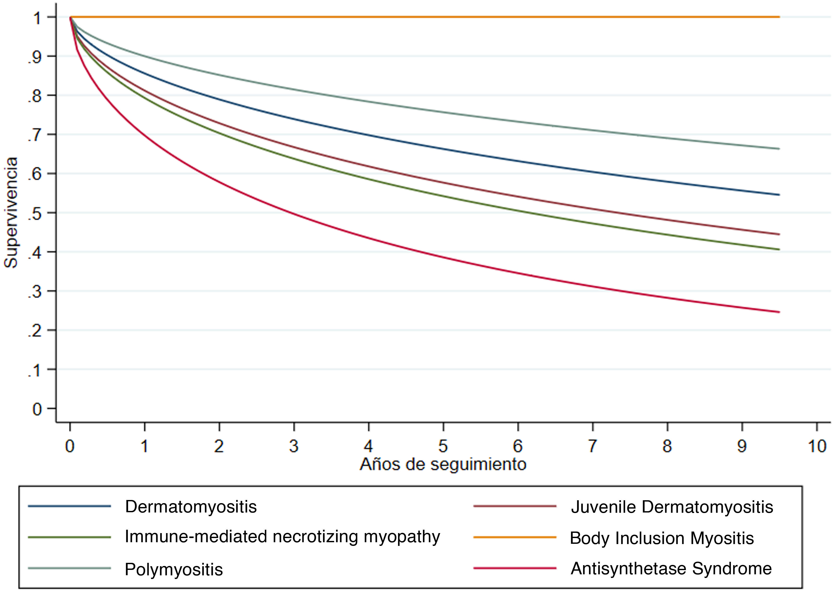
ResultsA total of 278 patients followed for a mean of 6.60 years were included. Of these, 70.9% were women, with a median age of 57 (IQR 40–66) years. Dermatomyositis (60.1%) and polymyositis (28.1%) were the most frequent phenotypes. Relapse-free survival at one year was 86.3%, and at five years, 68.3%, with a median relapse-free time of 13.8 years. By phenotype, the lowest median relapse-free survival rates were observed for antisynthetase syndrome at 3 years, immune-mediated necrotizing myopathy at 6 years, and juvenile dermatomyositis at 7.3 years.
ConclusionRelapse-free survival is highly heterogeneous according to the clinical phenotype of idiopathic inflammatory myopathy, being lowest in patients with antisynthetase syndrome.
Las recaídas de la enfermedad en miopatías inflamatorias idiopáticas (MII) tienen un peso relevante en la carga de la enfermedad. No obstante, los estudios colombianos con seguimientos a largo plazo son escasos.
ObjetivoEstimar la supervivencia libre de recaída de la enfermedad a diez años en pacientes con miopatía inflamatoria idiopática atendidos en un centro médico especializado en enfermedades autoinmunes con presencia en diferentes ciudades de Colombia.
Materiales y métodosSeguimiento retrospectivo a una cohorte basada en registros médicos. Se incluyeron todos los pacientes con diagnóstico de miopatía inflamatoria idiopática atendidos entre enero de 2012 y junio de 2022. Como variable de desenlace se definió el tiempo a la recaída. Adicionalmente, se caracterizaron variables demográficas, clínicas, de laboratorio y de tratamiento. Los análisis se realizaron mediante regresión de supervivencia de Weibull para datos censurados en intervalo.
ResultadosSe incluyeron 278 pacientes, seguidos en promedio por 6,60 años, 70,9% mujeres, con edad mediana de 57 años (rango intercuartil 40–66). La dermatomiositis (60,1%) y la polimiositis (28,1%) fueron los fenotipos más frecuentes. La supervivencia libre de recaída a un año fue de 86,3% y a cinco años de 68,3%, con una mediana de tiempo libre de recaída de 13,8 años. Por fenotipo, las menores medianas de supervivencia libre de recaída se observaron para síndrome antisintetasa, con 3 años, miopatía necrosante inmunomediada con 6 años y dermatomiositis juvenil con 7,3 años.
ConclusiónLa supervivencia libre de recaída es muy heterogénea, según el fenotipo clínico de la miopatía inflamatoria idiopática; es menor en pacientes con síndrome antisintetasa.
Idiopathic inflammatory myopathies (IIMs) are a heterogeneous group of diseases characterized by immune-mediated muscle damage. This group includes dermatomyositis (DM), polymyositis (PM), overlap myositis (OS), inclusion body myositis (IBM), and immune-mediated necrotizing myopathy (INM) [1]. Their incidence ranges from 2.1 to 7.7 cases per million inhabitants, with a 10-year mortality rate of 31%, with cardiorespiratory disorders and malignancies being the main causes of death [2]. As it is a chronic disease, comorbidities and relapses are frequent, increasing the medical and economic burden [3,4].
The definition of relapse in IIMs is not clear. In practice, a relapse is diagnosed when symptoms recur or worsen after improvement with initial treatment [5]. Recent studies indicate that only 20–40% of patients with PM or DM achieve clinical remission, while 60–80% experience a clinical course with recurrent relapses. Remissions without control medication are rare, except in juvenile dermatomyositis (jDM) [6,7].
Several factors have been identified as predictors of relapse and adverse outcomes such as death, treatment resistance, and disease progression. These factors include advanced age, a long period between symptom onset and treatment, cardiac or pulmonary involvement, the presence of underlying malignancies, extensive skin affection, myositis-specific antibodies, as well as dysphonia and dysphagia at diagnosis. These factors have been derived from studies with heterogeneous populations and do not include data specific to the Colombian population [5].
In Colombia, the understanding of autoimmune muscular diseases is in its early stages. While some studies describe the situation in the country’s major cities, none specifically address the risk of relapse and its progression over time.
This study aimed to estimate the 10-year relapse-free survival in IIM subjects treated at a medical center specializing in autoimmune diseases with presence in different cities in Colombia.
MethodsThe report follows the recommendations of the STROBE guide for observational studies [8].
Design and contextA retrospective observational follow-up study was conducted on a cohort of patients diagnosed with IIM who were treated between January 2012 and June 2022 at Artmedica, a medical center specializing in autoimmune diseases with locations in seven Colombian cities: Medellin, Rionegro, Manizales, Pereira, Armenia, Cali, and Tunja. The follow-up period includes the time from the time of diagnosis until the occurrence of relapse, the date of the last assessment, or up to 10 years of follow-up.
ParticipantsIndividuals with a confirmed diagnosis of inflammatory myopathy according to the Bohan and Peter criteria were included [9]. Subjects diagnosed with toxic, infectious, endocrine, or metabolic myopathy, as well as patients with incomplete data, were excluded. Potentially eligible subjects were initially identified using the ICD-10 code corresponding to inflammatory myopathy (G72.8, M33.0, M33.1, M33.2, M33.9). Subsequently, five Internal Medicine fellows applied the inclusion and exclusion criteria by analyzing the complete electronic health record.
Variables and sources of informationThe outcome variable was the occurrence of relapse, as determined by the treating rheumatologist during follow-up. Additionally, information was collected on treatment, treatment response, and the need for therapy change due to refractoriness or relapse, such as persistent muscle weakness and elevated CK despite immunosuppressive treatment with at least two or more drugs at maximum tolerated doses administered sequentially or concomitantly, identified by the rheumatologist during consultation, or the appearance of new symptoms that may or may not be associated with enzyme elevation, without any other identifiable cause [9].
Regarding the subject’s features, demographic (age, sex), clinical (type of myopathy, manifestations, comorbidities), paraclinical (antibodies, biopsy), and treatment variables (medications used) were extracted. The researchers used a standardized data collection form.
Biases and sample sizeTo mitigate information bias, researchers extracted data from medical records using a standardized form in Google Forms. The institution's clinical information group conducted training and standardization. Additionally, the data extraction process was audited. Variable definition and the extraction process were supervised by a rheumatologist. To reduce selection bias, patient identification was performed in two stages: searching for potentially eligible individuals in administrative records and confirming eligibility criteria by reviewing medical records.
Statistical analysisDescriptive analysis of patients’ characteristics was performed using frequencies and percentages for categorical variables and median and interquartile range (IQR) for quantitative variables. These results are presented according to the clinical phenotype of IIM. Relapse-free survival by phenotype was modeled using interval-censored Weibull survival regression. The cumulative survival function is presented graphically, along with median relapse-free times. The Weibull model was used because it obtained the best Akaike and Bayesian goodness-of-fit indices. Exploratory analyses of factors associated with relapse-free time were also performed and expressed as hazard ratios (HR) with 95% confidence intervals (95% CI) and p-values. The significance level was set at p < 0.05. Analyses were performed using Stata version 16.1 (College Station, TX, USA).
Ethical considerationsThe protocol of this study was approved by the Institutional Ethics Committee for Research in Human Beings of the Universidad CES (AE-960) as research without risk, in accordance with Resolution 8430 of 1993. Upon admission, informed consent was obtained from the patients for the use of their medical history data under conditions of confidentiality and anonymity for research purposes.
ResultsParticipantsOf the 460 potentially eligible subjects, 278 were included and followed for an average of 6.60 years. By municipality of care, 66.9% of the patients were from the Medellin site, 8.3% from Pereira, 5.0% from Manizales, 4.3% from Tunja, 4.0% from Rionegro, 3.6% from Armenia, 3.2% from Monteria, 1.8% from Cali, 1.4% from Apartado, and 1.1% from Pasto.
Descriptive dataOf the total of individuals included, 70.9% were women, and the median age at diagnosis was 57 years (IQR 40–66). DM was the most frequent clinical phenotype, observed in 60.1% of patients, followed by PM, observed in 28.1%. Other phenotypes were less frequent: JDM (4.7%), INM (4.0%), antisynthetase syndrome (ASS) (1.8%), and IBM (1.4%).
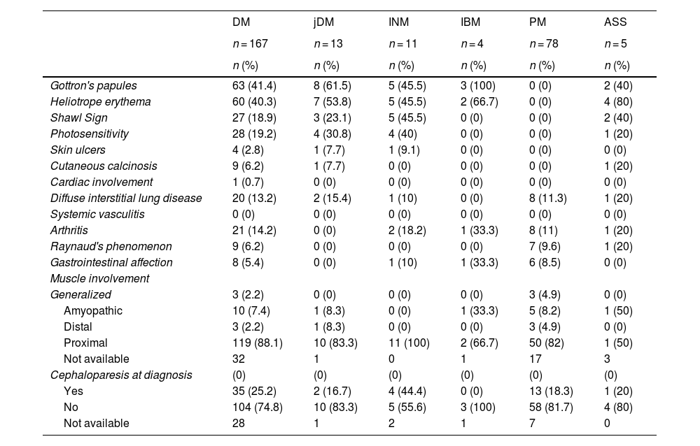
Clinical featuresTable 1 depicts the clinical characteristics according to clinical phenotype. The most common clinical signs varied with the phenotype. Interstitial lung disease was present in 13.2% of DM cases and in 15.4% of JDM cases. Regarding the distribution of muscle involvement, most cases presented proximal involvement, and cephaloparesis at the time of diagnosis was observed in 25.2% of DM cases and in 16.7% of JDM cases.
Clinical characteristics, according to phenotype of idiopathic inflammatory myopathy (n = 278).
| DM | jDM | INM | IBM | PM | ASS | |
|---|---|---|---|---|---|---|
| n = 167 | n = 13 | n = 11 | n = 4 | n = 78 | n = 5 | |
| n (%) | n (%) | n (%) | n (%) | n (%) | n (%) | |
| Gottron's papules | 63 (41.4) | 8 (61.5) | 5 (45.5) | 3 (100) | 0 (0) | 2 (40) |
| Heliotrope erythema | 60 (40.3) | 7 (53.8) | 5 (45.5) | 2 (66.7) | 0 (0) | 4 (80) |
| Shawl Sign | 27 (18.9) | 3 (23.1) | 5 (45.5) | 0 (0) | 0 (0) | 2 (40) |
| Photosensitivity | 28 (19.2) | 4 (30.8) | 4 (40) | 0 (0) | 0 (0) | 1 (20) |
| Skin ulcers | 4 (2.8) | 1 (7.7) | 1 (9.1) | 0 (0) | 0 (0) | 0 (0) |
| Cutaneous calcinosis | 9 (6.2) | 1 (7.7) | 0 (0) | 0 (0) | 0 (0) | 1 (20) |
| Cardiac involvement | 1 (0.7) | 0 (0) | 0 (0) | 0 (0) | 0 (0) | 0 (0) |
| Diffuse interstitial lung disease | 20 (13.2) | 2 (15.4) | 1 (10) | 0 (0) | 8 (11.3) | 1 (20) |
| Systemic vasculitis | 0 (0) | 0 (0) | 0 (0) | 0 (0) | 0 (0) | 0 (0) |
| Arthritis | 21 (14.2) | 0 (0) | 2 (18.2) | 1 (33.3) | 8 (11) | 1 (20) |
| Raynaud's phenomenon | 9 (6.2) | 0 (0) | 0 (0) | 0 (0) | 7 (9.6) | 1 (20) |
| Gastrointestinal affection | 8 (5.4) | 0 (0) | 1 (10) | 1 (33.3) | 6 (8.5) | 0 (0) |
| Muscle involvement | ||||||
| Generalized | 3 (2.2) | 0 (0) | 0 (0) | 0 (0) | 3 (4.9) | 0 (0) |
| Amyopathic | 10 (7.4) | 1 (8.3) | 0 (0) | 1 (33.3) | 5 (8.2) | 1 (50) |
| Distal | 3 (2.2) | 1 (8.3) | 0 (0) | 0 (0) | 3 (4.9) | 0 (0) |
| Proximal | 119 (88.1) | 10 (83.3) | 11 (100) | 2 (66.7) | 50 (82) | 1 (50) |
| Not available | 32 | 1 | 0 | 1 | 17 | 3 |
| Cephaloparesis at diagnosis | (0) | (0) | (0) | (0) | (0) | (0) |
| Yes | 35 (25.2) | 2 (16.7) | 4 (44.4) | 0 (0) | 13 (18.3) | 1 (20) |
| No | 104 (74.8) | 10 (83.3) | 5 (55.6) | 3 (100) | 58 (81.7) | 4 (80) |
| Not available | 28 | 1 | 2 | 1 | 7 | 0 |
Frequencies (n) and percentages (%) are presented based on complete data.
DM: Dermatomyositis; jDM: juvenile Dermatomyositis; INM: Immune-mediated Necrotizing Myopathy; IBM: Inclusion Body Myositis; PM: Polymyositis; ASS: Antisynthetase Syndrome.
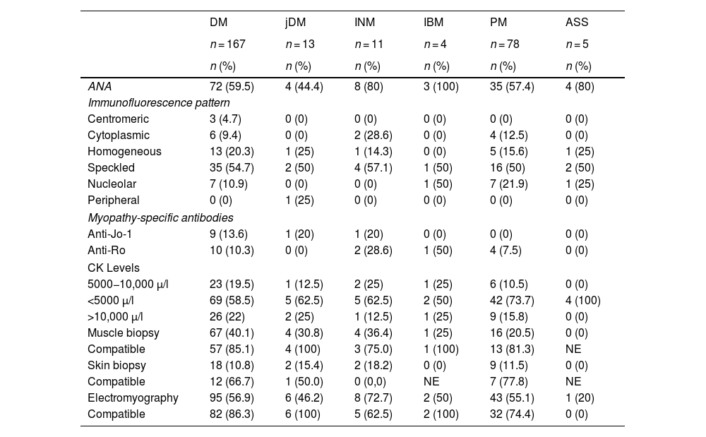
Table 2 presents the serological features and other laboratory findings according to clinical phenotype. Antinuclear antibodies (ANA) were detected with variability in the different types of myopathies: 59.5% in DM, 44.4% in jDM, 80% in INM, 100% in IBM, 57.4% in PM, and 80% in ASS. The most frequent immunofluorescence patterns for ANA were speckled and homogeneous.
Serological features and other laboratories, according to phenotype of idiopathic inflammatory myopathy (n = 278).
| DM | jDM | INM | IBM | PM | ASS | |
|---|---|---|---|---|---|---|
| n = 167 | n = 13 | n = 11 | n = 4 | n = 78 | n = 5 | |
| n (%) | n (%) | n (%) | n (%) | n (%) | n (%) | |
| ANA | 72 (59.5) | 4 (44.4) | 8 (80) | 3 (100) | 35 (57.4) | 4 (80) |
| Immunofluorescence pattern | ||||||
| Centromeric | 3 (4.7) | 0 (0) | 0 (0) | 0 (0) | 0 (0) | 0 (0) |
| Cytoplasmic | 6 (9.4) | 0 (0) | 2 (28.6) | 0 (0) | 4 (12.5) | 0 (0) |
| Homogeneous | 13 (20.3) | 1 (25) | 1 (14.3) | 0 (0) | 5 (15.6) | 1 (25) |
| Speckled | 35 (54.7) | 2 (50) | 4 (57.1) | 1 (50) | 16 (50) | 2 (50) |
| Nucleolar | 7 (10.9) | 0 (0) | 0 (0) | 1 (50) | 7 (21.9) | 1 (25) |
| Peripheral | 0 (0) | 1 (25) | 0 (0) | 0 (0) | 0 (0) | 0 (0) |
| Myopathy-specific antibodies | ||||||
| Anti-Jo-1 | 9 (13.6) | 1 (20) | 1 (20) | 0 (0) | 0 (0) | 0 (0) |
| Anti-Ro | 10 (10.3) | 0 (0) | 2 (28.6) | 1 (50) | 4 (7.5) | 0 (0) |
| CK Levels | ||||||
| 5000−10,000 µ/l | 23 (19.5) | 1 (12.5) | 2 (25) | 1 (25) | 6 (10.5) | 0 (0) |
| <5000 µ/l | 69 (58.5) | 5 (62.5) | 5 (62.5) | 2 (50) | 42 (73.7) | 4 (100) |
| >10,000 µ/l | 26 (22) | 2 (25) | 1 (12.5) | 1 (25) | 9 (15.8) | 0 (0) |
| Muscle biopsy | 67 (40.1) | 4 (30.8) | 4 (36.4) | 1 (25) | 16 (20.5) | 0 (0) |
| Compatible | 57 (85.1) | 4 (100) | 3 (75.0) | 1 (100) | 13 (81.3) | NE |
| Skin biopsy | 18 (10.8) | 2 (15.4) | 2 (18.2) | 0 (0) | 9 (11.5) | 0 (0) |
| Compatible | 12 (66.7) | 1 (50.0) | 0 (0,0) | NE | 7 (77.8) | NE |
| Electromyography | 95 (56.9) | 6 (46.2) | 8 (72.7) | 2 (50) | 43 (55.1) | 1 (20) |
| Compatible | 82 (86.3) | 6 (100) | 5 (62.5) | 2 (100) | 32 (74.4) | 0 (0) |
Frequencies (n) and percentages (%) are presented based on complete data. The percentages of compatible biopsies and electromyography are based on the total number of patients for whom this test was requested.
ANA: antinuclear antibodies; CK: creatine phosphokinase; DM: Dermatomyositis; jDM: juvenile Dermatomyositis; INM: Immune-mediated Necrotizing Myopathy; IBM: Inclusion Body Myositis; PM: Polymyositis; ASS: Antisynthetase Syndrome.
CK levels showed significant variations among the different types of myopathies. For example, in DM, 19.5% of patients had levels between 5000 and 10,000 µL, 58.5% had levels below 5000 µL, and 22% had levels above 10,000 µL. These proportions are comparable to those in jDM, where 12.5% had levels between 5000 and 10,000 µL, 62.5% had levels below 5000 µL, and 25% had levels above 10,000 µL. In INM, 25% had levels between 5000 and 10,000 µL, 62.5% had levels below 5000 µL, and 12.5% had levels above 10,000 µL. In IBM, a uniform distribution was observed across the categories. In PM, 10.5% had levels between 5000 and 10,000 µL, 73.7% had levels below 5000 µL, and 15.8% had levels above 10,000 µL. Finally, in SAS, all individuals had levels below 5000 µL.
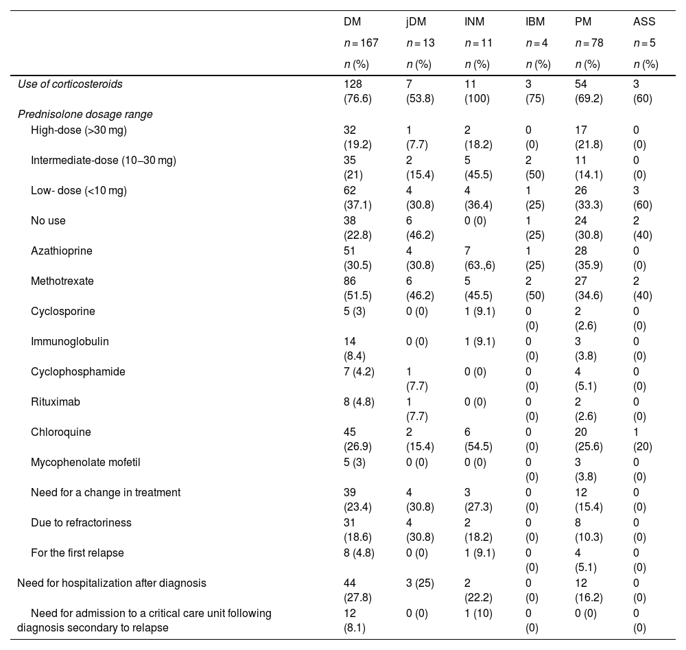
Treatment and response to therapyTable 3 presents data on treatment and response according to clinical phenotype. Regarding corticosteroid use, variability in prednisolone doses was observed among the different types of myopathies. In DM, the most used dose was intermediate (in 37.1% of cases), followed by low (in 21%). With respect to immunosuppressants, the most frequently used medication in DM and PM was azathioprine, while methotrexate was the most frequently employed in INM.
Treatment and response, according to idiopathic inflammatory myopathy phenotype (n = 278).
| DM | jDM | INM | IBM | PM | ASS | |
|---|---|---|---|---|---|---|
| n = 167 | n = 13 | n = 11 | n = 4 | n = 78 | n = 5 | |
| n (%) | n (%) | n (%) | n (%) | n (%) | n (%) | |
| Use of corticosteroids | 128 (76.6) | 7 (53.8) | 11 (100) | 3 (75) | 54 (69.2) | 3 (60) |
| Prednisolone dosage range | ||||||
| High-dose (>30 mg) | 32 (19.2) | 1 (7.7) | 2 (18.2) | 0 (0) | 17 (21.8) | 0 (0) |
| Intermediate-dose (10−30 mg) | 35 (21) | 2 (15.4) | 5 (45.5) | 2 (50) | 11 (14.1) | 0 (0) |
| Low- dose (<10 mg) | 62 (37.1) | 4 (30.8) | 4 (36.4) | 1 (25) | 26 (33.3) | 3 (60) |
| No use | 38 (22.8) | 6 (46.2) | 0 (0) | 1 (25) | 24 (30.8) | 2 (40) |
| Azathioprine | 51 (30.5) | 4 (30.8) | 7 (63.,6) | 1 (25) | 28 (35.9) | 0 (0) |
| Methotrexate | 86 (51.5) | 6 (46.2) | 5 (45.5) | 2 (50) | 27 (34.6) | 2 (40) |
| Cyclosporine | 5 (3) | 0 (0) | 1 (9.1) | 0 (0) | 2 (2.6) | 0 (0) |
| Immunoglobulin | 14 (8.4) | 0 (0) | 1 (9.1) | 0 (0) | 3 (3.8) | 0 (0) |
| Cyclophosphamide | 7 (4.2) | 1 (7.7) | 0 (0) | 0 (0) | 4 (5.1) | 0 (0) |
| Rituximab | 8 (4.8) | 1 (7.7) | 0 (0) | 0 (0) | 2 (2.6) | 0 (0) |
| Chloroquine | 45 (26.9) | 2 (15.4) | 6 (54.5) | 0 (0) | 20 (25.6) | 1 (20) |
| Mycophenolate mofetil | 5 (3) | 0 (0) | 0 (0) | 0 (0) | 3 (3.8) | 0 (0) |
| Need for a change in treatment | 39 (23.4) | 4 (30.8) | 3 (27.3) | 0 (0) | 12 (15.4) | 0 (0) |
| Due to refractoriness | 31 (18.6) | 4 (30.8) | 2 (18.2) | 0 (0) | 8 (10.3) | 0 (0) |
| For the first relapse | 8 (4.8) | 0 (0) | 1 (9.1) | 0 (0) | 4 (5.1) | 0 (0) |
| Need for hospitalization after diagnosis | 44 (27.8) | 3 (25) | 2 (22.2) | 0 (0) | 12 (16.2) | 0 (0) |
| Need for admission to a critical care unit following diagnosis secondary to relapse | 12 (8.1) | 0 (0) | 1 (10) | 0 (0) | 0 (0) | 0 (0) |
Frequencies (n) and percentages (%) are presented based on complete data.
DM: Dermatomyositis; jDM: Juvenile Dermatomyositis; INM: Immune-mediated Necrotizing Myopathy; IBM: Inclusion Body Myositis; PM: Polymyositis; ASS: Antisynthetase Syndrome.
Concerning the need to change treatment due to refractoriness or relapse, the following proportions were observed according to the type of myopathy: 46.2% in jDM, 29.3% in DM, 23.1% in PM, 20% in ASS, and 18.2% in INM. As for treatment refractoriness, the frequencies were as follows: 30.8% in jDM, 18.6% in DM, 18.2% in INM, and 10.3% in PM. Regarding the need for hospitalization after diagnosis, the rates were 27.8% for DM, 25% for jDM, 22.2% for INM, and 16.2% for PM.
OutcomeFig. 1 shows the cumulative survival by clinical myopathy phenotype. The lowest median relapse-free survival was observed for ASS at 3 years, followed by INM at 6 years, and jDM at 7.3 years. For the remaining phenotypes, cumulative survival did not reach 50% at 10 years of follow-up. No relapses were observed among the 4 patients with IBM. In all individuals, relapse-free survival was estimated at 86.3% at 1 year and 68.3% at 5 years, with a median relapse-free survival time of 13.8 years.
Exploratory analyses
When analyzing all included IIM patients, only an association between anti-Jo1 antibody positivity and time to relapse was identified (HR = 2.9; 95% CI, 1.3–6.7). When performing analyses by phenotype subgroups, additional features were found among patients with DM. In these subjects, time to relapse was shorter among individuals with refractoriness (HR = 2.0; 95% CI, 1.1–4.1) and arthritis (HR = 2.0; 95% CI, 1.1–4.4).
DiscussionIt is essential to highlight the scarcity of studies on myopathies in Latin American populations, despite their significant clinical and socioeconomic relevance, as well as their impact on health systems. Our study contributes to the existing literature, offering additional data on follow-up and relapses in these subjects [10,11].
Concerning the demographic characteristics of our cohort, a predominance of females (70.9%) was observed, consistent with reports from various cohorts [12,13]. DM was the most prevalent clinical phenotype, observed in 60.1%, followed by PM (28.1%), jDM (4.7%), INM (4.0%), IBM (1.4%), and ASS (1.8%). This predominance of DM and PM, widely described in the literature, reflects similar distribution patterns of these disorders in populations studied globally. Unfortunately, myositis-specific antibody (MSA) results were only available for a minority of patients, preventing more precise phenotyping of the entire spectrum of inflammatory myopathies and enabling more accurate differentiation of myopathy subtypes [14].
Since the diagnosis of most patients was confirmed at a time when the availability of the specific antibody panel in the country was very low, clinical and electromyographic parameters predominated, and muscle biopsy was used to a lesser extent due to the difficulty in the opportunity to perform it. On average, 30.5% of individuals underwent muscle biopsy, of which 72.3% showed abnormalities suggestive of an inflammatory myopathy. However, muscle biopsy can be negative due to patchy involvement or prior glucocorticoid use, and its use is not essential; even a negative result does not rule out the disease [15].
Electromyography was performed in an average of 50% of the population, with myopathic findings supporting the diagnosis in 83.5% of patients. This was due to its greater availability and sensitivity in detecting muscle abnormalities such as increased insertional activity and spontaneous activity with fibrillations, predominantly without nerve conduction abnormalities, among other factors [16]. Similarly, low use of skin biopsies was demonstrated, given that the clinical findings are very characteristic, especially in DM. However, compatible histopathological findings helped rule out other disorders with a similar appearance when the clinician had doubts. In the current cohort of 18 biopsies performed in patients with DM, 33.3% were compatible with IIM.
In our study, relapse-free survival was 86.3% at one year and 68.3% at five years, with most relapses occurring within the first three years. These results are like those reported in other series where relapse rates range from 35% to 65%, depending on the observation period. For example, Agarwal et al. reported that, in the follow-up of 53 patients with a mean observation period of 65±43 months, 65% experienced at least one relapse and 32% suffered multiple relapses, the first occurring between three and 69 months, with a mean of 17.5±14.4 months, after the start of treatment, and with a relapse-free survival rate of approximately 85% at one year and close to 30% at five years. However, it is highlighted that most events occurred in the first 2 years. Additionally, it is mentioned that most relapses (25/33) took place during the withdrawal of the initial treatment [17].
Similarly, Ponyi et al. found in their series of 35 subjects followed for 78 months that 37.1% experienced at least one relapse, while only 2.9% showed a chronic course of multiple relapses. Strikingly, relapse-free survival at 12 months was again around 80% and at 60 months 65%, with most relapses occurring within the first 2 years [18]. Likewise, in the series by Phillips et al., which included 50 individuals and further distinguished between PM, DM, and overlap syndrome, with a follow-up of up to 13 years (mean 2.4, 4.7, and 4.9 years, respectively), an average relapse rate of 60% was found, slightly more than half occurring within the first 2 years after treatment initiation, but relapses can occur up to slightly beyond 4 years; of these, 33% occurred during corticosteroid withdrawal and the remaining occurred during the stable phase of the disease or in the absence of treatment [19].
These data reflect a similar pattern to that found in our cohort, but all patients relapsed while on treatment. However, due to the nature of the data collection, it cannot be determined whether a factor as important as adherence played a role. Furthermore, this highlights the idea that the first four years after diagnosis may be the most critical period and one that requires close attention due to the likelihood of relapse.
When we take as a reference other studies with a follow-up period as long as ours, we find the series by Janardana et al., who followed 175 subjects for 12 years and report a relapse-free survival of 10.08 years, which is moderately lower than ours (13.8 years); however, it should be clarified that this is an estimate and not such a precise data derived from the CIs of both studies [7].
Due to the small number of events found in our cohort, it was difficult to identify factors associated with relapse; only anti-Jo1 positivity in the context of ASS was found to be associated with a higher risk of developing a relapse over time. However, this is important because it provides a little more clarity regarding the predictors of sustained remission rather than relapse. Thus, Watanabe et al. showed that in the presence of SMA antibodies, including anti-MDA5, anti-NXP2, anti-SAE, anti-SRP, anti-TIF1y, and SAS antibodies (not exclusively anti-Jo1), there is a lower probability of achieving sustained remission, a phenomenon that we observed in our study, even though we only had the anti-Jo1 result available and not the results for the other SMA antibodies [20]. In the current cohort, a low proportion of subjects had myopathy panel data, partly because most were diagnosed before 2022, when this diagnostic tool was only available in practice. However, these findings underscore the need to increase the use of this diagnostic tool for better characterization of myopathy subtypes and to establish more accurate prognoses, even in individuals who have already been diagnosed based on clinical criteria.
The study faced several limitations that must be taken into account when interpreting the results. The main limitation is the retrospective nature of the study, as the data were obtained from secondary sources by trained medical personnel from standardized records in an institutional medical history system. Furthermore, a specific sample size was not calculated; instead, all patients available in the databases were included. Due to significant data loss resulting from the extraction of data from previous databases, calculating some statistical associations would not have served the purpose of the study.
Another limitation is the restricted availability and high cost of MSA, which prevented all patients from undergoing these tests, potentially affecting the accuracy of diagnosis, prognosis, and follow-up. Furthermore, although the data were collected from different centers, they all belonged to the same institution, thus excluding other institutions in the country that also treat these types of patients.
Exploratory analyses should be treated with caution due to the small sample size and limited number of relapse events. Finally, treatment adherence was not consistently monitored, preventing a definitive assessment of its impact as a risk factor for clinical relapses. These limitations underscore the need for cautious interpretation of the findings and suggest areas for improvement in future research.
ConclusionsThis study highlights the importance of understanding the long-term behavior of inflammatory myopathies in the Colombian population. A significant proportion of patients experienced frequent relapses, with a median relapse-free interval of 13.8 months. The factors described, including specific clinical characteristics and the presence of myositis-specific antibodies, are crucial for risk stratification and the development of personalized treatment. Furthermore, relapses during treatment withdrawal underscore the need for careful management strategies and continuous monitoring. This study contributes to the understanding of these diseases in local contexts and underscores the need for further research to optimize treatment protocols and improve clinical outcomes in patients with inflammatory myopathies.
CRediT authorship contribution statementJCD and NP conceived the study, and DFRG performed the analyses. All authors contributed to the design, data acquisition, and interpretation; PAG, AEC, MAGD, MMM, and MPR wrote the first draft. All authors contributed to the critical review of the intellectual content and approved the version presented.
FinancingNone.
Data availabilityData are accessible through the corresponding author with prior authorization from Artmedica and the Ethics Committee for Human Research at Universidad CES, after assessment of the motivations and conditions of use.
None.


