

metricas
Revista Colombiana de Reumatología (English Edition) Are ANCA antibodies necessary in the definition of interstitial pneumonia with a...
Journal Information
Vol. 32. Issue 4.
Pages 299-422 (October - December 2025)
Cite
Cite
Share
Download PDF
More article options
Visits
1546
Vol. 32. Issue 4.
Pages 299-422 (October - December 2025)
Case report
Full text access

Are ANCA antibodies necessary in the definition of interstitial pneumonia with autoimmune features? Illustrative case presentation

¿Los anticuerpos ANCA son necesarios en la definición de la neumonía intersticial con características autoinmunes? Presentación de un caso ilustrativo
Visits
1546
Andrea Cabra Sierra
Corresponding author
ancabras@unal.edu.co

Corresponding author.
, Jorge Lechuga-Ortiz, Yimy Medina Velásquez, Luis Javier Cajas Santana
Department of Internal Medicine, Faculty of Medicine, Universidad Nacional de Colombia, Bogotá, Colombia
This item has received
Article information
Abstract
Full Text
Bibliography
Download PDF
Statistics
Figures (3)
fig0005
fig0010
fig0015
Tables (1)
Table 1. Profile of autoimmunity studies of the patient.
Tables
Abstract

We present the case of an adult female patient with a diagnosis of IPAF (Interstitial pneumonia with autoimmune features) based on serological and morphological domains according to the diagnostic consensus, in whom the additional positivity of an autoantibody not considered until now within the serological domain is highlighted, namely antineutrophil cytoplasmic antibodies (ANCA)-C. The patient failed multiple treatments. In the medical literature, there is growing interest in defining the usefulness of including ANCA as a significant autoantibody in the diagnosis of IPAF. This is the first case reported in Colombia.

Keywords:
Interstitial lung disease
Antibodies
Antineutrophil cytoplasmic
IPAF
Resumen

Se presenta el caso de una paciente adulta con diagnóstico de IPAF (interstitial pneumonia with autoinmune features) basado en dominios serológico y morfológico, según el consenso diagnóstico, en quien se destaca la positividad adicional de un autoanticuerpo no considerado hasta el momento dentro del dominio serológico como lo es el anticuerpo anticitoplasma de neutrófilos (ANCA)-C. La paciente tuvo falla en múltiples tratamientos. En la literatura médica hay un creciente interés por definir la utilidad de incluir los ANCA como autoanticuerpos útiles en el diagnóstico de IPAF. Este es el primer caso reportado en Colombia.


Palabras clave:
Enfermedad pulmonar intersticial
Anticuerpos
Anticitoplasma de neutrófilos
IPAF
Full Text
Clinical case

A 44-year-old woman referred to rheumatology (from pulmonology) for two years of cough with whitish sputum and progressive exertional dyspnea (2/4 mMRC [modified Medical Council Research] at the time of rheumatological evaluation). After ruling out other causes of idiopathic interstitial lung disease (ILD), the patient was referred from pulmonology with a diagnosis of interstitial pneumonia with autoimmune features (IPAF) (clinical domain: does not present; serological domain: antinuclear antibodies [ANA] 1:80 AC-4 pattern; morphological domain: unclassifiable interstitial tomographic pattern [Fig. 1]). The patient had transbronchial cryobiopsy with organizing pneumonia. Infections were ruled out by bronchoalveolar lavage studies (Gram, ZN, India ink, KOH stains, cultures for common germs and Mycobacterium tuberculosis, and polymerase chain reaction for M. tuberculosis), as well as bleeding and malignancy by cytology.

Figure 1.

High-resolution CT scan of the chest of the patient with findings of interstitial lung disease (unclassifiable usual interstitial pneumonia pattern). Diffuse distribution of reticular opacities without apico-basal gradient, random micronodules in upper lobes, and bronchiolectasis.

As antecedents, the patient presented hypothyroidism treated with levothyroxine and denied occupational or exposure history. On the review by systems, she denied orthopnea, paroxysmal nocturnal dyspnea, edema, chest pain, nocturnal snoring, fever, Raynaud's phenomenon, bleeding, alopecia, photosensitivity, ulcers, muscle weakness, dry symptoms, arthralgias, and morning stiffness. On physical examination, she presented fine bilateral pulmonary rales (he had no cutaneous lesions, digital hypocratism, or abnormalities in vital signs), and the lung ultrasonography showed granular pleura and B lines (Fig. 2).

Figure 2.

Lung ultrasound of the patient with findings of pulmonary interstitial disease. The red arrows point to B lines, while the granular pleural line is indicated with a yellow arrow.

The autoimmunity studies are described in Table 1. The pulmonary function tests (PFTs) included: spirometry, dated December 23, 2021, with forced vital capacity (FVC) of 74%, and forced expiratory volume in one second (FEV) of 83%. Control of November 9, 2022 with FVC: 53%, and FEV1: 63%. Diffusing capacity of lungs for carbon dioxide (DLCO) adjusted for alveolar volume on December 23, 2021 of 72%, and on November 9, 2022 of 49%. Until now she had received 6 months of prednisolone 1 mg/kg/day and azathioprine 50 mg every 12 h, without symptomatic improvement.

Table 1.

Profile of autoimmunity studies of the patient.

Autoimmunity studies
Positive  Negative 
ANA: 1:80 fine granular nuclear pattern (AC-4)Rheumatoid factor, anti-cyclic citrullinated peptide 
Anti MPO, anti-PR3 (ELISA method) 
Anti-Ro, anti-La, anti-RNP, anti-Sm 
Anti-neutrophil cytoplasmic antibodies × indirect immunofluorescence: cytoplasmic pattern (ANCA-C) in 1:40 dilutionsAnti-double stranded DNA 
Anti-Scl70 
Specific panel for myositis (16 antibodies: Mi2α, Mi2β, TIF1γ, MDA5, NXP2, SAE1, Ku, PM/Scl75, PM/Scl75, Jo1, SRP, PL7, PL12, EJ, OJ, Ro52) 

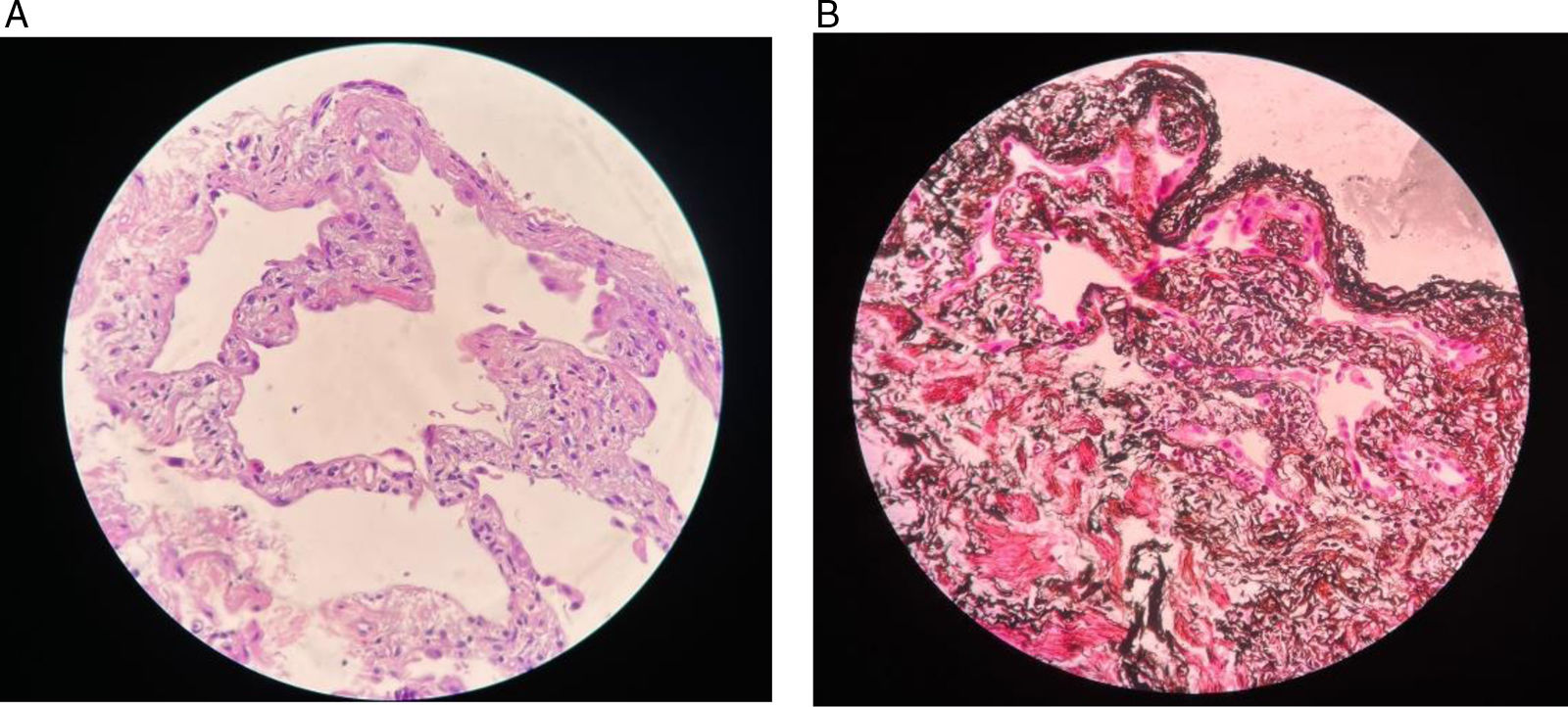
Rheumatology considered that the patient had IPAF, without ANCA vasculitis, since there was no evidence of small or medium-vessel vasculitis. And she only presented ANCA-C 1: 40 dilutions as a manifestation of such vasculitis. She has been presenting FVC and DLCO with progressive detriment and dyspnea without improvement, despite treatment with steroids and azathioprine. The case was presented to a multidisciplinary team (rheumatology-pulmonology-pathology-radiology), where cryobiopsy was reconsidered as non-specific interstitial pneumonia-fibrotic phase with acute inflammation (Fig. 3) + bronchiolectasis.

Figure 3.

Histopathological images of the patient with a finding of non-specific interstitial pneumonia. Representation of the pulmonary parenchyma with homogeneous thickening of the interalveolar septa without distortion of the architecture, and with slight lymphoplasmacytic inflammatory infiltrate. The fibrotic component that was seen in other areas of the tissue under study is not represented in the microscopy photographs provided by pathology.

Due to progressive deterioration of PFT and persistent dyspnea, monthly intravenous cyclophosphamide (CYC) 0.75 g/m2 of body surface area was ordered. In control after the third dose of CYC, there was an increase in dyspnea (3/4 mMRC), FVC 50% and DLCO-AV 47%. For this reason, and in view of the worsening of her ILD, without improvement with steroids, azathioprine, CYC and pulmonary rehabilitation, nintedanib 150 mg every 12 h was indicated. The patient developed diarrhea as a side effect, so it was reduced to 100 mg every 12 h with better tolerance. After 3 months with nintedanib, the patient did not present improvement in dyspnea, which is why she was referred to the lung transplant board on a multidisciplinary basis.

Discussion

IPAF is a relatively recent concept with areas for discussion.1 In this particular case, the selection of autoantibodies in the IPAF classification criteria is highlighted, since only some were taken into account, which do not represent all those involved in the immune-mediated pathology of ILD.2–4 An example of the above is the present case of IPAF, since the patient had ANA (included in the serological domain) and ANCA-C (not included in such domain). This brings to discussion the role that ANCAs may have in IPAF, since their presence in patients with vasculitis is related to a higher probability of interstitial lung involvement (mainly in the form of usual interstitial pneumonia). In addition, it is necessary to make clarifications about the ANA-ANCA relationship.5–8:

  • The probable interference of ANA in the measurement of ANCA is described, but in the form of ANCA-P not of ANCA-C (pattern presented by the patient).

  • ANCAs as false positives in patients with ANA are usually associated with other specific autoantibodies: antiSM, antiRNP or antiDNA, which were negative in the patient of the case.

  • The presence of ANA and other antibodies such as antiDNA has been described in subjects with ANCA vasculitis with frequencies of up to 50% for ANA, but mainly in cases of ANCA-P/MPO vasculitis.

ILD may begin early, on average 2.2 years before the formal diagnosis of ANCA vasculitis, and it can be seen more frequently in patients older than 60 years and with anti-MPO.9–11 It is not found described in that was reviewed whether there are cut-off points of ANCA titers to consider a higher or lower risk of ILD.

The sera from patients with ANCA-C react against proteinase-3 (PR-3) in 80–90% of cases, and may also react against other antigens such as MPO, elastase, cationic protein 57 or cathepsin G in the remaining 10–20% of sera. ANCA-P sera typically react against MPO in 40–70% of cases; the rest react against elastase, cathepsin G, azurocidin, lactoferrin, or lysozyme.12 It is noteworthy that the patient, despite being positive for ANCA-C, has not had any vasculitic manifestations during her follow-up that would allow her to be classified as such.

The prevalence of ILD in ANCA vasculitis has been described with ranges between 2.7% (Europe) and 39.1% (Asia), a situation that may reflect ethnic differences or differences in the search for the disease.13,14 In parallel, it has also been described that subjects initially considered to have pulmonary fibrosis have later developed ANCA vasculitis, which could reinforce the theory that ILD may be an infrequent, early and isolated manifestation of vasculitic activity limited to the lung.15 The presentation of ILD and ANCA (without a diagnosis of vasculitis) is found in the literature as scarce reports.16–18

The importance of an adequate interpretation of ANCAs in ILD lies in the fact that, once the complete picture is established (26% of cases with ANCA vasculitis develop ILD), it is known that: a) mortality can be up to 22% for positive ANCA-C and 16% for those positive ANCA-P; b) ANCA-C and anti-MPO usually have progressive fibrosing phenotype, and c) ANCA-P are more related to progressive inflammatory and fibrosing phenotypes.19 Given the relevance of ANCA in ILD, their consideration in the IPAF criteria may be meritorious, as well as in the follow-up of these patients due to the possibility of evolving to vasculitis. Also noteworthy is the usefulness of lung ultrasound for the diagnosis of IPAF and other ILDs, as it is practical, reproducible, low cost and entails low radioactive exposure, as well as the usefulness of multidisciplinary teams that are consolidated as the gold standard for the comprehensive management of ILDs.20–22

Conclusion

Information is provided to the growing data on the relationship between ANCA (without a diagnosis of vasculitis) and IPAF, a situation that should be further explored given the probable need to include ANCAs within the serological domain of IPAF, due to their association with progression to vasculitis in this type of patients.

CRediT authorship contribution statement

Jorge Andrés Lechuga Ortiz: Collection of information of the clinical case, schematic organization of the structure of the clinical case, writing of the clinical case, obtention of case report image and their respective informed consents, topic review of the clinical case (ANCAs and relationship with interstitial lung disease), spelling and semantic review.

Andrea Inés Cabra Sierra: Engagement and outreach of the patient of the clinical case, collection of informed consent from the patient in the case, presentation of the case to the medical ethics committee of the Hospital Universitario Nacional de Colombia, writing of the clinical case, topic review of the clinical case (interstitial pneumonia with some autoimmune features), spelling and semantic review, completion and submission of electronic formats for presentation in ASOREUMA Journal and submission of the body of the document.

Yimy Medina Velásquez: Engagement and outreach of the patient of the clinical case, review of the form of the body of the case report, in-depth review of the case report, selection of the scientific journal to be submitted, thematic review of ANCAs and their relationship with interstitial lung disease.

Luis Javier Cajas Santana: Obtention of images of the case report and their respective informed consents, revision of the form of the body of the case report, in-depth thematic review of ANCAs and their relationship with interstitial pneumonia with some autoimmune features.

Ethical considerations

This case report has the respective informed consent of the patient for the handling of the clinical information and the respective publication, respecting the confidentiality and anonymity of their data. It has been considered that, as it is a medical case report, it requires a concept by a health research ethics committee (see annex at the end of this document), once the signature of the informed consent is obtained directly from the patient. This report does not contain personal information or images that would allow the identification of the patient.

Funding

The authors state that the preparation of this case report was financed with the authors’ own resources.

Declaration of competing interest

The authors of this case report declare that they do not present a conflict of interest for the publication of the document.

References
[1]
A. Fischer, K.M. Antoniou, K.K. Brown, J. Cadranel, T.J. Corte, R.M. Du Bois, et al.
An official European Respiratory Society/American Thoracic Society research statement: interstitial pneumonia with autoimmune features.
Eur Respir J, 46 (2015), pp. 976-987
[2]
E. Wilfong, G. Schroeder, A. Barnado, A. Fernandez-Codina, S. Shiboski, L. Chung, et al.
Myositis and systemic sclerosis spectrum IPAF patients are more likely to respond favorably to immunosuppression [abstract].
[3]
J. Graham, I. Bauer Ventura, C.A. Newton, S. Frankel, J. Joerns, J.H. Ryu, et al.
Myositis-specific antibodies identify a distinct interstitial pneumonia with autoimmune features phenotype.
[4]
A.S. Jee, S. Adelstein, J. Bleasel, T.J. Corte, C. Grainge, P.M. Hopkins, et al.
Role of autoantibodies in the diagnosis of connective-tissue disease ILD (CTD-ILD) and interstitial pneumonia with autoimmune features (IPAF).
J Clin Med, 6 (2017), pp. 51
[5]
M. Mahto, N. Rai, P.R. Das, R. Srivastava, A. Ahuja, A. Thakur, et al.
Interference of anti-nuclear antibodies on determination of anti-neutrophil cytoplasmic antibodies in patients suspected of vasculitis: a case series.
[6]
H. Yamada.
ANCA: associated lung fibrosis.
Semin Respir Crit Care Med, 32 (2011), pp. 322-327
[7]
X. Zhao, Q. Wen, Y. Qiu, F. Huang.
Clinical and pathological characteristics of ANA/anti-dsDNA positive patients with antineutrophil cytoplasmic autoantibody-associated vasculitis.
Rheumatol Int, 41 (2021), pp. 455-462
[8]
S. Deka, D. Kalita, U.S. Rekha, P. Mahanta, D. Rani, R. Shankar, et al.
interference of antinuclear antibody (ANA) in indirect immunofluorescence assay (IIFA)-based perinuclear antineutrophil cytoplasmic antibody (pANCA) interpretation.
Autoimmune Dis, 2022 (2022),
[9]
M.A. Alba, L.F. Flores-Suárez, A.G. Henderson, J.H. Stone, E.M. Miloslavsky.
Interstitial lung disease in ANCA vasculitis.
Autoimmun Rev, 16 (2017), pp. 722-729
[10]
B. Doliner, K. Rodríguez, S.B. Montesi, A. Doran, K. Batra, D. Feller-Kopman, et al.
Interstitial lung disease in ANCA associated vasculitis: associated factors, radiographic features and mortality.
Rheumatology, 62 (2023), pp. 716-725
[11]
N. Kishore, N. Gupta, A. Dhar, R. Agarwal, A. Chakrabarti, J.C. Suri, et al.
Interstitial lung disease with usual interstitial pneumonia pattern preceding the presentation of ANCA-associated vasculitis by 4 years: coincidence or correlation?.
Breathe, 14 (2018), pp. e105-10
[12]
M. García-Castro, F.J. López-Longo, M.D. Casas, J.M. Pego-Reigosa, V. Rodriguez-Valverde, M.A. Gonzalez-Gay, et al.
¿Cuándo solicitar los anticuerpos anticitoplasma de neutrófilo?.
Sem Fund Esp Reumatol, 9 (2008), pp. 235-239
[13]
K.E. Sada, M. Yamamura, M. Harigai, T. Fujii, Y. Takasaki, K. Amano, et al.
Classification and characteristics of Japanese patients with antineutrophil cytoplasmic antibody-associated vasculitis in a nationwide, prospective, inception cohort study.
Arthritis Res Ther, 16 (2014), pp. R101
[14]
N. Arulkumaran, N. Periselneris, G. Gaskin, N. Strickland, P.W. Ind, A.J. Rice, et al.
Interstitial lung disease and ANCA associated vasculitis: a retrospective observational cohort study.
Rheumatology, 50 (2011), pp. 2035-2043
[15]
Y. Katsumata, Y. Kawaguchi, H. Yamanaka.
Interstitial lung disease with ANCA associated vasculitis.
Clin Med Insights Circ Respir Pulm Med, 9 (2015), pp. 51-56
[16]
N. Kagiyama, N. Takayanagi, T. Kanauchi, T. Ishiguro, Y. Sugita, T. Yanagisawa, et al.
Antineutrophil cytoplasmic antibody-positive conversion and microscopic polyangiitis development in patients with idiopathic pulmonary fibrosis.
BMJ Open Respir Res, 2 (2015),
[17]
M. Ando, E. Miyazaki, T. Ishii, T. Hirano, T. Kadowaki, T. Kumagai, et al.
Incidence of myeloperoxidase anti-neutrophil cytoplasmic antibody positivity and microscopic polyangitis in the course of idiopathic pulmonary fibrosis.
Respir Med, 107 (2013), pp. 608-615
[18]
G. Foulon, P. Delaval, D. Valeyre, B. Wallaert, V. Cottin, M.P. Debray, et al.
ANCA-associated lung fibrosis: analysis of 17 patients.
Respir Med, 102 (2008), pp. 1392-1398
[19]
S. Juman, A. Oldroyd, N. Chaudhuri, W. Wallace, G. Jenkins, E. Renzoni.
AB0593 Interstitial lung disease associated with ANCA positivity: a retrospective analysis.
Ann Rheum Dis, 78 (2019), pp. 1757-1758
[20]
A. Falcetta, S. Leccardi, E. Testa, E. Zampogna, A. Bellone, L. Richeldi, et al.
The role of lung ultrasound in the diagnosis of interstitial lung disease.
[21]
Y. Wang, W. Xie, S. Zheng, R. Guo, J. Liu, X. Zhang, et al.
Serum B-cell activating factor and lung ultrasound B-lines in connective tissue disease related interstitial lung disease.
Front Med (Lausanne), 9 (2022),
[22]
E. De Lorenzis, S.L. Bosello, F. Varone, G. Sgalla, C. Ciarlo, S. Iavazzo, et al.
Multidisciplinary evaluation of interstitial lung diseases: new opportunities linked to rheumatologist involvement.
Diagnostics (Basel), 10 (2020), pp. 664
Copyright © 2025. Asociación Colombiana de Reumatología
asdasdasd
Article options
Tools