Buscar en
Revista Colombiana de Reumatología
Toda la web
Inicio Revista Colombiana de Reumatología Condromatosis sinovial en la vaina del flexor propio del primer dedo del pie: un...
Journal Information
Vol. 31. Issue 1.
Pages 80-83 (January - March 2024)
Share
Share
Download PDF
More article options
Visits
2834
Vol. 31. Issue 1.
Pages 80-83 (January - March 2024)
Informe de caso
Condromatosis sinovial en la vaina del flexor propio del primer dedo del pie: un hallazgo infrecuente
Synovial chondromatosis in the flexor hallucis longus of the foot: A rare finding
Visits
2834
Irene Garrido Márquez
Corresponding author
igamar26@gmail.com

Autor para correspondencia.
, Laura Fernández Navarro, Marta Revelles Paniza
Servicio de Radiodiagnóstico, Hospital Universitario Clínico San Cecilio, Granada, España
This item has received
Article information
Abstract
Full Text
Bibliography
Download PDF
Statistics
Figures (2)
Resumen

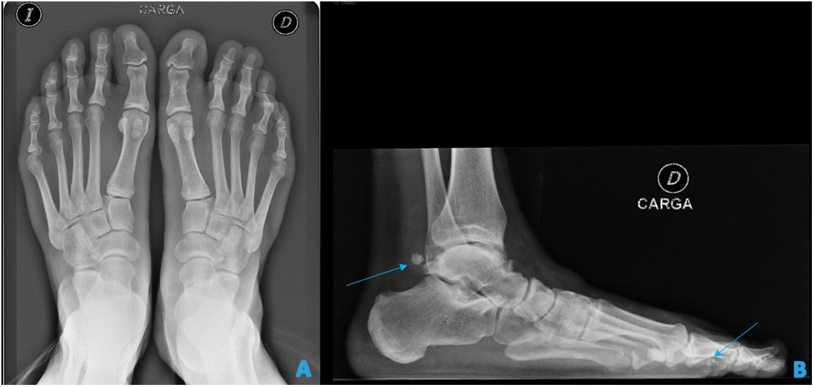
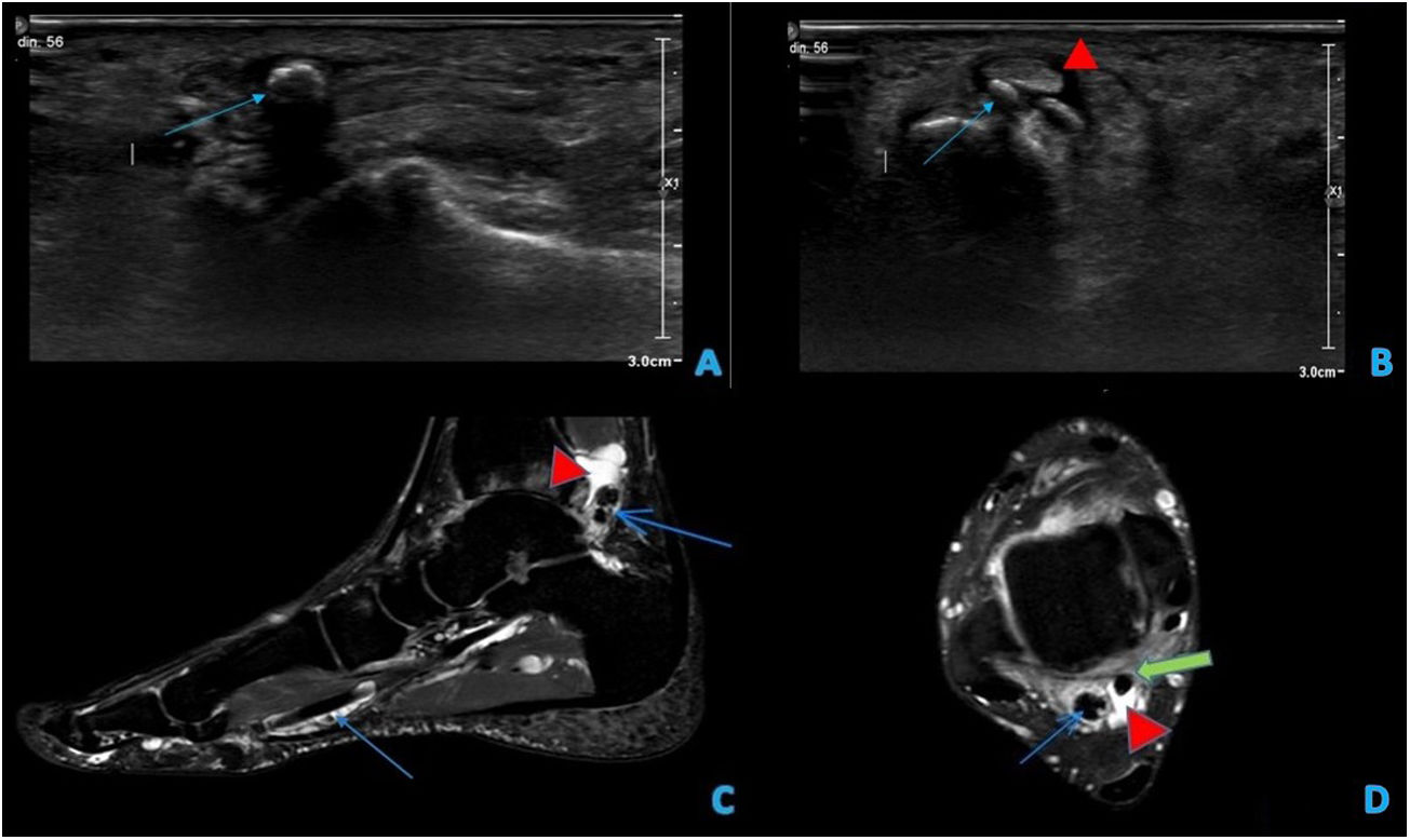
La condromatosis sinovial es una lesión poco frecuente que se caracteriza por la metaplasia cartilaginosa de la membrana sinovial con la habitual formación de cuerpos libres osteocartilaginosos y que típicamente afecta grandes articulaciones. La localización en el tobillo y en el pie es poco frecuente y, a su vez, muy rara en la vaina sinovial de los tendones del pie. El diagnóstico de esta entidad poco reconocida es de gran importancia porque es una condición progresiva que conlleva un riesgo sustancial de la recidiva local. Las pruebas de imagen, como la TC o la RM, ayudan a identificar los hallazgos característicos y la localización exacta para así orientar al traumatólogo en la cirugía. Su tratamiento definitivo es quirúrgico, mediante resección completa de la sinovial (sinovectomía).

Palabras clave:
Condromatosis sinovial
Pie
Flexor hallucis longus
Ecografía
Resonancia magnética
Abstract

Synovial chondromatosis is an uncommon lesion characterized by cartilaginous metaplasia of the synovial layer with the usual formation of free osteocartilaginous bodies and typically involving large joints. Location in the ankle and foot is rare and, in turn, very rare in the synovial sheath of the foot tendons. The diagnosis of this little recognized entity is of great importance because it is a progressive condition that carries a substantial risk of local recurrence. Imaging tests such as CT or MRI help to identify the characteristic findings and the exact location to guide the orthopaedic surgeon in surgery. Its definitive treatment is surgical, through complete resection of the synovium (synovectomy).

Keywords:
Synovial chondromatosis
Foot
Flexor hallucis longus
Ultrasound
Magnetic resonance imaging

Article

These are the options to access the full texts of the publication Revista Colombiana de Reumatología
Subscriber
Subscriber

If you already have your login data, please click here .

If you have forgotten your password you can you can recover it by clicking here and selecting the option “I have forgotten my password”
Purchase
Purchase article

Purchasing article the PDF version will be downloaded

Price 19.34 €

Purchase now
Contact
Phone for subscriptions and reporting of errors
From Monday to Friday from 9 a.m. to 6 p.m. (GMT + 1) except for the months of July and August which will be from 9 a.m. to 3 p.m.
Calls from Spain
932 415 960
Calls from outside Spain
+34 932 415 960
E-mail
Article options
Tools
Quizás le interese:
10.1016/j.rcreu.2021.05.003
No mostrar más