Buscar en
Revista Científica de la Sociedad Española de Enfermería Neurológica (English ed.)
Toda la web
Inicio Revista Científica de la Sociedad Española de Enfermería Neurológica (Englis... Local and intracraneal complications derived from cerebral arteriography
Journal Information
Visits
86
Original article
Full text access
Available online 25 June 2022
Local and intracraneal complications derived from cerebral arteriography
Complicaciones locales e intracraneales derivadas de la arteriografía cerebral
Visits
...
Mònica Serrano Clerenciaa,b, Almudena Sánchez-Gómeza,b, Anna Grau Ginera,b, Laura Cardete Moralesa,b, Sergi Amaro Delgadoa,b,
Corresponding author
samaro@clinic.cat

Corresponding author.
a Instituto de Neurociencias, Servicio de Neurología, Hospital Clínic de Barcelona, Barcelona, Spain
b Instituto de Neurociencias, Maeztu Center, Universidad de Barcelona, Barcelona, Spain
Received 19 November 2021. Accepted 15 February 2022
Article information
Abstract
Full Text
Bibliography
Download PDF
Statistics
Figures (1)
Abstract
Background

Although cerebral angiography is the reference technique for the diagnosis and treatment of cerebrovascular diseases, it is not without complications.

Objectives

To determine the incidence and risk factors of complications derived from therapeutic cerebral angiography (TCA), as well as to assess the risk factors associated with the appearance of local and intracranial complications.

Methodology

Retrospective cross-sectional study on TCAs carried out in 2018 on admission to the Stroke Unit of the Hospital Clínic de Barcelona. The study was approved by the centre’s ethics committee. Demographic, clinical, analytical, and procedure-derived variables were collected. All patients older than 18 years undergoing TCA were included. Patients undergoing diagnostic cerebral arteriography and/or with a hospital stay less than 24h were excluded. The Mann-Whitney U test was used for the comparison of quantitative variables and Pearson’s Chi-squared test for the qualitative variables.
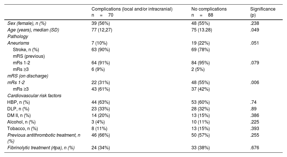
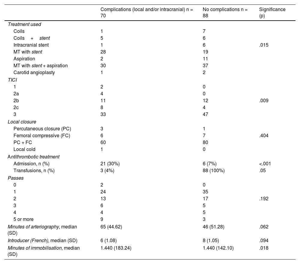
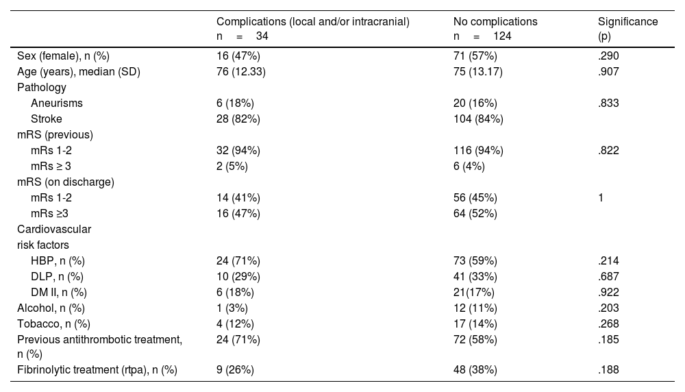
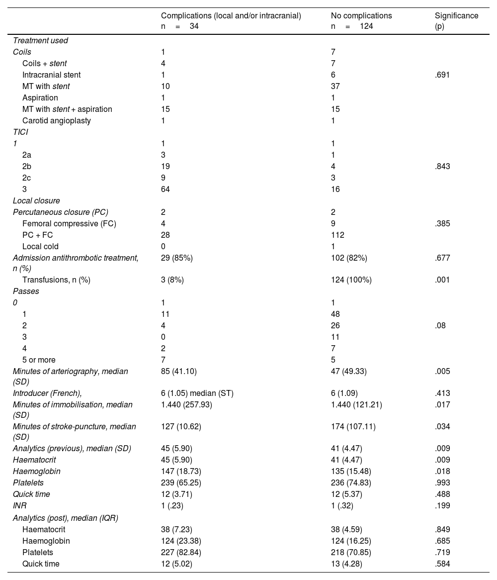
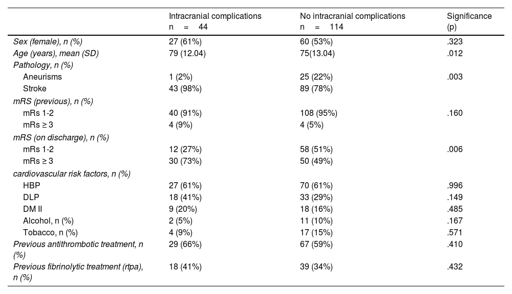
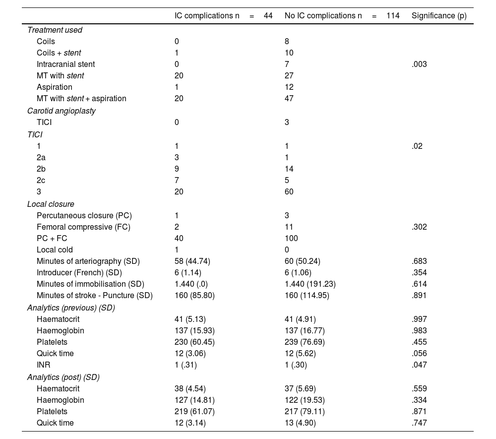
Results

Complications were frequent, occurring in almost half of the sample (44%). A longer duration of arteriography was associated with an increase in local complications (p=.005). Intracranial complications occurred in 33% of the patients who underwent mechanical thrombectomy and were associated with older age (p=.012), stent use (associated or not with aspiration) (p=.003), and complete recanalization (p=.02), as well as having a worse functional status at discharge (p=.006).

Conclusions

Complications derived from ACT are frequent. Their importance and incidence must be known to detect those subjects at higher risk for developing them due to their functional implications and increased hospital stay.

Keywords:
Cerebral angiography
Postoperative complications
Thrombectomy
Embolization
Therapeutic
Intracranial aneurysm
Resumen
Fundamento

La arteriografía cerebral es la técnica de referencia para el diagnóstico y tratamiento de enfermedades cerebrovasculares. No obstante, no está exenta de complicaciones.

Objetivo

Determinar la incidencia de las complicaciones derivadas de la arteriografía cerebral terapéutica (ACT) y evaluar cuáles son los factores de riesgo asociados a la aparición de complicaciones tanto locales como intracraneales.

Metodología

Estudio retrospectivo transversal sobre las ACT realizadas en el año 2018 que ingresaban en la Unidad de Ictus del Hospital Clínic de Barcelona. El estudio fue aprobado por el comité ético del centro. Se recopilaron variables demográficas, clínicas, analíticas y derivadas del procedimiento. Se incluyeron todos los pacientes mayores de 18 años sometidos a una ACT durante el 2018. Se excluyeron los pacientes sometidos a arteriografías cerebrales diagnósticas y con una estancia hospitalaria menor a 24horas. Se utilizó el test U de Mann Whitney para la comparación de variables cuantitativas y el test Chi cuadrado de Pearson para las cualitativas.

Resultados

Las complicaciones han sido frecuentes, llegándose a producir casi en la mitad de la muestra (44%). Se ha asociado la mayor duración de la arteriografía con un aumento de las complicaciones locales (p=0,005). Las complicaciones intracraneales ocurrieron en un 33% de los pacientes sometidos a trombectomía mecánica y se asociaron con presentar una edad más avanzada (p=0,012), el uso del stent (asociado o no a aspiración) (p=0,003) y una recanalización completa (p=0,02) así como el comportar un peor estado funcional al alta (p=0,006).

Conclusiones

Las complicaciones derivadas de la ACT son frecuentes. Se debe conocer su importancia e incidencia para poder detectar a aquellos sujetos con mayor riesgo a su desarrollo por sus implicaciones funcionales y de aumento de estancia hospitalaria.

Palabras clave:
Angiografía
Complicaciones posoperatorias
Trombectomía
Embolización terapéutica
Aneurisma intracraneal
Full Text
Introduction

Cerebral arteriography or cerebral angiography has become the gold standard technique for the diagnosis and treatment of many cerebrovascular diseases, especially in the acute treatment of ischmic stroke,1,2 and for the treatment of cerebral aneurysms.3

Femoral artery access is commonly used, as it allows the use of larger catheters,4 although radial access can also be used. The femoral puncture is performed next to the femoral head, an area that is easy to compress after treatment is completed.5 The duration of cerebral angiography depends on whether it is a diagnostic arteriography, where it is usually less than one hour, or several hours in the case of complex acute ischaemic stroke treatment. At the end of the procedure, the catheter is removed and haemostasis is performed by manual compression of the femoral artery or percutaneous closure of the puncture site.5 Both the access technique and the femoral puncture closure depend on the complications arising from this intervention.4

The main advantage of this technique is that it allows accurate, real-time assessment of cerebral circulation, and in turn, offers the possibility of performing occlusive therapeutic procedures in the case of cerebral aneurysms or arteriovenous malformations and recanalisation in ischaemic strokes.

Despite being a safe technique, it is an invasive procedure that is not exempt from complications, ranging from local to systemic, which can lead to high morbidity and mortality.1,5–8 The most frequent complications reported in the published literature are local haemorrhages. One of the most common is the appearance of an inguinal haematoma at the puncture site,9 followed by retroperitoneal haematoma, which may require blood transfusions or surgical evacuation.5,8 Other local complications include the appearance of a pseudoaneurysm or arteriovenous fistulas due to alterations in the integrity of the blood vessels, ischaemia of the limb due to vessel occlusion, 9 and even local infections.10

There are several factors associated with reported complications: a low body mass index,9,10 the use of antithrombotics,9 advanced age, female sex,10 larger caliber of the devices used in angiography9,10 and and performing therapeutic versus diagnostic arteriography, among others.9

Regarding complications specifically related to aneurysmal embolisations, there is a risk of arterial perforation during treatment of ruptured aneurysms compared to delayed treatment of incidentally diagnosed cerebral aneurysms.8 Regarding endovascular treatment of stroke, intracranial haemorrhage, either haemorrhagic transformation in the infarct core or subarachnoid haemorrhage, is the most serious complication that can be related to this procedure, as it can aggravate the functional and even vital prognosis of the patient.11 It can occur during the procedure or up to 72h after the treatment and can be symptomatic (worsening of neurological status) or asymptomatic. Many patients undergoing mechanical thrombectomy receive fibrinolytic therapy, which could be a risk factor, although there is insufficient scientific evidence.12 Although cerebral arteriography or angiography is a technique that has been perfected over the years, peripheral vascular complications related to the puncture site and secondary intracranial complications are still observed. All this, in addition to being related to a functional and vital risk for the patient, entails a longer hospital stay and, consequently, a significant increase in costs.

Since we found insufficient information in the published literature on the complications derived from cerebral arteriography, we proposed to retrospectively study all subjects in our centre who required at least one therapeutic cerebral arteriography (mainly endovascular treatment of large-vessel ischaemic strokes, but also endovascular treatment of cerebral aneurysms) to analyse the rate of complications and, in turn, to study the possible triggering factors. It has been considered a priori that the duration of the procedure, the use of different devices, age and associated antithrombotic treatment could be some of them.

The presence of both local and intracranial complications derived from the performance of therapeutic cerebral arteriography can lead to clinical worsening (from mild to severe) in patients and cause an increase in hospital stay. For these reasons, the aim of this study was to determine the incidence and risk factors of complications related to therapeutic cerebral arteriography in the Stroke Unit (IU) of Hospital Clínic.

MethodologyStudy design

Retrospective cross-sectional study in which the medical records of patients who underwent therapeutic cerebral arteriography between 1 January 2018 and 31 December 2018 at our centre were evaluated. We used two databases collected by the Neurology and Angioradiology Department of Hospital Clínic: one with all patients who underwent aneurysmal embolisation and the other with all mechanical thrombectomies performed between 1 January 2018 and 31 December 2018. Recruitment was by non-randomised purposive sampling. Patients over 18 years of age who were admitted to the Stroke Unit after undergoing therapeutic cerebral arteriography, either for mechanical stroke treatment or aneurysmal embolisation, were included. Patients who had undergone diagnostic arteriography and patients from other referral hospitals who stayed less than 24h in our centre were excluded.

For the collection of variables, a questionnaire prepared by the researchers on an Excel sheet was used. Information was collected on sociodemographic variables (age, sex), clinical variables (cardiovascular risk factors, degree of independence, anticoagulant or antiplatelet therapy, and analytical values related to haemogram and coagulation prior to treatment). Treatment-related variables were also collected (fibrinolytic treatment, time from stroke onset to arterial puncture in mechanical thrombectomy [MT], duration of the procedure, technique used for endovascular treatment, introducer calibre, degree of reperfusion in MT and complications during admission).

To assess the degree of independence, the modified Rankin scale (mRs) was used, where 0 is completely independent, 5 completely dependent and 6 deaths.13 To assess the degree of reperfusion, the Thrombolysis in Cerebral Infarction scale (TICI) was used, which describes the degree of arterial recanalisation, with 0 being no recanalisation and 3 complete recanalisation.14

Complications were classified as local, referring to the puncture site, and intracranial, highlighting haemorrhagic transformations or subarachnoid haemorrhages after the procedure.

Statistical methods

All qualitative variables were presented as absolute and relative frequencies, and quantitative variables as medians and standard deviation. Differences between groups of quantitative variables were performed with the Mann Whitney U test and those of qualitative variables were compared with Pearson's χ2 test. All analyses were two-tailed with a significance threshold set at .05. Statistical analysis of the results was performed with the SPSS for IOS package (version 21 for IOS; IBM Corp., Armonk, NY, USA).

Ethical considerations

The project was approved by the Ethics Committee of Hospital Clínic. Because the study collected retrospective data, informed consent was not required from participants. However, personal identification codes were encrypted to maintain the anonymity of the study subjects. The team involved only worked with anonymised data.

Results

A total of 158 individuals were identified who underwent therapeutic cerebral arteriography with the inclusion and exclusion criteria described above. Of these, 132 (84%) subjects required cerebral arteriography for MT because they had an ischaemic stroke with large vessel occlusion, and the other 26 (16%) individuals required cerebral arteriography for cerebral aneurysm embolisation (AE). Of the total sample, there were 87 women (55%) and 71 men (45%). The mean age was 73 years and the standard deviation (SD) 12.95.

Analysis of total complications

Of the total sample, there were 78 complications, 34 (44%) local (local haematoma, deep haematoma and bleeding) and 44 (56%) intracranial complications (haemorrhagic transformation or subarachnoid haemorrhage). Only 8 (5%) individuals had both complications, so 70 subjects (44%) had some type of complication.

The mean age of the individuals who had a complication (local or intracranial) was 74.5 years (ST=12.27). Of these, 39 were women and 31 men. There were no differences in the distribution of cardiovascular risk factors (high blood pressure [HBP], dyslipidaemia [DLP], type II diabetes mellitus [DM II], smoking and alcohol), between the total complication and non-complication groups. As for the association between prior antithrombotic and fibrinolytic therapy, there was no difference between the two groups (p=.225 and p=.676, respectively). Patients who received antithrombotic treatment during admission had a higher complication rate than those who did not (p<.001).

Patients treated with stented and non-stented MT with or without aspiration were associated with a higher rate of complications (p=.015) (Table 1). Those with complete recanalisation or TICI 3 were associated with a higher number of complications than those with incomplete recanalisation or no recanalisation (p=.009). Arteriography duration minutes, introducer calibre and lower limb immobilisation minutes did not differ significantly between the two groups. Pre- and post-arteriography blood count and coagulation parameters were not associated with the occurrence of complications. Finally, in relation to transfusions and disability at discharge, as expected, there was a statistically significant higher rate of complications in transfused patients and those with unfavourable disability (p=.05 and p=.006, respectively) (Tables 1 and 2).

Table 1.

Differences between complication and non-complication groups.

  Complications (local and/or intracranial) n=70  No complications n=88  Significance (p) 
Sex (female), n (%)  39 (56%)  48 (55%)  .238 
Age (years), median (SD)  77 (12,27)  75 (13.28)  .049 
Pathology       
Aneurisms  7 (10%)  19 (22%)  .051 
Stroke, n (%)  63 (90%)  69 (78%)   
mRS (previous)       
mRs 1-2  64 (91%)  84 (95%)  .079 
mRs ≥3  6 (9%)  2 (5%)   
mRS (on discharge)       
mRs 1-2  22 (31%)  48 (55%)  .006 
mRs ≥3  43 (61%)  37 (42%)   
Cardiovascular risk factors       
HBP, n (%)  44 (63%)  53 (60%)  .74 
DLP, n (%)  23 (33%)  28 (32%)  .89 
DM II, n (%)  14 (20%)  13 (15%)  .386 
Alcohol, n (%)  3 (4%)  10 (11%)  .225 
Tobacco, n (%)  8 (11%)  13 (15%)  .393 
Previous antithrombotic treatment, n (%)  46 (66%)  50 (57%)  .255 
Fibrinolytic treatment (rtpa), n (%)  24 (34%)  33 (38%)  .676 

DLP, Dyslipidaemia; DM II, Diabetes Mellitus type 2; HBP, High blood pressure; mRs, Modified Rankin scale; rtpa, Fibrinolytic treatment; SD, Standard Deviation.

Table 2.

Differences between complications and non-complications group related to arteriography.

  Complications (local and/or intracranial) n = 70  No complications n = 88  Significance (p) 
Treatment used
Coils   
Coils+stent   
Intracranial stent  .015 
MT with stent  28  19   
Aspiration  11   
MT with stent + aspiration  30  37   
Carotid angioplasty   
TICI
 
2a   
2b  11  12  .009 
2c   
33  47   
Local closure
Percutaneous closure (PC)   
Femoral compressive (FC)  .404 
PC + FC  60  80   
Local cold   
Antithrombotic treatment
Admission, n (%)  21 (30%)  6 (7%)  <.001 
Transfusions, n (%)  3 (4%)  88 (100%)  .05 
Passes
 
24  35   
13  17  .192 
 
 
5 or more   
Minutes of arteriography, median (SD)  65 (44.62)  46 (51.28)  .062 
Introducer (French), median (SD)  6 (1.08)  8 (1.05)  .094 
Minutes of immobilisation, median (SD)  1.440 (183.24)  1.440 (142.10)  .018 

FC, Femoral Compressive; MT, Mechanical Thrombectomy; PC, Percutaneous closure; SD, Standard Deviation; TICI, Arterial reperfusion scale.

Analysis of local complcations

The mean age of the individuals who presented with a local complication was 72.65 years. Of these, 16 were women and 18 men. There were no differences in the distribution of cardiovascular risk factors (HBP, DM II, LDL, alcohol and smoking) between the groups who had experienced a local complication and those who had not. Neither was prior antithrombotic treatment (p=.185) nor fibrinolytic treatment (p=.188) associated with local complications. Analytically, higher haematocrit (p=.009) and previous haemoglobin (p=.018) were associated with local complications.

For arteriography values, neither the endovascular treatment used (p=.691), nor the TICI (p=.843) nor the number of passes (p=.08) were associated with more local complications. On the other hand, the faster the stroke treatment (stroke-to-arterial puncture time), the more local complications occurred (p=.034) (Tables 3 and 4). Finally, the longer the duration of arteriography, the higher the rate of local complications (p=.005) (Fig. 1).

Table 3.

Differences between groups of local complications and non-local complications.

  Complications (local and/or intracranial) n=34  No complications n=124  Significance (p) 
Sex (female), n (%)  16 (47%)  71 (57%)  .290 
Age (years), median (SD)  76 (12.33)  75 (13.17)  .907 
Pathology       
Aneurisms  6 (18%)  20 (16%)  .833 
Stroke  28 (82%)  104 (84%)   
mRS (previous)       
mRs 1-2  32 (94%)  116 (94%)  .822 
mRs ≥ 3  2 (5%)  6 (4%)   
mRS (on discharge)       
mRs 1-2  14 (41%)  56 (45%) 
mRs ≥3  16 (47%)  64 (52%)   
Cardiovascular       
risk factors       
HBP, n (%)  24 (71%)  73 (59%)  .214 
DLP, n (%)  10 (29%)  41 (33%)  .687 
DM II, n (%)  6 (18%)  21(17%)  .922 
Alcohol, n (%)  1 (3%)  12 (11%)  .203 
Tobacco, n (%)  4 (12%)  17 (14%)  .268 
Previous antithrombotic treatment, n (%)  24 (71%)  72 (58%)  .185 
Fibrinolytic treatment (rtpa), n (%)  9 (26%)  48 (38%)  .188 

DLP, Dyslipidaemia; DM II, Diabetes Mellitus type 2; HBP, High blood pressure; mRs, Modified Rankin scale; rtpa, Fibrinolytic treatment; SD, Standard Deviation.

Table 4.

Differences between group of local complications and non-local complications related to arteriography.

  Complications (local and/or intracranial) n=34  No complications n=124  Significance (p) 
Treatment used       
Coils   
Coils + stent   
Intracranial stent  .691 
MT with stent  10  37   
Aspiration   
MT with stent + aspiration  15  15   
Carotid angioplasty   
TICI       
1   
2a   
2b  19  .843 
2c   
64  16   
Local closure       
Percutaneous closure (PC)   
Femoral compressive (FC)  .385 
PC + FC  28  112   
Local cold   
Admission antithrombotic treatment, n (%)  29 (85%)  102 (82%)  .677 
Transfusions, n (%)  3 (8%)  124 (100%)  .001 
Passes       
0   
11  48   
26  .08 
11   
 
5 or more   
Minutes of arteriography, median (SD)  85 (41.10)  47 (49.33)  .005 
Introducer (French),  6 (1.05) median (ST)  6 (1.09)  .413 
Minutes of immobilisation, median (SD)  1.440 (257.93)  1.440 (121.21)  .017 
Minutes of stroke-puncture, median (SD)  127 (10.62)  174 (107.11)  .034 
Analytics (previous), median (SD)  45 (5.90)  41 (4.47)  .009 
Haematocrit  45 (5.90)  41 (4.47)  .009 
Haemoglobin  147 (18.73)  135 (15.48)  .018 
Platelets  239 (65.25)  236 (74.83)  .993 
Quick time  12 (3.71)  12 (5.37)  .488 
INR  1 (.23)  1 (.32)  .199 
Analytics (post), median (IQR)       
Haematocrit  38 (7.23)  38 (4.59)  .849 
Haemoglobin  124 (23.38)  124 (16.25)  .685 
Platelets  227 (82.84)  218 (70.85)  .719 
Quick time  12 (5.02)  13 (4.28)  .584 

FC, Femoral compressive; INT, International Normalized Ratio; MT, Mechanical Thrombectomy; PC, Percutaneous closure; SD, Standard Deviation; TICI, arterial reperfusion scale.

Figure 1.

Box plot showing a higher proportion of patients with local complications (X-axis) in relation to more minutes of arteriography duration (Y-axis).

(0.06MB).
Analysis of intracraneal complications

The mean age of the individuals with intracranial complications was 76.84 years, significantly older than those without intracranial complications (p=.012). Of these, 27 were female and 17 were male. Intracranial complications occurred in 97.8% of MTs, accounting for 33% of all MTs and there was only one case (2.2%) of intracranial complications (p=.003) in AE (secondary subarachnoid haemorrhage).

There were no significant differences in sex or cardiovascular risk factors between those with and without intracranial complications. There were also no significant differences in having received thrombolytic treatment prior to arteriography (Tables 5 and 6).

Table 5.

Differences between intracranial complications and non-intracranial complications groups.

  Intracranial complications n=44  No intracranial complications n=114  Significance (p) 
Sex (female), n (%)  27 (61%)  60 (53%)  .323 
Age (years), mean (SD)  79 (12.04)  75(13.04)  .012 
Pathology, n (%)
Aneurisms  1 (2%)  25 (22%)  .003 
Stroke  43 (98%)  89 (78%)   
mRS (previous), n (%)
mRs 1-2  40 (91%)  108 (95%)  .160 
mRs ≥ 3  4 (9%)  4 (5%)   
mRS (on discharge), n (%)
mRs 1-2  12 (27%)  58 (51%)  .006 
mRs ≥ 3  30 (73%)  50 (49%)   
cardiovascular risk factors, n (%)
HBP  27 (61%)  70 (61%)  .996 
DLP  18 (41%)  33 (29%)  .149 
DM II  9 (20%)  18 (16%)  .485 
Alcohol, n (%)  2 (5%)  11 (10%)  .167 
Tobacco, n (%)  4 (9%)  17 (15%)  .571 
Previous antithrombotic treatment, n (%)  29 (66%)  67 (59%)  .410 
Previous fibrinolytic treatment (rtpa), n (%)  18 (41%)  39 (34%)  .432 

DLP, Dyslipidaemia; DM II, Diabetes Mellitus type 2; HBP, High blood pressure; mRs, Modified Rankin scale; rtpa, Fibrinolytic treatment; SD, Standard Deviation.

Table 6.

Differences between intracranial complications (IC) and non-intracranial complications (IC) group related to arteriography.

  IC complications n=44  No IC complications n=114  Significance (p) 
Treatment used
Coils   
Coils + stent  10   
Intracranial stent  .003 
MT with stent  20  27   
Aspiration  12   
MT with stent + aspiration  20  47   
Carotid angioplasty
TICI   
TICI
.02 
2a   
2b  14   
2c   
20  60   
Local closure
Percutaneous closure (PC)   
Femoral compressive (FC)  11  .302 
PC + FC  40  100   
Local cold   
Minutes of arteriography (SD)  58 (44.74)  60 (50.24)  .683 
Introducer (French) (SD)  6 (1.14)  6 (1.06)  .354 
Minutes of immobilisation (SD)  1.440 (.0)  1.440 (191.23)  .614 
Minutes of stroke - Puncture (SD)  160 (85.80)  160 (114.95)  .891 
Analytics (previous) (SD)
Haematocrit  41 (5.13)  41 (4.91)  .997 
Haemoglobin  137 (15.93)  137 (16.77)  .983 
Platelets  230 (60.45)  239 (76.69)  .455 
Quick time  12 (3.06)  12 (5.62)  .056 
INR  1 (.31)  1 (.30)  .047 
Analytics (post) (SD)
Haematocrit  38 (4.54)  37 (5.69)  .559 
Haemoglobin  127 (14.81)  122 (19.53)  .334 
Platelets  219 (61.07)  217 (79.11)  .871 
Quick time  12 (3.14)  13 (4.90)  .747 

FC, Femoral compressive; INT, International Normalized Ratio; MT, Mechanical Thrombectomy; PC, Percutaneous closure; SD, Standard Deviation; TICI, Arterial reperfusion scale.

In endovascular treatment, there was a higher rate of intracranial complications in patients treated with stent associated or not with aspiration (p=.003) and those with complete recanalisation (p=.02). There were no differences in the time from stroke to treatment, in the duration of the procedure or in the calibre of the introducer, nor in the minutes of immobilisation. There were only analytical differences in pre-treatment INR, which was higher in those with complications (p=.047).

Discussion

Complications of arteriography have been frequent, occurring in almost half of the patients. Neither local nor intracranial complications were associated with cardiovascular risk factors, antithrombotic treatment prior to admission or fibrinolytic treatment. Regarding the combined treatment between alteplase thrombolysis and mechanical thrombectomy, studies are ongoing to evaluate the potential effect and risk of intracranial complications of applying both treatments simultaneously.12

Although the published literature on the benefit and safety of mechanical thrombectomy describes the local complications of the procedure, the risk factors associated with them are not extensively analysed or determined.15 Our main finding in relation to local complications is the association with a longer duration of arteriography, without linking the calibre of the introducer or the type of closure used with their occurrence. Other studies have also failed to document the association of local complications with the size of the introducer in patients undergoing mechanical thrombectomy.16

The preference for femoral access in mechanical thrombectomy is due to the larger calibre of the vessel and the need to use larger introducers and devices. Brachial, radial or direct carotid accesses are the alternatives when femoral access is not possible.17 Radial access has been associated in a small cohort of vertebrobasilar strokes with a lower incidence of local complications (haematomas, pseudoaneurysms) and shorter endovascular procedure time.18 Although there is a selection bias in this study because it is limited to posterior territory strokes and a very small number of patients (eight), if confirmed in randomised clinical trials, comparing the different vascular accesses, it could be a valid alternative at least in these cases. The fact that local complications were associated with longer immobilisation time is considered a selection bias, since patients with local complications required more rest time.

With regard to intracranial complications in relation to mechanical thrombectomy, we have found 33% of them to occur, although they are anecdotal in aneurysmal embolisations. Our data are similar to those obtained in the literature for mechanical thrombectomy, with a frequency between 33% and 47%.19,20 Intracranial complications cannot be analysed between the ischaemic stroke and aneurysm groups as they are distinct diseases. In addition, selection bias should be noted because arteriography for the treatment of aneurysms is scheduled, whereas all reperfusion treatments in ischaemic strokes are urgent.

In terms of the device used, stent retriever has been associated with a higher rate of intracranial complications vs. isolated aspiration,19,20 although a recent meta-analysis found no difference in intracranial complications between the two techniques, with no difference in efficacy and prognosis at three months between the two.21

In relation to recanalisation, intracranial complications are significantly associated with complete recanalisation (TICI 3). In general, good recanalisation (TICI 2b, 2c preferably) or complete recanalisation is associated with a good prognosis. Some studies have found the opposite association to our results, with complete recanalisation being a protective factor for intracranial haemorrhagic complications.22 As expected, complications result in a worse prognosis at discharge (measured by mRS scores of 3 or more), data already known in the literature.23

The main limitation of the study is the retrospective nature of the data, both in terms of losses and validity. Also, the single-centre, cross-sectional nature of the study limits the extrapolation of the data, as well as the collection of data in reference to the times evaluated. Nevertheless, the main strengths of the study would be that it is a considerable sample of subjects collected in a tertiary hospital with an important activity of therapeutic cerebral arteriography. The fact of describing the incidence and risk factors associated with the complications of this procedure (mainly local) brings new information to the scientific literature that has been little represented so far.

In conclusion, a large percentage of the patients studied had some complication, with local complications being related to a longer duration of the procedure. Intracranial complications occurred in MTs and were mainly associated with age and the type of treatment used, leading to a worse functional prognosis at discharge.

Therefore, in nursing care after therapeutic cerebral arteriography, special attention should be paid to puncture sites in patients who have undergone a long-lasting procedure, and to possible neurological clinical changes in patients, mainly elderly, who have undergone mechanical thrombectomy.

Financing

Spanish Society of Neurological Nursing. Research Award. XXVIII Annual Congress, 2021.

Conflict of interests

The authors have no conflict of interests to declare.

References
[1]
J. Krupinsky, X. Ustrell, M. Font, A. Arboix.
Guies mèdiques de la societat catalana de neurologia. Protocol de tractament. Diagnòstic i tractament de les malalties vasculars cerebrals.
[2]
Z. Khan, P. Nattanamai, P. Keerthivaas, C.R. Newey.
An evaluation of complications in femoral arterial sheaths maintained post-neuroangiographic procedures.
[3]
M.S. Pearl, C. Torok, J. Wang, E. Wyse, M. Mahesh, P. Gailloud.
Practical techniques for reducing radiation exposure during cerebral angiography procedures.
J Neurointerv Surg., 7 (2015), pp. 141-145
[4]
D. Dencker, F. Pedersen, T. Engstrøm, L. Køber, S. Højberg, M.B. Nielsen, et al.
Major femoral vascular access complications after coronary diagnostic and interventional procedures: a Danish register study.
Int J Cardiol., 202 (2016), pp. 604-608
[5]
V. Alakbarzade, A.C. Pereira.
Cerebral catheter angiography and its complications.
Pract Neurol., 18 (2018), pp. 393-398
[6]
S.H. Akpinar, G. Yilmaz.
Periprocedural complications in endovascular stroke treatment.
[7]
S. Valiee, M. Fathi, N. Hadizade, D. Roshani, P. Mahmoodi.
Evaluation of feasibility and safety of changing body position after transfemoral angiography: a randomized clinical trial.
J Vasc Nurs., 34 (2016), pp. 106-115
[8]
M.C. Davis, J.P. Deveikis, M.R. Harrigan.
Clinical presentation, imaging, and management of complications due to neurointerventional procedures.
Semin Intervent Radiol., 32 (2015), pp. 98-107
[9]
R. Grandhi, H. Kanaan, A. Shah, G. Harrison, C. Bonfield, T. Jovin, et al.
Safety and efficacy of percutaneous femoral artery access followed by Mynx closure in cerebral neurovascular procedures: a single center analysis.
J Neurointerv Surg., 6 (2014), pp. 445-450
[10]
G. Berga, P. Murillo, A. Márquez, G. Casajús.
Complicaciones vasculares en el paciente sometido a procedimientos cardiovasculares percutáneos.
Enferm Cardiol, 58-59 (2013), pp. 56-61
[11]
P.B. García Jurado, E. Roldán Romero, M.E. Pérez Montilla, R. Valverde Moyano, I.M. Bravo Rey, F. Delgado Acosta, et al.
Incidence, prognosis, prediction of haemorrhagic transformation after revascularisation for stroke.
Neurol (Engl Ed), 36 (2021), pp. 589-596
[12]
B.C.V. Campbell.
Thrombolysis and thrombectomy for acute ischemic stroke: Strengths and synergies.
Semin Thromb Hemost, 43 (2017), pp. 185-190
[13]
J.P. Broderick, O. Adeoye, J. Elm.
Evolution of the modified Rankin scale and its use in future stroke trials.
Stroke, 48 (2017), pp. 2007-2012
[14]
D.M. Heiferman, N.C. Pecoraro, A.W. Wozniak, K.C. Ebersole, L.M. Jimenez, M.R. Reynolds, et al.
Reliability of the modified TICI score among endovascular neurosurgeons.
AJNR AM J Neuroradiol, 41 (2020), pp. 1441-1446
[15]
T.G. Jovin, A. Chamorro, E. Cobo, M.A. de Miquel, C.A. Molina, A. Rovira, et al.
Thrombectomy within 8 hours after symptom onset in ischemic stroke.
N Eng J Med, 372 (2015), pp. 2296-2306
[16]
V.A. Shah, C.O. Martin, A.M. Hawkins, W.E. Holloway, S. Junna, N. Akhtar.
Groin complications in endovascular mechanical thrombectomy for acute ischemic stroke: a 10-year single center experience.
J Neurointerv Surg, 8 (2016), pp. 568-570
[17]
J.S. Balami, P.M. White, P.J. McMeekin, G.A. Ford, A.M. Buchan.
Complications of endovascular treatment for acute ischemic stroke: Prevention and management.
Int J Stroke, 13 (2018), pp. 348-361
[18]
M. Oselkin, S.R. Satti, S.H. Sundararajan, D. Kung, R.W. Hurst, B.A. Pukenas.
Endovascular treatment for acute basilar thrombosis via a transradial approach: Initial experience and future considerations.
Interv Neuroradiol, 24 (2018), pp. 64-69
[19]
J. Kaesmacher, M. Kaesmacher, C. Maegerlein, C. Zimmer, A.S. Gersing, S. Wunderlich, et al.
Hemorrhagic transformations after thrombectomy: risk factors and clinical relevance.
Cerebrovasc Dis, 43 (2017), pp. 294-304
[20]
S. Jiang, A. Fei, Y. Peng, J. Zhang, Y.-R. Lu, H.-R. Wang, et al.
Predictors of outcome and hemorrhage in patients undergoing endovascular therapy with Solitaire Stent for acute ischemic stroke.
[21]
M. Boulanger, B. Lapergue, F. Turjman, E. Touzé, R. Anxionnat, S. Bracard, et al.
First-line contact aspiration vs stent-retriever thrombectomy in acute ischemic stroke patients with large-artery occlusion in the anterior circulation: systematic review and meta-analysis.
Interv Neuroradiol, 25 (2019), pp. 244-253
[22]
L.A. Slater, J.M. Coutinho, J. Gralla, R.G. Nogueira, A. Bonafé, A. Dávalos, et al.
TICI and age: what’s the score?.
AJNR Am J Neuroradiol, 37 (2016), pp. 838-843
[23]
C. Qin, K. Shang, S.-B. Xu, W. Wang, Q. Zhang, D.-S. Tian.
Efficacy and safety of direct aspiration versus stent-retriever for recanalization in acute cerebral infarction: a PRISMA-compliant systematic review and meta-analysis.
Medicine (Baltimore), 97 (2018),

Please cite this article as: Serrano Clerencia M, Sánchez-Gómez A, Grau Giner A, Cardete Morales L, Amaro Delgado S. Complicaciones locales e intracraneales derivadas de la arteriografía cerebral, Rev Cient Soc Esp Enferm Neurol. 2022. https://doi.org/10.1016/j.sedene.2022.02.001

Copyright © 2022. Sociedad Española de Enfermería Neurológica
Article options
Tools
es en pt

¿Es usted profesional sanitario apto para prescribir o dispensar medicamentos?

Are you a health professional able to prescribe or dispense drugs?

Você é um profissional de saúde habilitado a prescrever ou dispensar medicamentos