


Complications in right heart catheterization are almost all access-site related. Forearm veins may be a target to reduce access-site complications during the procedure. However, data regarding wide application of this technique is scarce.
MethodsThis is a case-series that reports our first experiences in right heart catheterization through the antecubital approach.
ResultsWe attempted to perform right heart catheterization in 20 patients using antecubital approach on January 2016. The antecubital approach was successful in 19 (95.0%) cases. All venous access were obtained with ultrasound guidance. Simultaneous right and left heart catheterization was performed in 12 cases (60.0%). Left heart catheterization was performed through right radial artery in 11 cases (91.7%) and through the right brachial artery in 1 case (8.3%). Antecubital access was obtained through the basilic vein in 18 (94.7%) cases and through the cephalic vein in 1 (5.3%) case.
ConclusionsRight heart catheterization through the antecubital fossa veins appears to be feasible and safe. Further controlled studies are required to establish the best access site to perform right heart catheterization.
As complicações no cateterismo cardíaco direito estão quase sempre relacionadas ao local de acesso. As veias do antebraço podem ser um alvo para reduzir tais complicações durante o procedimento. No entanto, dados relativos à ampla aplicação desta técnica são escassos.
MétodosSérie de casos que relata nossas primeiras experiências com o cateterismo cardíaco direito por acesso venoso antecubital.
ResultadosTentamos realizar o cateterismo cardíaco direito em 20 pacientes com abordagem antecubital em janeiro de 2016. A abordagem antecubital foi bem-sucedida em 19 casos (95,0%). Todos os acessos venosos foram obtidos guiados por ultrassonografia. Os cateterismos cardíacos direito e esquerdo foram realizados simultaneamente em 12 casos (60,0%). O cateterismo cardíaco esquerdo foi realizado através da artéria radial direita em 11 casos (91,7%), e da artéria braquial direita em 1 caso (8,3%). O acesso antecubital foi obtido pela veia basílica em 18 (94,7%) casos, e pela veia cefálica em 1 (5,3%) caso.
ConclusõesO cateterismo cardíaco direito através das veias da prega antecubital parece ser viável e seguro. Outros estudos controlados são necessários para estabelecer o melhor local de acesso para realizar o cateterismo cardíaco direito.
In 1929, Werner Forssmann performed the first human cardiac catheterization, accessing his own right heart through a left antecubital vein.1 Since then, much has evolved and right heart catheterization has become an important tool in the precise evaluation of several conditions, such as congenital heart disease, pulmonary vascular disease, intracardiac shunts, valvular heart disease and heart failure.
Right heart catheterizations are currently performed predominantly through the femoral or internal jugular veins. Although infrequent, complications are almost all site access related, including inadvertent puncture of an adjacent artery or hematomas resulting from inadequate compression of the proximal veins (femoral, jugular or subclavian).2,3 The femoral approach is also associated with longer stay at the hospital before discharge.4 As a parallel to the radial approach in left heart catheterization, forearm veins may be a target to reduce access-site complications during right heart catheterizations and also a potentially more comfortable manner to access the right heart. On the other hand, data regarding wide application of this technique is scarce.
Recently, the widespread use of ultrasound imaging to guide venous access has minimized complications associated with these procedures.5 In this setting, ultrasound guidance may facilitate access to antecubital veins, such as the basilic or cephalic veins, allowing successful performance of right heart catheterizations through this alternative access. The aim of this study is to report our first experience in right heart catheterizations through ultrasound-guided antecubital vein approach.
MethodsOn January 2016, we attempted to perform right heart catheterization procedures through the antecubital vein approach. If the patient required simultaneous left cardiac catheterization, such as coronary angiography, we were able to perform the left cardiac catheterization via the radial artery and the right cardiac catheterization via the antecubital fossa vein.
Patients that had been referred for right heart catheterization came to the catheterization laboratory after an 8-hour fast and provided written Informed Consent. Evaluation of the right antecubital fossa veins was performed by a portable ultrasound machine (Sonosite™, Fujifilm, Tokyo, Japan) with tourniquet application to the upper arm. The brachial artery was used as a landmark, followed by identification of both the cephalic and basilic veins. Preference was given to basilic approach, since it is generally the larger antecubital vein.
Under local anesthesia and guided by ultrasound visualization, the selected vein was punctured with a 21 gauge needle. A 0.018-inch guidewire was advanced through the vessel, followed by the insertion of a 10cm, 5 F sheath. Next, the 5 F sheath was exchanged to a 10cm, 7 F sheath through a 0.035 inch guidewire. Under fluoroscopy, a 7 F Swan-Ganz catheter was advanced until the pulmonary artery and hemodynamic evaluations were performed. After removal of the catheter, the sheath was withdrawn. Manual compression was performed for 5minutes and the puncture site was dressed. Patients were discharged 10minutes after the procedure, unless left heart catheterization had also been performed.
Continuous variables are summarized as mean ± standard deviation, while categorical variables are presented as absolute numbers and percentages.
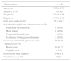
ResultsWe attempted to perform right heart catheterization in 20 patients through antecubital approach. The procedure was successful in 19 (95.0%) cases (Table 1).
Clinical and procedural characteristics.
| Characteristics | n = 20 |
|---|---|
| Age, years | 56.2 ± 15.9 |
| Male sex, n (%) | 10 (50) |
| Weight, kg | 71.4 ± 17.5 |
| Height, m | 1.65 ± 0.90 |
| Body mass index, kg/m2 | 26.2 ± 6.2 |
| Indication for right heart catheterization, n (%) | |
| Pulmonary hypertension | 6 (30.0) |
| Heart failure | 6 (30.0) |
| Congenital heart disease | 4 (20.0) |
| Evaluation for lung transplantation | 4 (20.0) |
| Success in antecubital approach, n (%) | 19 (95.0) |
| Vein access, n (%) | |
| Basilic vein | 18 (94.7) |
| Cephalic vein | 1 (5.3) |
| Fluoroscopic time, minutes | 5.4 ± 3.6 |
| Complications, n (%) | 0 (0) |
The only unsuccessful case occurred due to inadvertent puncture of the right brachial artery. In this specific case, as it was proposed to perform both right and left heart catheterizations, the left heart procedure was performed through brachial access (5 F sheath) and right heart catheterization was performed through the right internal jugular vein.
Simultaneous right and left heart catheterization was required in 12 (60.0%) cases. Left heart catheterization was performed through the right radial artery in 11 (91.7%) cases and through the right brachial artery in 1 (8.3%) case. The most common indications for right heart catheterization were heart failure and evaluation for pulmonary hypertension.
The mean fluoroscopic time was 5.0 ± 3.4minutes. Antecubital approach was obtained through the basilic vein in 18 (94.7%) cases and through the cephalic vein in 1 (5.3%) case. Among individuals who underwent single right heart catheterization, mean fluoroscopic time was 2.9 ± 1.3minutes. There were no procedure-related complications.
DiscussionThe present study reinforces that right heart catheterization through the antecubital veins is feasible and safe. Also, when concomitant left heart catheterization is needed, the combination of antecubital vein and radial artery approaches might be very useful, since left and right heart chambers may be reached with limited manipulation to the arm and forearm.
The antecubital approach was the first venous access site used for heart catheterization.1 Preference has, however, shifted to proximal vessels, such femoral and internal jugular veins. The widespread adoption of ultrasound guidance has minimized vascular complications in this setting.5 Its use is recommended by most medical, surgical, and anesthesia societies.5,6
Previous retrospective studies7 have demonstrated successful performance of right heart catheterization through the antecubital approach without ultrasound guidance. Most of these studies, however, included only individuals in which a superficial antecubital intravenous line had already been successfully obtained prior to entrance in the cath lab, which might represent a selection bias. Although experienced nurses may achieve comparably high success rates in obtaining intravenous lines at antecubital fossa,8 ultrasound guidance for accessing the vessels through micropuncture technique may offer some advantages.9
First, it broadens the use of the antecubital fossa vein approach to a larger group of individuals, since it sets aside the requirement of visualization or palpation of a suitable vein after tourniquet application. Second, ultrasound guidance may offer a safer way to access antecubital veins, which becomes even more advantageous in fully anticoagulated patients. Third, ultrasound guidance offers the operator the possibility to identify the vein with the greatest caliber in the forearm and to choose this vessel as the access site. This is important because one of the most common reasons for unsuccessful attempts to perform right heart catheterization through this approach is the impossibility to advance guidewires in small veins.
Harwani et al.10 demonstrated 100% success in performing right heart catheterization through antecubital fossa vein approach, with ultrasound-guided procedures. In our series, our success rate was 95%. Our only unsuccessful case was related to inadvertent puncture of the brachial artery, probably driven by our lack of experience in the early stages of the procedure.
The above-cited study,10 in a retrospective analysis of 1,130 cases, demonstrated that the antecubital approach is associated with fewer complications than the internal jugular vein approach for both right heart catheterization and for right heart biopsy. Patients that were submitted to interventions in both access sites (internal jugular vein and antecubital fossa vein) reported less anxiety and pain with the forearm approach. Moreover, the antecubital vein approach was reported as the preferential choice of access by these individuals. In this study, venous accesses were obtained under ultrasound guidance and their protocol for performing right heart catheterization through the antecubital approach was similar to our own.
Shah et al.2 demonstrated that in comparison to the femoral vein approach, the antecubital vein approach was associated with shorter fluoroscopic time for right heart catheterization. This result indicates that the forearm approach may reduce radiation exposure times for both patients and operators. In our study, mean fluoroscopic time was 5.4 ± 3.6minutes and for single right heart catheterization it was 2.9 ± 1.3minutes.
LimitationsOur study has several limitations. First, it is a single-center and small sample sized study. Second, it is an observational study and, as a case-series, there is no control group. Third, we did not assess the procedure duration time, neither the time required for attainment of venous access. Fourth, this was our early experience with this procedure. In this setting, our results need to be seen with caution and in an exploratory fashion.
On the other hand, the study has strengths. To our knowledge, this is the first report of right heart catheterization through the antecubital fossa vein approach under ultrasound-guided venous puncture in South America. Our study suggests that when both right and left heart catheterizations are required, combination of antecubital vein and radial artery approaches might be considered. Also, our high success and absence of complications encourage further studies with larger sample sizes and with a more powerful design.
ConclusionsOur study emphasizes that performance of right heart catheterization through the antecubital fossa vein approach appears to be feasible and safe. Further controlled studies are required to establish the best access site to perform right heart catheterization.
Funding sourcesNone declared.
Conflicts of interestThe authors declare no conflicts of interest.
Peer review under the responsibility of Sociedade Brasileira de Hemodinâmica e Cardiologia Intervencionista.