


Acute respiratory infection (ARI) is one of the principal causes of morbidity worldwide, with respiratory viruses being common etiological agents. Among them, endemic human coronaviruses (hCoVs) including CoV-229E, CoV-OC43, CoV-NL63, and CoV-HKU1 can cause mild ARI but are usually not evaluated in the clinical setting. The aim of this work was to determine the prevalence of all respiratory pathogens, with the focus placed on endemic hCoVs in the pre-pandemic period. Circulating species, clinical associations and coinfections with other respiratory pathogens were evaluated in 510 immunocompetent patients (children and adults) with ARI using the FilmArray® Respiratory Panel (BioFire/bioMérieux). A total of 399 children (252 outpatients and 147 hospitalized) and 111 adult outpatients were enrolled in the pre-pandemic period (2008–2010 and 2016). Endemic hCoVs were the third and fifth more frequently detected viruses among adults and outpatient children, respectively, with an overall frequency close to 10%. The most prevalent species were CoV-OC43 (42.8%) and CoV-HKU1 (40.5%), followed by CoV-NL63 (19.0%) and CoV-229E (4.8%). Tachypnea, wheezing and chest indrawing were more frequent in hospitalized children compared to outpatients. All adult patients presented with symptoms of a common cold. Endemic hCoVs were detected year-round, primarily between June and November. Our results highlight their clinical relevance, and the need to include endemic hCoVs in routine screening. In the post-pandemic period, further long-term surveillance is needed for understanding the epidemiology of endemic hCoVs and their evolution, as a tool to anticipate the possible emergence of new species.
Las infecciones respiratorias agudas (IRA) son una de las principales causas de morbilidad a nivel mundial, y los virus respiratorios se encuentran entre sus agentes etiológicos. Entre ellos, los coronavirus endémicos (hCoV) CoV-229E, CoV-OC43, CoV-NL63 y CoV-HKU1 pueden causar IRA leve, aunque habitualmente no son evaluados en el entorno clínico. El objetivo de este trabajo fue determinar la prevalencia de los patógenos respiratorios, en particular, de hCoV endémicos, en períodos prepandémicos. Se evaluaron las especies circulantes, las asociaciones clínicas y las coinfecciones con otros patógenos respiratorios en 510 pacientes inmunocompetentes (niños y adultos) con IRA utilizando el panel respiratorio FilmArray®. Durante 2 períodos prepandemia (2008-2010 y 2016), se enrolaron 399 niños (252 ambulatorios y 147 hospitalizados) y 111 adultos ambulatorios. Los hCoV endémicos ocuparon el tercer lugar en los adultos y el quinto lugar en niños ambulatorios dentro de los virus más detectados, con una frecuencia general cercana al 10%. Las especies más prevalentes fueron CoV-OC43 (42,8%) y CoV-HKU1 (40,5%), seguidas de CoV-NL63 (19,0%) y CoV-229E (4,8%). Las manifestaciones clínicas taquipnea, sibilancias y tiraje fueron más frecuentes en niños hospitalizados que en ambulatorios. Todos los pacientes adultos presentaron un resfriado común. Se detectaron hCoV endémicos durante todo el año, mayoritariamente entre junio y noviembre. Nuestros resultados resaltan la relevancia clínica de los hCoV endémicos y la necesidad de incluirlos en exámenes de rutina. Para comprender su epidemiología y evolución pospandemia, es necesario realizar una exhaustiva vigilancia epidemiológica aplicando herramientas que permitan anticipar la emergencia de nuevas especies.
Acute respiratory infection (ARI) is one of the principal causes of morbidity at all ages and worldwide. ARIs are usually more severe among children, the elderly and immunocompromised patients. Lower ARIs (ALRI) are the third cause of mortality in children25.
Respiratory viruses are the most frequent etiology of ARI in children. Classical respiratory viruses, including respiratory syncytial virus (RSV), influenza (Flu) A and B, parainfluenza (PIV) and adenovirus (ADV) were initially diagnosed by immunofluorescence and/or cell culture. Molecular methods significantly improved diagnosis due to a higher analytical sensitivity, achieved in less time. In addition, PCR assays permitted the detection of other respiratory viruses, such as human rhinovirus (HRV), human bocavirus (hBoV) and endemic human coronaviruses (hCoVs) that were not effectively detected by conventional methods2,21.
PCR assays using point-of-care formats were developed to simultaneously detect viral and bacterial respiratory pathogens within 2h. The FilmArray® Respiratory Panel (FA-RP) (BioFire/bioMérieux) can simultaneously detect 17 respiratory viruses (including the classical respiratory viruses, human metapneumovirus (hMPV), HRV and endemic hCoVs) and three respiratory bacteria (Bordetella pertussis, Mycoplasma pneumoniae, and Chlamydophila pneumoniae)20.
Coronaviruses, members of the Coronaviridae family, are enveloped viruses with a large, positive, single-stranded RNA genome of 27–32kb. They are classified into four genera (alpha, beta, gamma, and delta). Identification in humans date back to the 1960s and only alpha and beta coronaviruses are known to infect humans5.
To date, seven hCoVs have been described. CoV-229E and CoV-NL63 are alpha coronaviruses while HCoV-HKU1, HCoV-OC43, MERS-CoV, SARS-CoV-1 and SARS-CoV-2 (responsible for the recent COVID-19 pandemic) are beta coronaviruses37.
Endemic hCoVs including CoV-229E, CoV-OC43, CoV-NL63, and CoV-HKU1 cause mild ARI and common colds in adults and children, but can occasionally cause bronchiolitis and pneumonia as well. They are most frequently detected during the winter, and commonly in coinfection, with a detection rate of 2–20%, depending on the study setting19.
CoV-OC43 and CoV-229E were isolated in cell culture in 1965 and 1967, respectively, from patients with respiratory illnesses in the USA. CoV-OC43 was first discovered by using a human embryonic tracheal organ culture (OC) technique, while CoV-229E was first isolated from a secondary human kidney tissue culture with a rare cytopathic effect10,24.
With the advances in molecular diagnosis, two more endemic hCoVs were described. CoV-NL63 was almost simultaneously described by two independent groups in The Netherlands in 2004, from two pediatric patients with pneumonia and bronchiolitis, respectively8,17. In 2005, CoV-HKU1 was described by Woo et al.38 from a 71-year-old man with pneumonia in China.
Highly pathogenic hCoVs include severe acute respiratory syndrome (SARS) and Middle East respiratory syndrome (MERS), caused by SARS-CoV-1 and MERS-CoV, respectively. First described in China in 2003 and in the Middle East in 2012, SARS-CoV-1 and MERS-CoV had case mortality rates of ∼9.6% and ∼34%, respectively. In December, 2019, SARS-CoV-2 was identified in Wuhan, China and rapidly expanded worldwide, causing the COVID-19 pandemic6,40,42. This renewed interest in all hCoVs and their potential for serological cross-protection was evaluated1,33. Further studies are needed to understand the burden of endemic hCoV infections in our community.
Endemic hCoVs are prevalent, but usually not evaluated in the clinical setting. Therefore, their clinical impact and epidemiology are underestimated. So the aim of this work was to determine the prevalence of all respiratory pathogens, with special focus placed on endemic hCoVs in the pre-pandemic period. Endemic hCoV circulating species, clinical associations and coinfections with other respiratory pathogens were evaluated in immunocompetent patients with ARI using a multiplex PCR assay.
Materials and methodsStudy populationAs part of a larger observational, descriptive, cross-sectional study of ARI, samples were obtained from children and adults with ARI who attended the Emergency Department at Centro de Educación Médica e Investigaciones Clínicas (CEMIC) University Hospital, Buenos Aires, Argentina during 2008–2010 and 2016.
Clinical and demographic data were recorded by the attending physician using a specially designed form. Collected information included demographic data, vaccination schedule, personal clinical history, symptoms and presumptive diagnosis. In hospitalized patients, data on length of stay (LOS), oxygen therapy, admission to an intensive care unit and outcome were recorded. Informed consent was signed in all cases, by a parent/tutor (for children) or the patient.
Inclusion criteria were: patients with ARI aged between 2 months and 6 years of age for children and older than 18 years old for adults were included. A maximum of 5 patients with ARI per day were randomly selected to be tested by a multiplex PCR assay. Symptom onset within the preceding 7 days was considered acute respiratory infection (ARI). For this analysis, only participants with complete medical records who were tested using a multiplex PCR assay were included.
ARI was defined as the presence of at least two of the following signs or symptoms: fever/history of fever, cough, tachypnea, wheezing, difficulty breathing, diffuse or focal signs at auscultation or presumptive diagnosis of bronchiolitis, influenza-like illness, bronchitis, or pneumonia.
Exclusion criteria included congenital cardiac disease, neurological or genetic disorders, cancer, HIV, immunosuppression or solid organ or hematopoietic stem cell transplantation.
Viral detectionRespiratory samples studied were nasopharyngeal swabs, obtained in viral transport media. Samples were tested within an hour of arrival using the FilmArray® Respiratory Panel (RP-FA). This commercial multiplex PCR can detect 17 viruses (RSV, FluA H1, H1-2009, H3, FluB, AdV, PIV 1-4, HRV/enterovirus HRV/EV, hMPV, hBoV and CoV-OC43, CoV-229E, CoV-NL63, CoV-HKU1), and three bacteria commonly associated with ARI (B. pertussis, M. pneumoniae, and C. pneumoniae) within 2h. Samples were tested according to the manufacturer's instructions (BioFire/bioMérieux).
Coinfection was defined as a sample with a positive test result for two or more respiratory pathogens.
Statistical analysesData were stored in a digital-supported database. Values were expressed as percentages for categorical variables or as median and interquartile range (IQR) for continuous variables. Data were analyzed in GraphPad Prism software (version 5; GraphPad software, La Jolla, CA, USA), using the Chi-square or Fisher's exact test for comparing categorical variables and the Mann–Whitney–Wilcoxon test for numeric variables. Statistical significance was assumed for p values less than 0.05.
Ethical approvalThe study protocol was reviewed and approved by the CEMIC Institutional Review Board (No. 00001745) and was conducted in accordance with the Declaration of Helsinki.
ResultsA total of 510 immunocompetent patients with ARI were enrolled at CEMIC University Hospital. Specifically, 399 children (252 outpatients and 147 hospitalized) were enrolled in two periods (June 2008–June 2010 and April–September 2016) and 111 adult outpatients were enrolled in one period (April–September 2016).
Among the children, the median age in months was 10.0 (IQR: 3.0–17.0) for hospitalized and 15.0 (IQR: 7.0–28.2) for outpatients. Most hospitalized (83.0%, 122/147) and outpatient (69.0%, 174/252) children were younger than 2 years of age. Male percentage (∼58.0%) was similar in both groups. Flu vaccination in children aged 6–24 months old was more frequent in outpatients (42.5%, 54/127) than in hospitalized children (24.3%, 17/70) (p=0.013). In the adult population, the median age in years was 45 (IQR: 35.0–63.5), 53.1% (59/111) were male and all were outpatients. In adults over 65 years old, the reported flu vaccination rate was 63.0% (17/27) (Table 1).
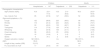
Demographic and clinical characteristics in 510 patients with acute respiratory infection (children n=399; adults n=111).
| Children | Adults | |||||
|---|---|---|---|---|---|---|
| Hospitalizedn=147 | Outpatientn=252 | Outpatientn=111 | ||||
| Demographic characteristics | ||||||
| Agea [median (IQR)] | 10.0 | (3.0–17.0) | 15.0 | (7.0–28.2) | 45.0 | (35.0–63.5) |
| Sex, male [n (%)] | 85 | (57.8) | 147 | (58.3) | 52 | (46.8) |
| Signs and symptoms, n (%) | ||||||
| Cough | 132 | (89.8) | 225 | (89.3) | 99 | (89.2) |
| Rhinitis | 119 | (81.0) | 233 | (92.5) | 84 | (75.7) |
| Fever | 93 | (63.3) | 153 | (60.7) | 38 | (34.2) |
| Tachypnea | 114 | (77.6) | 120 | (47.6) | 21 | (19.8) |
| Wheezing | 85 | (57.8) | 105 | (41.7) | 21 | (19.8) |
| Chest indrawing | 96 | (65.3) | 65 | (25.8) | 3 | (2.7) |
| Cyanosis | 16 | (10.9) | 4 | (1.6) | 0 | (0.0) |
| Apnea | 6 | (4.1) | 2 | (0.8) | 0 | (0.0) |
| Oxygen saturation, median (IQR) | 92.0 (90.0–95.0) | n/a | 98.0 (97.0–98.0) | |||
| Length of stay, median (IQR) | 3 (2–5) | n/a | n/a | |||
| Flu vaccination [n (%)]b | 17 | (24.3) | 54 | (42.5) | 17 | (63.0) |
IQR: interquartile range; n/a: not applicable.
Tachypnea, wheezing and chest indrawing were more frequent in hospitalized children compared to outpatients (77.6% vs 47.6%, 57.8% vs 41.7% and 65.3% vs 25.8%, respectively). Fever percentage (∼60.0%) was similar in both groups. In hospitalized children, 147/399 (36.8%), median LOS was 3 (IQR: 2–5) days. Oxygen therapy was required in over half of the cases (59.2%, 87/147). Median oxygen saturation at admission was 93.0% (IQR: 91.5–95.5). In the adults, frequent clinical manifestations included cough (89.2%), rhinitis (75.7%), and tachypnea (19.8%). Fever was reported in 34.2% (38/111) of cases. Median oxygen saturation was 98.0% (IQR: 97.0–98.0) (Table 1).
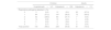
A respiratory pathogen was detected in 98.0% (144/147) of hospitalized children and 94.8% (239/252) of outpatient children using the RP-FA. A single pathogen was detected in most cases (57.2% for hospitalized and 58.3% for outpatients), followed by coinfections with two pathogens (28.6% for hospitalized and 31.7% for outpatients). In the hospitalized children, a quintuple viral coinfection was detected, along with three cases of bacterial etiology, always in coinfection with other viruses. In the adults, a viral pathogen was detected in 73.0% (81/111) of cases. A single pathogen was detected in 65.8% (73/111) of cases. Coinfections with two (6.3%) or three (0.9%) respiratory viruses were less frequently detected. No bacterial infection was detected in adults (Table 2).
Number of respiratory pathogens detected by RP-FA in 510 patients with ARI (children n=399; adults n=111).
| Children | Adults | |||||
|---|---|---|---|---|---|---|
| Hospitalizedn=147 | Outpatientn=252 | Outpatientn=111 | ||||
| Respiratory pathogens detected, n (%) | ||||||
| 0 | 3 | (2.0) | 13 | (5.2) | 30 | (27.0) |
| 1 | 84 | (57.2) | 147 | (58.3) | 73 | (65.8) |
| 2 | 42 | (28.6) | 80 | (31.7) | 7 | (6.3) |
| 3 | 14 | (9.5) | 10 | (4.0) | 1 | (0.9) |
| 4 | 3 | (2.0) | 2 | (0.8) | 0 | (0.0) |
| 5 | 1 | (0.7) | 0 | (0.0) | 0 | (0.0) |
| Total positive | 144 | (98.0) | 239 | (94.8) | 81 | (73.0) |
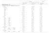
In the hospitalized children, HRV/EV was detected in 48.3% (71/147) of cases and half of them (36/71) were single pathogen infections. RSV was the second most frequent, detected in 32.0% (47/147) of children. Half of the cases (25/47) were coinfections. hBoV was detected in 17.0% (25/147) of cases, always in coinfection. AdV was detected in 15.0% (22/147) of the children and 81.8% of cases (18/22) were coinfections (Fig. 1a).

Bacterial etiology was documented in 3/147 (2.0%) of the hospitalized children with viral coinfection: 2/3 had C. pneumoniae (in coinfection with HRV/EV and triple coinfection with RSV and FluB) and 1/3 had B. pertussis (in coinfection with HRV/EV).
The patient with C. pneumoniae and HRV/EV had bronchitis, oxygen saturation at admission was 90% and LOS was 5 days. The patient with C. pneumoniae, RSV and FluB had bronchiolitis, oxygen saturation at admission was 96% and LOS was 3 days. The patient with B. pertussis and HRV/EV had bronchiolitis and conjunctivitis, LOS was 8 days with 6 days of oxygen therapy.
In the outpatient children, HRV/EV was detected in 40.8% (103/252) of the cases and half of them (52/103) were single pathogen infections. RSV was the second most frequent, detected in 26.2% (66/252) of the children. Over half of the cases, 55.9% (38/68), were coinfections. PIV3 was detected in 13.5% (34/252) of patients, mostly – 58.8% (20/34) – as the only pathogen detected. hBoV was detected in 12.7% (32/252) of cases, mostly – 90.2% (29/32) – in coinfection (Fig. 1b).
In the outpatient adults, HRV/EV was detected in 27.0% (30/111) of cases and in most of them, 76.7% (23/30), it was the only pathogen detected. The second most frequent was Flu A, detected in 19.8% (22/111) of cases, being the only pathogen detected in 77.3% (17/22) of them. RSV was detected in 9.0% (10/111) of cases, being the only pathogen detected in 90.0% (9/10) of them (Fig. 1c).
Endemic hCoVsOverall frequency of endemic hCoVs was 8.2% (42/510) using the RP-FA. Endemic hCoVs were detected year-round, primarily between June and November. Most cases, 33.3%, were detected in July. Across the studied populations, the four endemic species were detected, being the most prevalent species CoV-OC43 (42.8%) and CoV-HKU1 (40.5%), followed by CoV-NL63 (19.0%) and CoV-229E (4.8%).
In the children, endemic hCoVs were detected in 8.5% (34/399) of cases. Frequency was similar in outpatients and hospitalized children: 9.1% (23/252) and 7.5% (11/147), respectively (p=0.712). In adults, overall frequency was 7.2% (8/111) (Table 3).
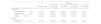
Endemic hCoV frequency and species characterization in 510 patients with acute respiratory infection (children n=399; adults n=111).
| All | Species | ||||
|---|---|---|---|---|---|
| hCoVs, n (%) | CoV-OC43 | CoV-HKU1 | CoV-NL63 | CoV-229E | |
| Children | |||||
| Hospitalized (n=147) | 11 (7.5) | 3 (27.2) | 5 (45.5) | 2 (18.2) | 1 (9.1) |
| Outpatienta (n=252) | 23 (9.1) | 12 (52.2) | 8 (34.8) | 5 (21.7) | 1 (4.3) |
| Total (n=399) | 34 (8.5) | 15 (3.7) | 13 (3.2) | 7 (1.7) | 2 (0.5) |
| Adults | |||||
| Outpatient (n=111) | 8 (7.2) | 3 (37.5) | 4 (50.0) | 1 (12.5) | 0 (0.0) |
In the hospitalized children, the most prevalent species were CoV-HKU1 (45.5%) and CoV-OC43 (27.2%), followed by CoV-NL63 (18.2%) and CoV-229E (9.1%). Among the outpatients, the most prevalent species was CoV-OC43 (52.2%), followed by CoV-HKU1 (34.8%), CoV-NL63 (21.7%) and CoV-229E (4.3%). Among the adults, the most prevalent species was CoV-HKU1 (50.0%), followed by CoV-OC43 (37.5%) and CoV-NL63 (12.5%). CoV-229E was not detected.
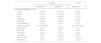
Cough and rhinitis percentages (∼90.0%) were similar in both groups. Tachypnea, fever, chest indrawing and wheezing were more frequent in hospitalized children compared to outpatients (90.9% vs 39.1%, 81.8% vs 60.9%, 72.7% vs 21.7% and 45.5% vs 34.8%, respectively) (Table 4). Most children, 53.0% (18/34), had bronchiolitis and 9.1% (1/11) of the hospitalized patients had pneumonia. Pharyngitis and laryngitis were only observed in outpatient children (21.7% and 4.3%, respectively). Median oxygen saturation was 93.0% (91.5–95.5) in hospitalized children, with a median LOS of 3 (IQR: 1.5–6.0) days. All the adult patients presented with a common cold and 12.5% (1/8) had pneumonia. Cough and fever percentages (∼88.0%) were similar. Most adults (75.7%) also had rhinitis and 12.5% had tachypnea (Table 4). Median oxygen saturation was 98.0% (IQR: 97.0–98.0).
Clinical characteristics associated with human coronaviruses in 42 patients with acute respiratory infection (children n=34; adults n=8).
| Children | Adults | ||
|---|---|---|---|
| Hospitalizedn=11 | Outpatientn=23 | Outpatientn=8 | |
| Clinical characteristics, n (%) | |||
| Cough | 10 (90.9) | 21 (91.3) | 7 (87.5) |
| Rhinitis | 10 (90.9) | 20 (87.0) | 6 (75.0) |
| Fever | 9 (81.8) | 14 (60.9) | 7 (87.5) |
| Tachypnea | 10 (90.9) | 9 (39.1) | 1 (12.5) |
| Wheezing | 5 (45.5) | 8 (34.8) | 0 (0.0) |
| Chest indrawing | 8 (72.7) | 5 (21.7) | 0 (0.0) |
| Oxygen saturationa | 93.0 (91.5–95.5) | n/a | 98.0 (97.0–98.0) |
| Bronchiolitis | 9 (81.8) | 9 (39.1) | n/a |
| Bronchitis | 0 (0.0) | 2 (8.7) | 0 (0.0) |
| Pharyngitis | 0 (0.0) | 5 (21.7) | 0 (0.0) |
| Laryngitis | 0 (0.0) | 1 (4.3) | 0 (0.0) |
| Common cold | 1 (9.1) | 5 (21.7) | 8 (100.0) |
| Pneumonia | 1 (9.1) | 0 (0.0) | 1 (12.5) |
IQR: interquartile range; n/a: not applicable.
In the children, most endemic hCoVs, 88.8% (30/34), were detected in coinfection with other viruses. Specifically, 90.9% (10/11) and 87.0% (20/23) of the cases in hospitalized and outpatient children were coinfections, respectively (Table 5).
Detection of hCoVs as a single pathogen or in coinfection with other respiratory viruses in 42 patients with ARI (children n=34; adults n=8).
| Children | Adults | ||
|---|---|---|---|
| Hospitalizedn=11 | Outpatientn=23 | Outpatientn=8 | |
| Single pathogen, n (%) | 1 (9.1) | 3 (13.0) | 7 (87.5) |
| Coinfection with other respiratory viruses, n (%) | |||
| Double | 4 (36.4) | 16 (69.6) | 0 (0.0) |
| HRV/EV | 0 (0.0) | 5 (21.7) | 0 (0.0) |
| hBoV | 0 (0.0) | 3 (13.0) | 0 (0.0) |
| hMPV | 3 (27.3) | 1 (4.3) | 0 (0.0) |
| RSV | 1 (9.1) | 2 (8.7) | 0 (0.0) |
| AdV | 0 (0.0) | 2 (8.7) | 0 (0.0) |
| FluA | 0 (0.0) | 1 (4.3) | 0 (0.0) |
| FluB | 0 (0.0) | 1 (4.3) | 0 (0.0) |
| PIV4 | 0 (0.0) | 1 (4.3) | 0 (0.0) |
| Triple | 3 (27.3) | 4 (17.4) | 1 (12.5) |
| hCoV+hBoV | 0 (0.0) | 1 (4.3) | 0 (0.0) |
| hCoV+PIV3 | 0 (0.0) | 1 (4.3) | 0 (0.0) |
| hCoV+hMPV | 0 (0.0) | 1 (4.3) | 0 (0.0) |
| RSV+HRV | 0 (0.0) | 1 (4.3) | 0 (0.0) |
| RSV+hBoV | 1 (9.1) | 0 (0.0) | 0 (0.0) |
| hMPV+hBoV | 1 (9.1) | 0 (0.0) | 0 (0.0) |
| hMPV+PIV3 | 1 (9.1) | 0 (0.0) | 0 (0.0) |
| FluA+HRV/EV | 0 (0.0) | 0 (0.0) | 1 (12.5) |
| Quadruple | 2 (18.2) | 0 (0.0) | 0 (0.0) |
| PIV3+hMPV+HRV/EV | 1 (9.1) | 0 (0.0) | 0 (0.0) |
| AdV+HRV/EV+hBoV | 1 (9.1) | 0 (0.0) | 0 (0.0) |
| Quintuple | 1 (9.1) | 0 (0.0) | 0 (0.0) |
| ADV+hMPV+HRV/EV+hBoV | 1 (9.1) | 0 (0.0) | 0 (0.0) |
In the hospitalized children, viral detection included double (36.4%), triple (27.3%), quadruple (18.2%) and quintuple (9.1%) coinfections with an endemic hCoV. Double coinfection with an endemic hCoV included hMPV (27.3%) and RSV (9.1%). hMPV was also detected in most cases (18.2%) of triple coinfection, in one of the two cases of quadruple coinfection and in the only case in our cohort that was positive for five respiratory viruses (Table 5).
Median LOS was 3.5 (IQR: 2.0–6.5) days for the 10 children with coinfections, and 10% (1/10) were transferred to the ICU for 8 days. Oxygen therapy was needed in 40.0% (4/10) of cases, for a median of 4.5 (IQR: 2.5–5.7) days. Most hospitalized patients with coinfections, 90.0% (9/10), had bronchiolitis and 10% (1/10) had pneumonia.
In the outpatient children, viral detection included double (69.6%) and triple (17.4%) coinfections. Other respiratory viruses detected in double coinfection with an endemic hCoV included HRV/EV and hBoV, in 21.7% (5/23) and 13.0% (3/23) of patients, respectively. Triple coinfections included two species of endemic hCoVs in 13.0% (3/23) of the outpatient cases (Table 5).
In the adults, most endemic hCoVs were detected as a single pathogen and only 12.5% (1/8) of patients had a coinfection. This was a triple coinfection, with FluA and HRV/EV (Table 5).
DiscussionIn recent years, endemic hCoVs have regained importance due to pandemics of zoonotic origin. These viruses were initially described in the mid-1960s, when two endemic species were detected in cell culture: CoV-OC43 and CoV-229E. In the early 2000s, two more endemic species were described using molecular biology assays: CoV-NL63 and CoV-HKU1. The first highly pathogenic hCoV outbreaks occurred in 2003 and 2012, caused by SARS-CoV-1 and MERS-CoV, respectively.
The COVID-19 pandemic, caused by a new highly pathogenic hCoV, started a global crisis, affecting the health care and socioeconomic development of the entire population. As with SARS-CoV-1 and MERS-CoV, but unlike the previous pandemic (caused by influenza in 2009), the highest mortality rate from COVID-19 was observed in older age groups4,13. This critical situation renewed interest in all hCoVs.
However, knowledge of the distribution, epidemiology, and symptoms associated with non-SARS hCoVs remains limited, with most of the research performed in countries from the Northern Hemisphere. In this study, respiratory samples from patients with ARI taken year-round in two pre-pandemic periods were evaluated in Buenos Aires, Argentina. hCoVs were detected in all studied years (2008–2010 and 2016). All four endemic species (CoV-229E, CoV-OC43, CoV-NL63, and CoV-HKU1) were detected. Importantly, endemic hCoVs were detected in children and adults in similar frequency (8.5% and 7.2%, respectively). This highlights hCoV capacity for infecting people of all ages, causing a short-lived immunity9. Memory B cells can persist several years but this humoral response shows not to be protective against re-infection32.
In our cohort, the overall frequency of endemic hCoVs was 8.2%. Similar overall frequencies of 10.6% and 7.6% were detected in the USA, from 2010 to 2018, and Japan, from 2010 to 2013, respectively23,26. In contrast, a retrospective study conducted from 2011 to 2012 in a different Argentinian region showed a lower overall frequency of 3.0%, when only screening for CoV-229E and CoV-OC4329.
Epidemiological studies on endemic hCoVs in adult populations are scarce, and reported detection frequencies vary. In our study, the frequency was 7.2% in adults with ARI. A nine-year study (2001–2010) in the neighboring country of Brazil also reported similar frequencies in adult patients (8%) and health care workers (10.5%) using a pancoronavirus RT-PCR assay3. However, a higher detection frequency (16.0%) was reported in Chinese adults in 2009–2011, using a different pancoronavirus assay, and in the Netherlands in 2017, using the RP-FA18,35. CoV-HKU1 was the most prevalent species, closely followed by CoV-OC43. A study in Hong Kong reported CoV-OC43 as the most prevalent species in hospitalized adults39. Remarkably, there were no cases of CoV-229E among adults in our study, but it was the most prevalent species in a Chinese study, with a frequency of 9.8%18.
Despite previous reports of hCoVs peaks overlapping with RSV and flu seasons16, most adult patients in our cohort had one of these viruses as the sole pathogen detected, suggesting the endemic hCoV caused their symptomatology. A greater sole pathogen detection for hCoVs was also reported in older patients in a comprehensive epidemiological analysis performed in Scotland27.
In our study, the overall pediatric frequency of endemic hCoVs was 8.5%. This is consistent with previous reports from children in the USA (8.6%) and Norway (9.1%)12,34. We detected similar frequencies in outpatients (9.1%) and hospitalized (7.5%) children. Accordingly, a slightly higher frequency in outpatient (10.8%) compared to hospitalized (8.2%) children was reported in the USA in a retrospective study from 2013 to 201434. The most prevalent species were CoV-OC43 and CoV-HUK1. Different studies also reported CoV-OC43 as one of the most prevalent species in children (up to 70%), but, interestingly, CoV-HKU1 was the less prevalent (7.8%) in Chinese children41.
In children, hCoVs were mostly detected in coinfection with other respiratory viruses, being HRV, RSV and hMPV the most common. A 1043 children study in the USA reported frequent hCoV coinfections (45.5% of cases) with RSV, hMPV and FluA, but they did not test for HRV15. A different study, including HRV testing, reported a clustering pattern in which HRV, AdV, RSV and hCoVs were simultaneously detected in children under 5 years old27. hBoV was among the most frequent pathogens detected in coinfection in our population and this is consistent with previous reports (25%)36. However, this virus is not currently screened for, as there is no clear association between hBoV and ARI22.
In the hospitalized children positive for an endemic hCoV, symptoms and LOS were affected by the presence of coinfections with other respiratory pathogens. In concordance with previous reports, patients with concomitant detection of viruses known to cause severe ARI (RSV, hMPV, PIV1–3) exhibited lower respiratory symptoms, such as tachypnea, chest indrawing, and longer hospital stays14,31. Conversely, in the outpatient children (regardless of the coinfection status) and adult patients, upper respiratory symptoms including cough and rhinitis were the most frequent. This is consistent with previous reports of mild symptoms caused by endemic hCoVs in outpatient children and adults18,34.
Notably, we registered two cases of pneumonia, in a hospitalized child (with a RSV coinfection) and an outpatient adult who required hospital admission. The 85-year-old patient presented an endemic hCoV as the sole pathogen detected, in agreement with previous reports of severe respiratory illness in older patients11.
In this study, endemic hCoV infections were detected year-round, and no difference in timing of season was observed between alpha and beta coronaviruses. We observed a higher number of infections during the winter and spring. Lower circulation of endemic hCoVs during the summer season was also reported for children under 2 years old in the UK, similar to our cohort30.
This study contributes to the knowledge of endemic hCoV prevalence and circulation patterns among Argentinian patients during the pre-pandemic period. In our cohort, endemic hCoVs were the third and fifth more detected viruses among adults and outpatient children, respectively, with an overall frequency close to 10%. This highlights their clinical relevance in the pre-pandemic era, and the need to include them in routine screening.
The limitations of this study were the retrospective nature of the analysis, and the single-center design. Larger prospective studies are needed to establish endemic hCoV epidemiology in the post-pandemic era, complemented with a multicenter approach to determine different geographical tendencies.
The development of in-house methods of detection is crucial to achieve this, as a cost-effective option to commercial methods. As part of the COVID-19 response, new detection methods were developed in record time, employing different respiratory samples, such as nasopharyngeal swabs and saliva7,28. Now, in the post-pandemic period, it is important to maintain these efforts for a better understanding of the impact of SARS-CoV-2 on the epidemiology of other respiratory viruses, including the endemic hCoVs.
Furthermore, the majority of new, emerging, or re-emerging diseases affecting individuals are of zoonotic origin, including COVID-19. Considering that human activities continue to negatively impact ecosystems, creating favorable conditions for the emergence and spread of diseases, an integrated strategy is indispensable to control future outbreaks.
One Health is a unifying approach, led by the World Health Organization, that aims to sustainably balance and optimize the health of individuals, animals and ecosystems. Infectious diseases disproportionately impact citizens of low- and middle-income countries and other vulnerable populations; therefore, incorporating endemic hCoV screening in these populations is clinically and epidemiologically relevant.
Furthermore, long-term surveillance is needed to understand the epidemiology of endemic hCoVs in our region, and their evolution. A comprehensive study on endemic hCoVs could be a useful tool to anticipate the possible emergence of new species.
FundingThis work was partially supported by a grant provided by Biofire/BioMérieux, USA. The sponsor had no involvement in the conduct of the study or the analysis of the data.
Conflict of interestsNone of the authors have a conflict of interest to disclose.
We would like to thank Dr. Jorge López Camelo for proofreading of the article.
