Buscar en
Radiología (English Edition)
Toda la web
Inicio Radiología (English Edition) The hilum of the lung: Two classical radiological signs to decipher it
Journal Information
Share
Share
Download PDF
More article options
Visits
10033
Article from a resident
The hilum of the lung: Two classical radiological signs to decipher it
El hilio pulmonar, dos signos radiológicos clásicos para descifrarlo
Visits
10033
T. Ludeñaa, A. Lozano-Samaniegob,
Corresponding author
aslozano@utpl.edu.ec

Corresponding author.
, S. Maldonadoc, F. Salasd
a Residente de Radiología, Hospital General Isidro Ayora, Loja, Ecuador
b Radiólogo, Docente de Radiología, Universidad Técnica Particular de Loja; Hospital General Isidro Ayora, Loja, Ecuador
c Estudiante, Titulación de Medicina Universidad Técnica Particular de Loja, Ecuador
d Médico, Analista de Provisión de Servicios, Ministerio de Salud Pública, Loja, Ecuador
This item has received
Received 27 February 2020. Accepted 07 September 2020
Article information
Abstract
Full Text
Bibliography
Download PDF
Statistics
Figures (9)
Show moreShow less
Abstract

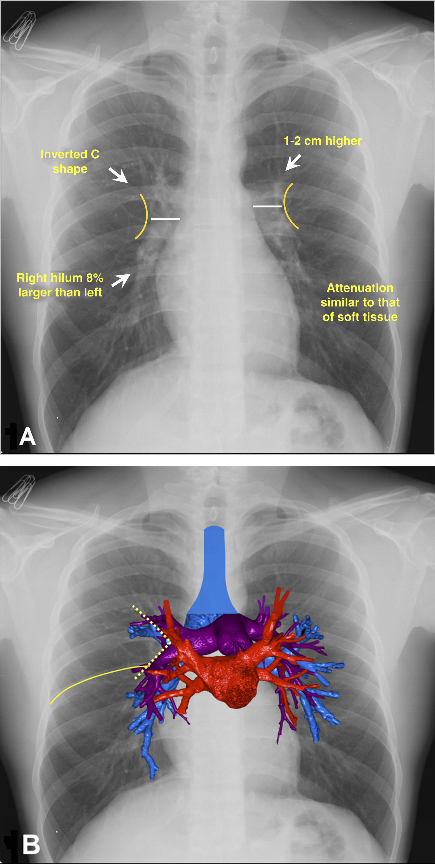
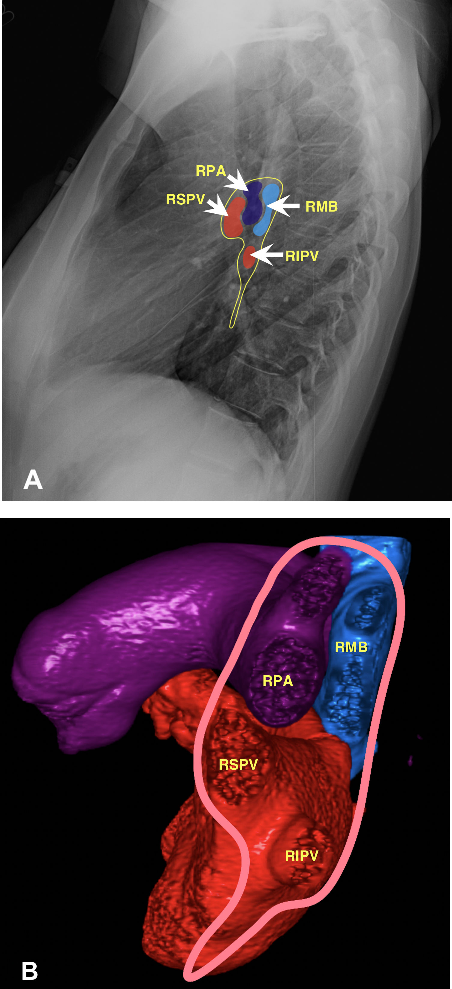
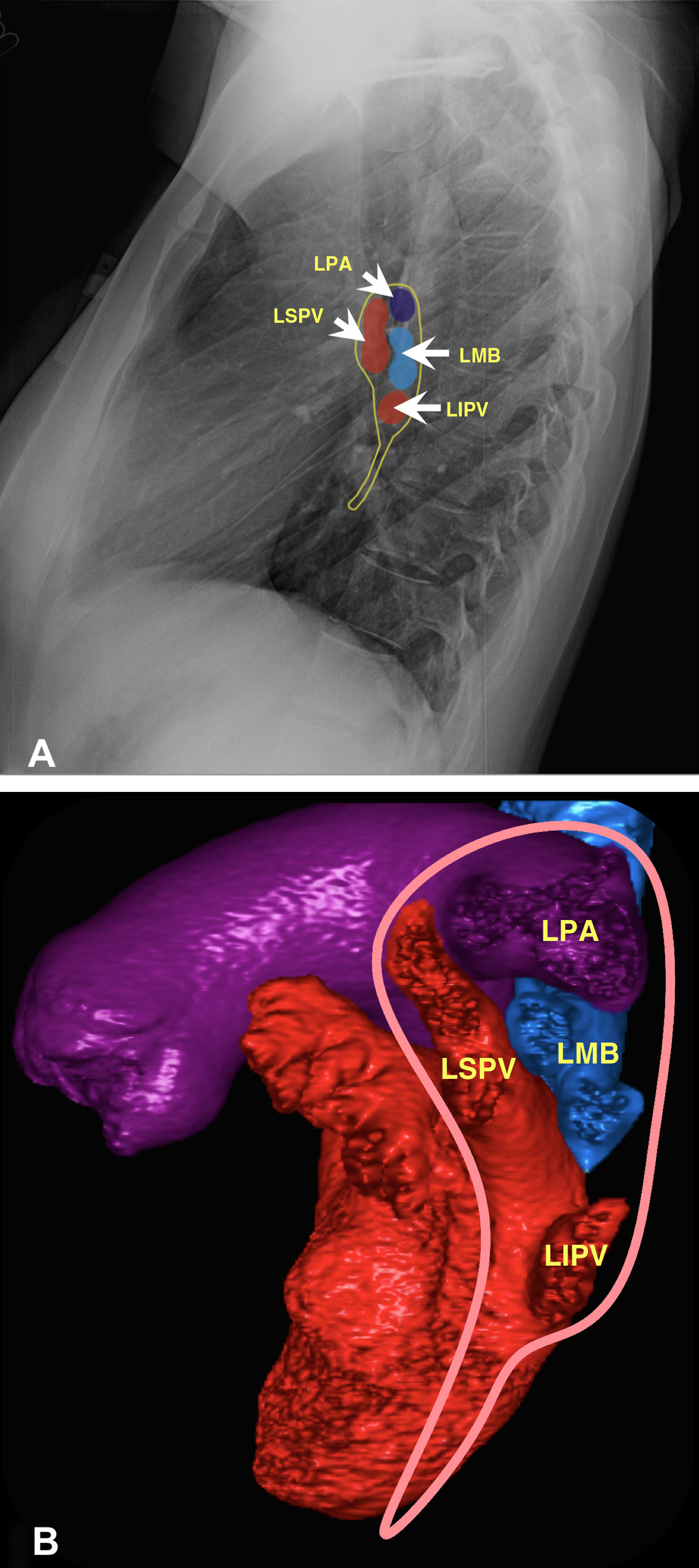
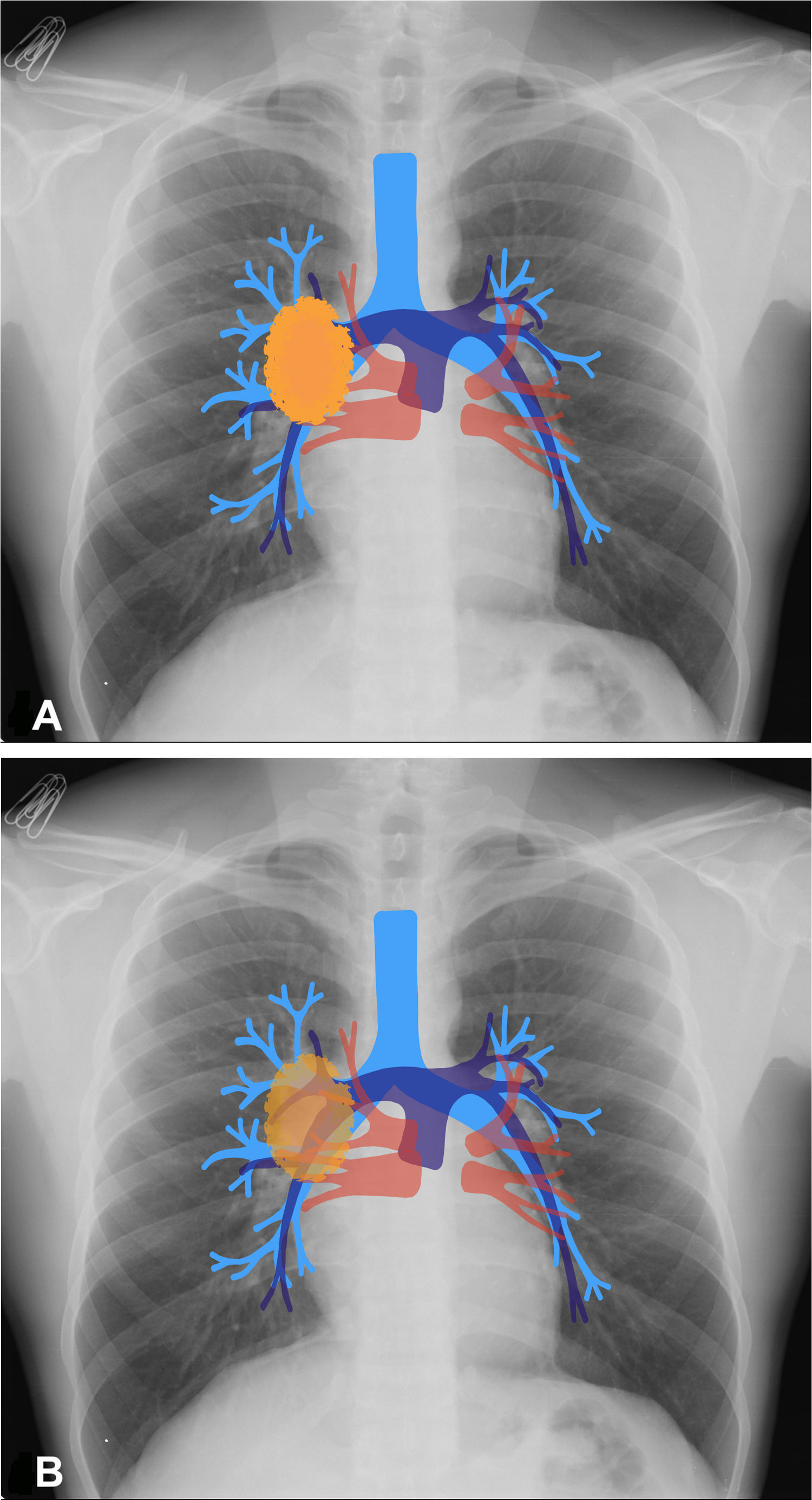
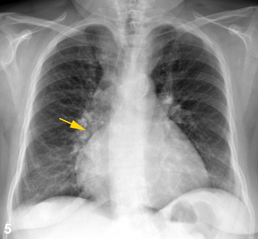
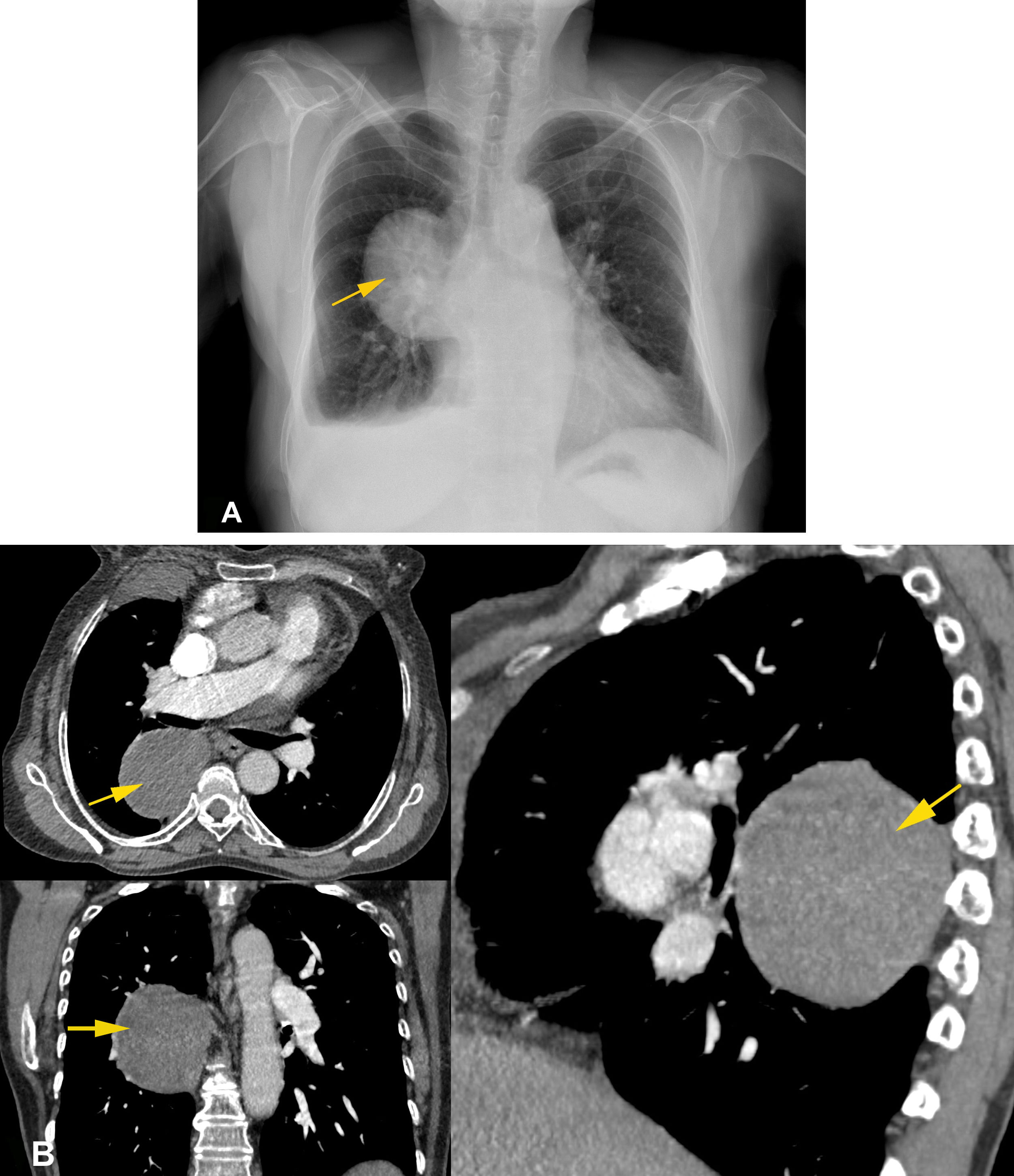
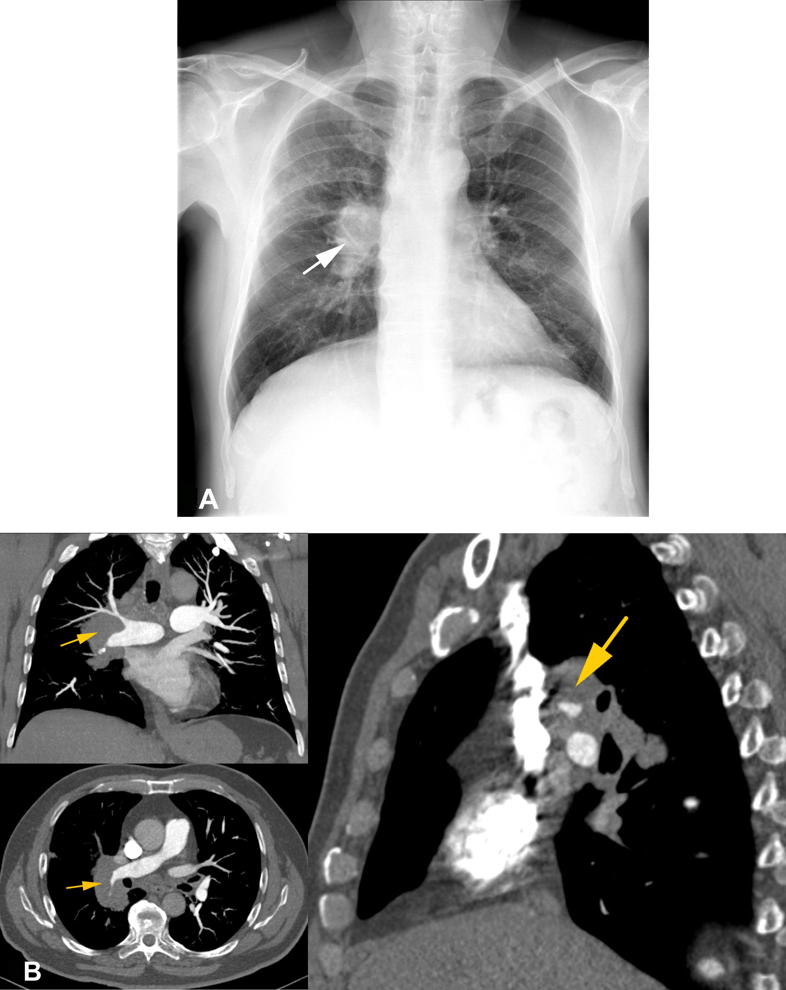
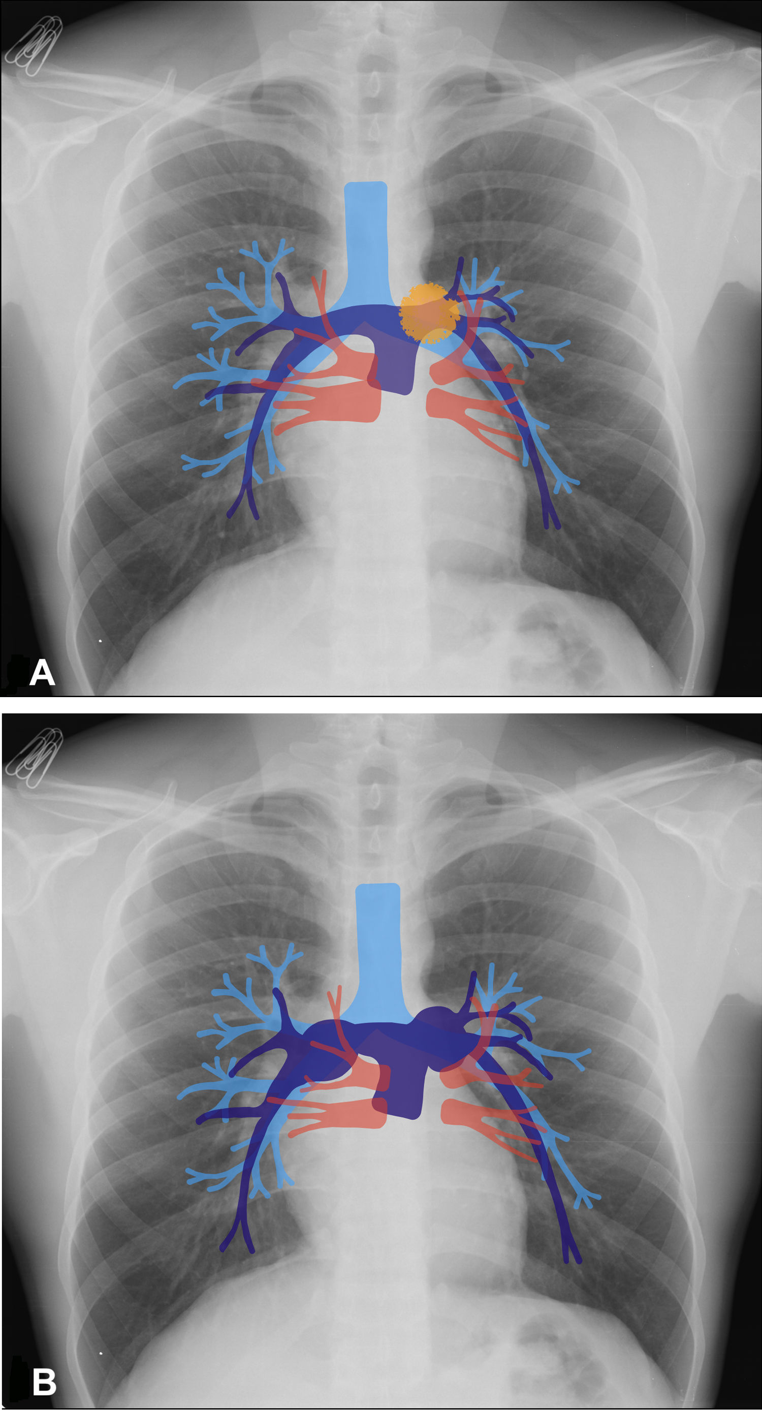
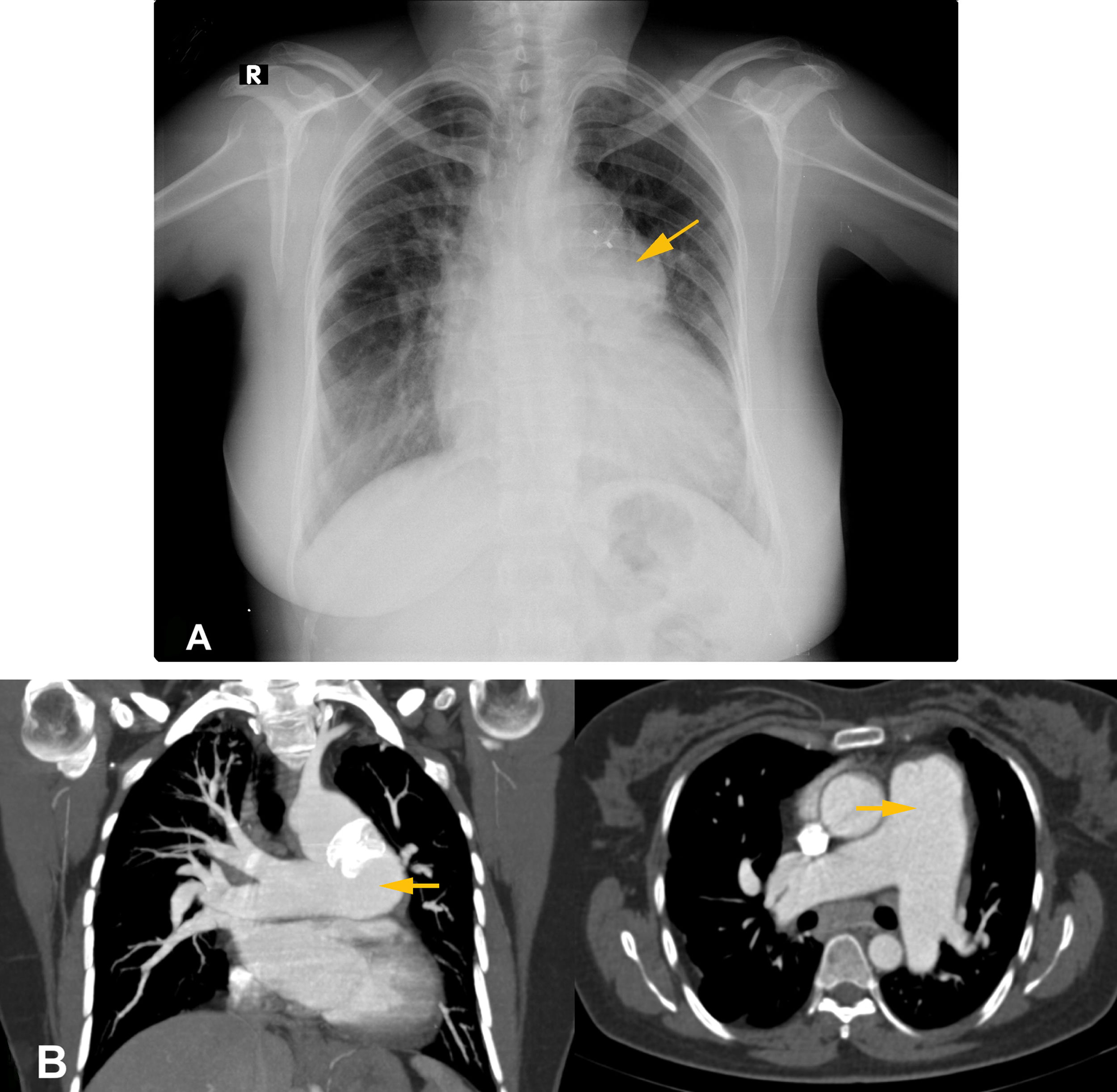
Assessing the hilum of the lung is a common challenge in daily practice because various structures converge in this complex anatomic region. Because chest X-rays are widely available and deliver relatively low doses of radiation, they continue to be the most common imaging test, although new imaging modalities have decreased the use of chest X-rays for differentiating between true abnormalities and superimposed lung opacities. This article reviews the literature and describes the principal anatomic relations of the lung hilum through illustrative cases to enable the two most important radiologic signs to be identified: “hilum overlay” and “hilum convergence”. In the initial imaging evaluation of patients with cardiothoracic disease, knowledge of these basic principles facilitates the three-dimensional location of lesions in a single-plane image, optimizing time and resources.

Keywords:
Thorax
Plain films
Interpreting X-rays
Computed tomography
Resumen

El análisis del hilio pulmonar es un reto frecuente en la práctica diaria, por tratarse de una región anatómica compleja donde confluyen varias estructuras. La radiografía de tórax, por su alta accesibilidad y baja dosis de radiación, se mantiene como la primera técnica de imagen solicitada, pese a que las nuevas modalidades han disminuido su uso en el momento de diferenciar verdaderas anormalidades de opacidades pulmonares superpuestas. Se realizó una revisión bibliográfica que ilustra mediante casos didácticos sus principales relaciones anatómicas, lo que permite identificar los signos radiológicos que revisten mayor importancia: “sobreposición hiliar” y “convergencia hiliar”. En la valoración inicial del paciente con patología cardiotorácica, tener conocimiento de estos principios básicos facilita localizar tridimensionalmente lesiones en una imagen planar, optimizando tiempo y recursos.

Palabras clave:
Tórax
Rayos X
Interpretación de
Imagen radiográfica
Tomografía computarizada

Article

These are the options to access the full texts of the publication Radiología (English Edition)
Subscriber
Subscriber

If you already have your login data, please click here .

If you have forgotten your password you can you can recover it by clicking here and selecting the option “I have forgotten my password”
Purchase
Purchase article

Purchasing article the PDF version will be downloaded

Price 19.34 €

Purchase now
Contact
Phone for subscriptions and reporting of errors
From Monday to Friday from 9 a.m. to 6 p.m. (GMT + 1) except for the months of July and August which will be from 9 a.m. to 3 p.m.
Calls from Spain
932 415 960
Calls from outside Spain
+34 932 415 960
E-mail
Article options
Tools
es en pt

¿Es usted profesional sanitario apto para prescribir o dispensar medicamentos?

Are you a health professional able to prescribe or dispense drugs?

Você é um profissional de saúde habilitado a prescrever ou dispensar medicamentos