


This study aimed to examine the clinical utility of the radiographic evaluation of the bicipital groove in predicting long head of biceps tendon (LHBT) pathology.
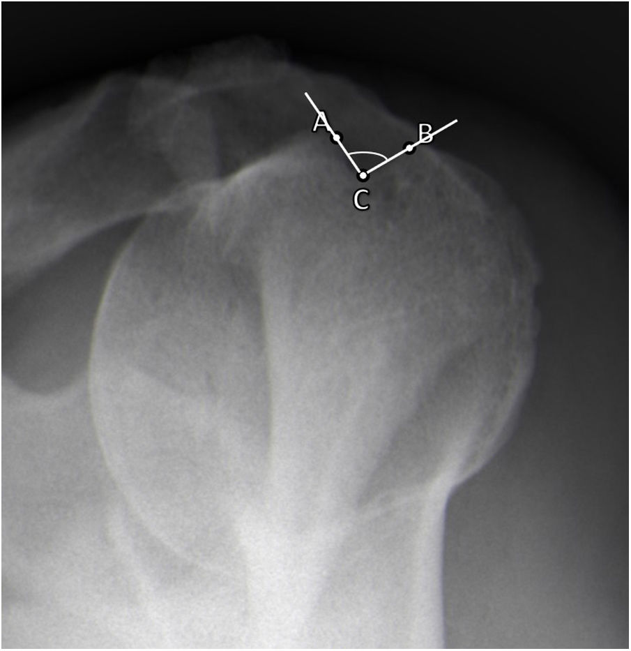
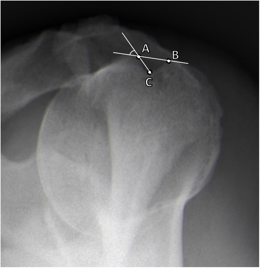
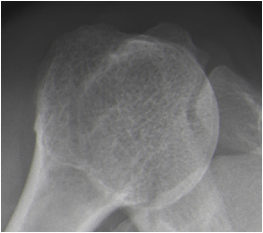
Material and methodsA prospective study was conducted, and sixty consecutive patients proposed to shoulder arthroscopic surgery were selected. Before surgery, a radiographic evaluation was performed with a supine and a Fisk radiograph. Most supine radiographs (>75%) were non-interpretable and were excluded from the study. As some Fisk radiographs (26.7%) were also non-interpretable, that left 44 interpretable radiographs in the study. These were measured for medial opening angle, total opening angle, width and depth of the bicipital groove. The radiographic measurements and the presence of LHBT pathology, as assessed at arthroscopy, were correlated.
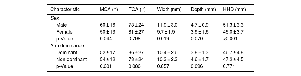
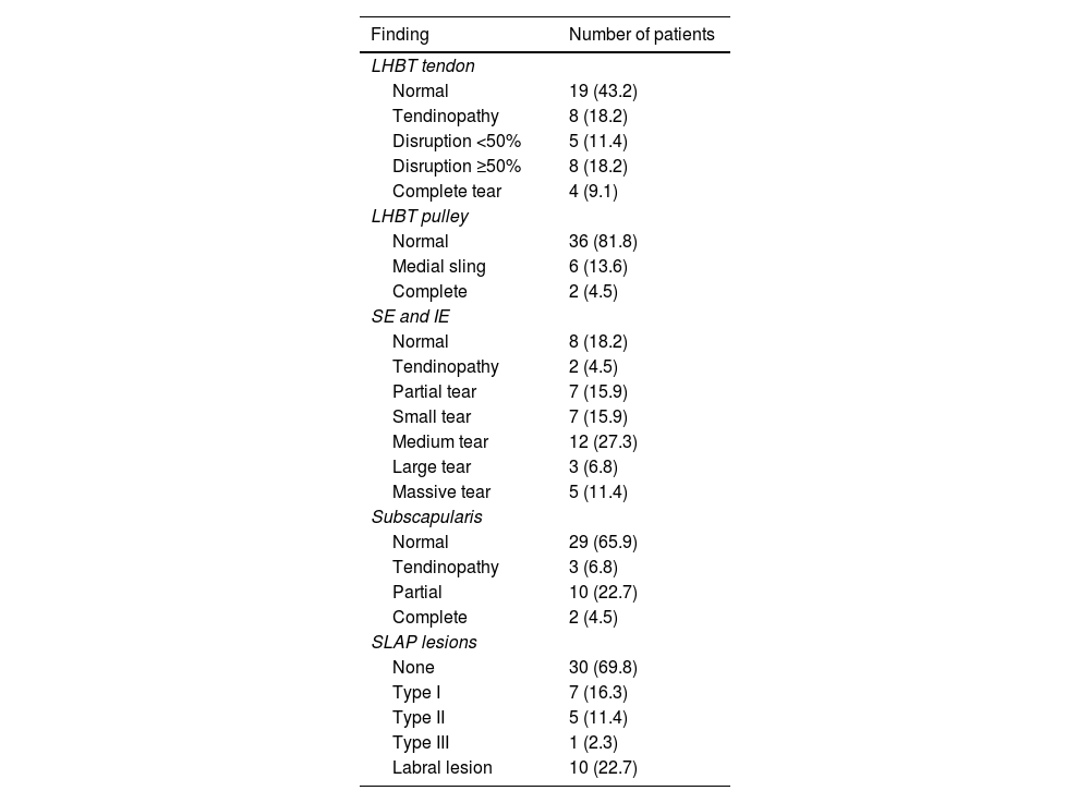
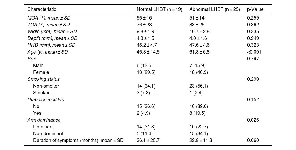
ResultsRadiographic evaluation of the bicipital groove showed a mean medial opening angle of 53 ± 15° (23–90), a mean total opening angle of 80 ± 26° (30–135), a mean width of 10.3 ± 2.5 mm (6–19) and a mean depth of 4.1 ± 1.5 mm (1–8). Men had higher medial opening angle (60 vs 50°, p = 0.044) and wider grooves (11.9 vs 9.7 mm, p = 0.019). Twenty-five patients (56.8%) were found to have an abnormal LHBT. No correlation was found between the radiographic measurements and LHBT pathology. Only age was correlated with a LHBT lesion (61.8 vs 46.3 years, p < 0.001).
ConclusionsOur results show that there is no correlation between radiographic morphologic evaluation of the bicipital groove and LHBT pathology.
El objetivo de este estudio es examinar la utilidad clínica de la evaluación radiográfica del surco intertubercular para pronosticar la patología del tendón de la porción larga del bíceps (TPLB).
Materiales y métodosSe llevó a cabo un estudio prospectivo y se seleccionaron 60 pacientes consecutivos propuestos para artroscopia de hombro. Antes de la cirugía, se realizó una evaluación radiográfica con una radiografía en decúbito supino y una radiografía con la técnica de Fisk. La mayoría de las radiografías realizadas en decúbito supino (>75%) no pudieron interpretarse y fueron excluidas del estudio. Como algunas radiografías con la técnica de Fisk (26,7%) tampoco pudieron interpretarse, quedaron 44 radiografías interpretables en el estudio. En ellas, se realizaron mediciones del ángulo de abertura medial, el ángulo de abertura total, la anchura y la profundidad del surco intertubercular. Se observó una correlación entre las mediciones radiográficas y la presencia de patología del TPLB, evaluadas en la artroscopia.
ResultadosLa evaluación radiográfica del surco intertubercular mostró un ángulo medio de abertura medial de 53 ± 15° (23−90°), un ángulo medio de abertura total de 80 ± 26° (30−135°), una anchura media de 10,3 ± 2,5 mm (6–19) y una profundidad media de 4,1 ± 1,5 mm (1–8). Los hombres tenían un ángulo de abertura medial mayor (60° frente a 50°, p = 0,044) y surcos más anchos (11,9 frente a 9,7 mm, p = 0,019). Había 25 (56,8%) pacientes con un TPLB anómalo. No se observó correlación entre las mediciones radiográficas y la patología del TPLB. Solo se observó correlación de la edad con una lesión del TPLB (61,8 frente a 46,3 años, p < 0,001).
ConclusionesNuestros resultados indican que no hay correlación entre la evaluación morfológica radiográfica del surco intertubercular y la patología del TPLB.

