Buscar en
Radiología (English Edition)
Toda la web
Inicio Radiología (English Edition) Mesenchymal hamartoma and undifferentiated embryonal sarcoma: Diagnostic keys th...
Journal Information
Share
Share
Download PDF
More article options
Visits
3
Radiology through images
Mesenchymal hamartoma and undifferentiated embryonal sarcoma: Diagnostic keys that radiologists need to know
Hamartoma mesenquimatoso y sarcoma embrionario indiferenciado: claves diagnósticas que el radiólogo debe conocer
Visits
3
M. Aineseder
Corresponding author
martina@aineseder.com

Corresponding author.
, M.A. Mestas Núñez, R. López Grove, M.L. Padilla, T.G. Kreindel
Servicio de Diagnóstico por Imágenes, Hospital Italiano de Buenos Aires, Buenos Aires, Argentina
This item has received
Received 19 May 2020. Accepted 17 September 2020
Article information
Abstract
Full Text
Bibliography
Download PDF
Statistics
Figures (7)
Show moreShow less
Tables (1)
Table 1. Comparison table of mesenchymal hamartoma, undifferentiated embryonal sarcoma and other differential diagnoses.
Resumen
Objetivo

Los tumores hepáticos son infrecuentes en la población pediátrica. Entre los más frecuentes se encuentran el hamartoma mesenquimatoso y el sarcoma embrionario indiferenciado, de diferente estirpe, pero con similitudes de imagen. El propósito de este artículo es repasar los hallazgos característicos en las imágenes y sus diagnósticos diferenciales. La ecografía es el método inicial para su estudio. La resonancia magnética y la tomografía computarizada son útiles para una mejor caracterización tumoral y planificación quirúrgica.

Conclusión

El radiólogo debe estar familiarizado con las características de imagen de las diferentes patologías y evaluarlas junto a la edad, antecedentes personales y análisis séricos del paciente.

Palabras clave:
Pediatría
Hígado
Tumor
Ultrasonografía
Tomografía
Resonancia magnética
Abstract
Objective

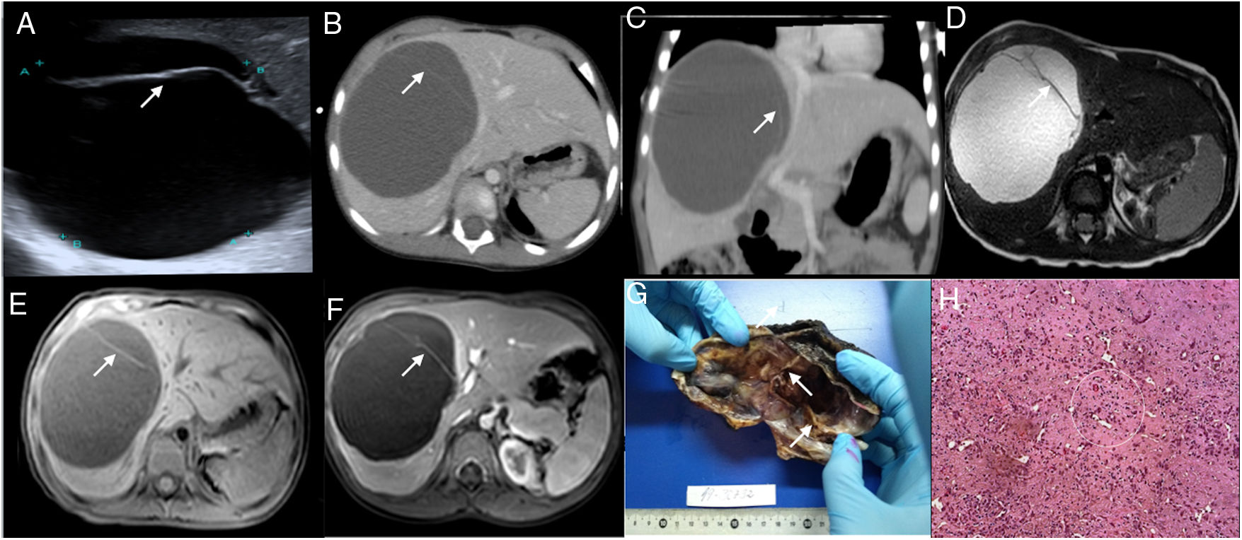
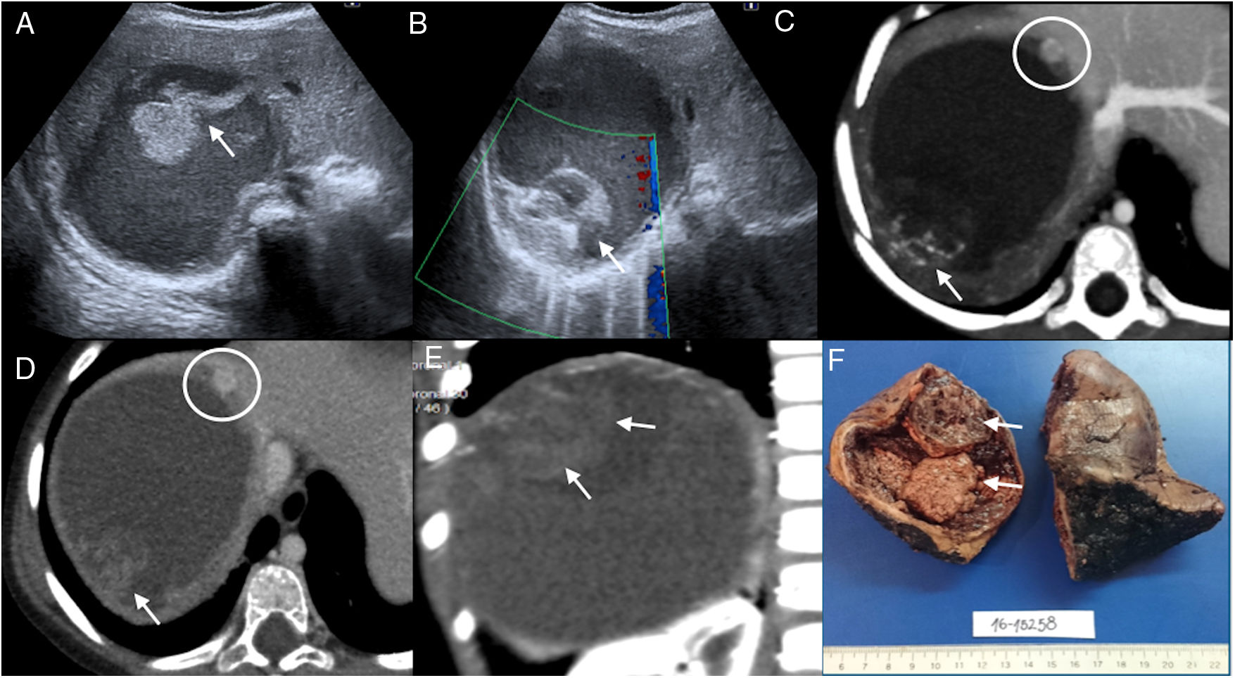
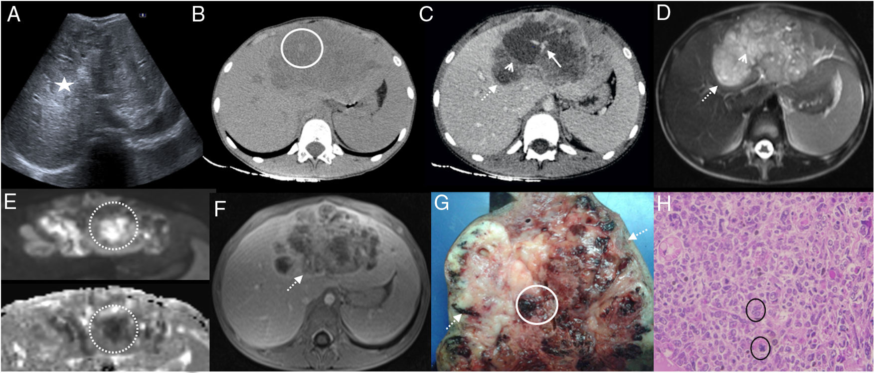
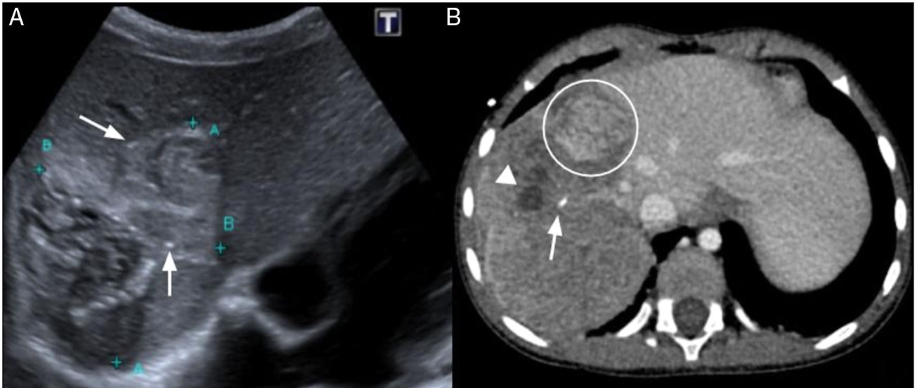
Hepatic tumors are uncommon in pediatric patients. Among the most common of these uncommon tumors are mesenchymal hamartoma and undifferentiated embryonal sarcoma, which have different origins but similar appearance on imaging studies. This paper reviews the characteristic findings and differential diagnosis of these entities. Ultrasonography is the first-line imaging test to study these tumors. Magnetic resonance imaging and computed tomography are useful for further characterizing the tumors and planning surgery.

Conclusion

Radiologists need to be familiar with the imaging findings of the different disease entities and to evaluate them together with the patient’s age, personal history, and bloodwork.

Keywords:
Pediatrics
Liver
Tumor
Ultrasonography
Computed tomography
Magnetic resonance imaging

Article

These are the options to access the full texts of the publication Radiología (English Edition)
Subscriber
Subscriber

If you already have your login data, please click here .

If you have forgotten your password you can you can recover it by clicking here and selecting the option “I have forgotten my password”
Purchase
Purchase article

Purchasing article the PDF version will be downloaded

Price 19.34 €

Purchase now
Contact
Phone for subscriptions and reporting of errors
From Monday to Friday from 9 a.m. to 6 p.m. (GMT + 1) except for the months of July and August which will be from 9 a.m. to 3 p.m.
Calls from Spain
932 415 960
Calls from outside Spain
+34 932 415 960
E-mail
Article options
Tools
es en pt

¿Es usted profesional sanitario apto para prescribir o dispensar medicamentos?

Are you a health professional able to prescribe or dispense drugs?

Você é um profissional de saúde habilitado a prescrever ou dispensar medicamentos