


In an era when it was not possible to achieve quality X-rays with short exposure times, the need to obtain chest images without movement led the French doctor Emré Hyacinthe Guilleminot to construct a machine that repeatedly emitted X-rays only when desired during the respiratory cycle. His aim was to create a satisfactory radiograph from multiple short bursts performed at the moment of inhalation or exhalation, based on Charles Bouchard's research on heart disease. He extended his idea to radiography of the heart, creating a system that enabled images to be obtained disassociating the phases of heartbeat. This article seeks to explain the basic functioning of these mechanisms, and to recover previous research papers that led to their creation. We will also retrieve biographical and personal data of the two people involved – one directly, the other indirectly – in these novel inventions.
En una época en que no era posible conseguir radiografías de calidad con tiempos de exposición cortos, la necesidad de obtener imágenes del tórax sin movimiento llevó al médico francés Emré Hyacinthe Guilleminot a construir un aparato que disparase rayos X de manera repetida únicamente en el momento deseado del ciclo respiratorio. Su objetivo fue, partiendo de las investigaciones sobre patologías cardíacas de Charles Bouchard, componer una radiografía satisfactoria a partir de múltiples disparos cortos realizados en el mismo instante de la inspiración o la espiración. Extendió su idea a la radiografía del corazón, creando un sistema que permitía obtener imágenes disociando las fases del latido. El presente artículo pretende explicar el funcionamiento básico de aquellos mecanismos, así como recuperar los trabajos de investigación previos que motivaron su creación. También rescatará datos biográficos y personales de las dos figuras involucradas – directamente uno, indirectamente el otro – en aquellas novedosas invenciones.
Systems for synchronising heart beat and breathing rhythms that are now so commonplace in magnetic resonance imaging (MRI) and computed tomography (CT) have their origin in the ingenuity of French scientist Hyacinthe Guilleminot who, in 1898, invented the first gating devices for radiological images. Exposure times in the early years of radiology were extremely long, and this meant that the quality of X-ray images of the chest were affected by the patient's breathing movements. Thanks to a series of advances in the manufacture of X-ray tubes, radiographic films and intensifying screens,1–3 by 1898 X-rays were already being performed in apnoea,4,5 although many researchers were not entirely satisfied with the results. In addition, the time of apnoea, sometimes up to 45s, was impossible for some patients. This is what prompted Dr Guilleminot, inspired by the groundbreaking work of his colleague Charles Bouchard on intrathoracic pathologies, to design two devices to obtain images of the thorax and the heart that were not affected by patient movement during the natural respiratory cycle. This he achieved by taking a sufficient number of short shots at a particular stage of the desired cycle, and thus obtained the exposure time needed for a valid image.
Today, more than 120 years after his invention, it is interesting to explain the basic principles of operation of those devices, and take a look at the earlier studies that led to Dr Guilleminot's breakthrough. First, we need to know a bit more about the two men involved in these novel inventions.
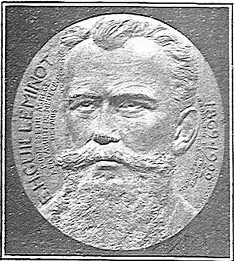
Dr GuilleminotEmré Hyacinte Guilleminot (07/06/1869, Laignes6 – 10/03/1922, Paris7) (Fig. 1) graduated from university with a degree in both law8 and medicine. In 1894, he worked as a medical intern in Le Charité (Paris) with Bouchard, with whom he struck up a fruitful professional and personal relationship. He later took a position as a professor of biological physics at the Faculty of Medicine in Paris, and between 1912 and 1913 was elected chairman of the Medical Radiology Society of Paris.9 He devoted part of his time to the study of dosimetry, publishing Fluoroscopic Radiometry10 in 1910 and inventing a dosimeter in 1917.11 He was also fascinated by electricity and its application in medicine, going on to write important papers on the subject, including Medical Electricity,12 a detailed treatise published in 1905. His research into this field earned him the 1917 Hébert Prize for the dissemination of electrical practise from the Academy of Sciences13 for the work The new horizons of science. Guilleminot was the first to come up with a system for obtaining chest X-rays during inspiration or expiration without the need for apnoea. Since this idea was inspired by the research of Dr Bouchard, his erstwhile teacher and later friend, into intrathoracic pathologies, we need to know more about Dr Bouchard and his contribution to science before embarking on a description of Guilleminot's inventions.
Dr Bouchard.")
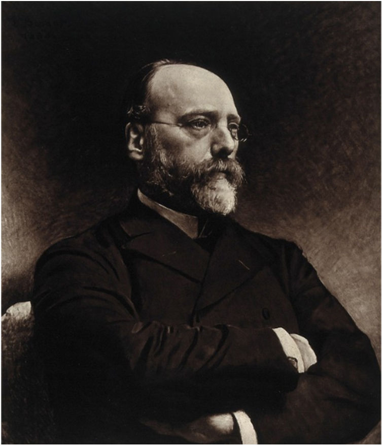
French physician Charles Jacques Bouchard (06/09/1837, Montier-en-Der14 – 28/10/1915, Lyon15) (Fig. 2) began his medical studies in 1857 in Lyon before continuing and concluding his studies in Paris, where in 1864 he worked under Jean-Martin Charcot in the Pitié-Salpêtrière hospital. He obtained his doctorate in 1866, and over the following years worked in the Parisian hospitals of Bicêtre, Lariboisière and Le Charité, among others. In 1879, he was appointed professor of General Pathology and Therapeutics of the Faculty of Medicine in Paris and in 1886 became a member of the Academy of Sciences, occupying the position of chairman between 1908 and 1909. In 1897, he was elected chairman of the French Biology Society. He was also co-founder of the journal Revue de la Tuberculose (1887) and founder of the Journal de Physiologie et de Pathologie générale (1899).16
.")
Following the discovery of X-rays at the end of 1895,17 Bouchard devoted himself to the study of these new rays and their application in medicine. In 1898, he commissioned Guilleminot to create an X-ray department in Le Charité,16 the second of its kind in a Parisian hospital,18 and headed by Guilleminot himself. Beginning his investigations in 1896, he soon made interesting observations about the intrathoracic organs with the help of the fluoroscopic screen. His first major discovery was the observation of pleural effusion.19 At a time when X-ray images were mainly positive, pleural effusion appeared as a dark area over the radiolucent lung. He also observed the contralateral displacement of the mediastinum in cases of complete unilateral pleural effusion, and its displacement to the affected side when resolution of pleural effusion led to pulmonary retraction. His observations were consistent with the results of routine clinical examinations, and he therefore considered radiological imaging to be a highly effective complementary study, and believed it to be even more accurate than physical examination in the diagnosis of mediastinal displacement. Acutely aware of the future promise of X-rays, during the presentation of these observations he commented “we have the right to think that examination with the aid of Röntgen rays will be as useful to (internal) medicine as it is to surgery”.19
Another of his discoveries was the visualisation of the signs of tuberculosis.20 In patients in whom the presence and extent of tuberculosis had previously been diagnosed by physical examination, radiological images were positive for tuberculosis in all cases except one. One case in particular encouraged Bouchard to continue his investigations: a patient presented cough and general signs suggestive of incipient tuberculosis, although sputum culture and physical examination were negative for the disease. However, radiological images showed positive signs that, a few days later, were confirmed by further auscultation and culture. In other cases, he identified a tracheobronchial lymphadenopathy; an ectopia cordis and a thoracic tumour diagnosed clinically as aortic aneurysms; several cardiac hypertrophies21; a bilobular tumour adjacent to the fourth dorsal vertebra, clinically diagnosed as oesophageal stenosis; and described the radiological signs of aortic regurgitation.22
Following his studies of the heart, in 1898 Bouchard reported his observations on certain movements of the right atrium that did not appear to correspond to the cardiac cycle itself. He had observed a shadow on the fluoroscopic screen that appeared to the right of the sternum and then disappeared. This was repeated several times, and he observed that it coincided with the descent and elevation of the liver during breathing: the shadow appeared on inspiration and disappeared on expiration. Bouchard concluded that this was caused by the isochronous dilatation of the right atrium on inspiration due to changes in intrathoracic pressure.23,24 Despite being able to visualise the phenomenon on the screen, he was unable to obtain separate radiographic images of the dilation and retraction of the atrium in order to compare and measure the movement. The X-ray would have to be taken with a very short exposure time and very high power at exactly the right stage of dilation or contraction, and this, despite the ongoing technological improvements, was impossible at that time. By 1898, exposure times had been reduced to enable shots to be taken in apnoea, although patients still needed to hold their breath for 30–45s.4,5 In addition to this, Bouchard needed the images to be obtained during the natural respiratory cycle, since he wanted to study the effect of this on the heart without forcing apnoea that artificially increased the lung volume and compressed or displaced the heart. It was this challenge in the embryonic field of radiology that Guilleminot decided to accept.
A rudimentary solutionIt should be noted that in 1897, the German military doctor Walter Stechow had attempted to obtain clear chest X-rays in different respiratory phases.25 His method consisted of placing a metal sheet in front of the X-ray tube that would move forward and backward during the long continuous exposure, allowing the rays to pass only at the desired moment of the respiratory cycle. This had to be repeated several times until the photographic plate had been sufficiently exposed. Obviously uncomfortable and impractical, Stechow's idea did not prosper, but served as inspiration for future researchers.
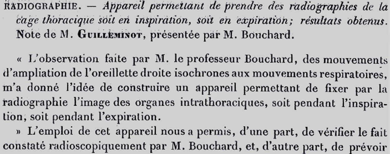
Guilleminot's respiratory synchronisationThe first device for synchronising respiratory movement to obtain clear chest X-rays was presented on 8 August 1898 at a meeting of the French Academy of Sciences26 (Fig. 3). Its inventor, Guilleminot, had written a note that Bouchard read before the assembled members: “Professor Bouchard's observation of the enlargement of the right atrium in synchronisation with respiratory movements gave me the idea of constructing an apparatus that would allow the image of the intrathoracic organs to be fixed on the X-ray during either inspiration or expiration” (Fig. 3). A promising introduction to describe the first ever device for synchronising (gating) respiratory movement in the history of radiology.

Heading of the publication containing the presentation of the respiratory gating device.26
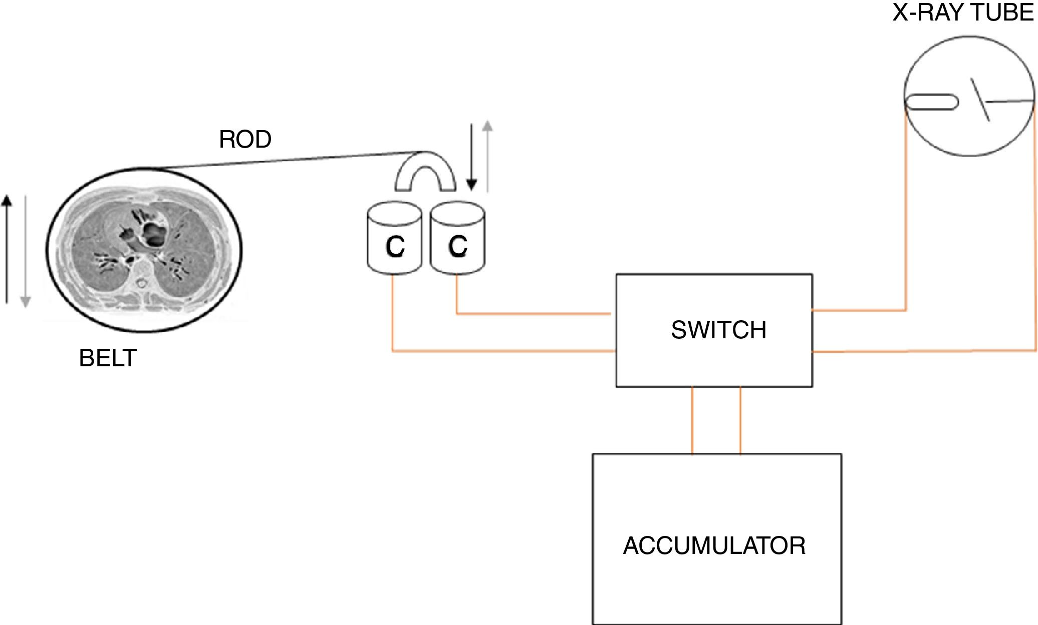
It is interesting to note that, despite its importance, Guilleminot's description of the apparatus was not detailed enough to fully understand how it worked. This rudimentary device consisted of two main parts: a belt adjusted to the size of the patient, and a switch on the X-ray generating circuit. The belt, which opened and closed the circuit, was a conventional leather belt that was placed around the lower part of the thorax with the buckle turned under the subject. The belt was cut in half, and a wooden plate was attached to each end. Contact between the plates was regulated by elastic bands. The tension of these could be adjusted to the abdomen of each patient, so that the plates could be easily separated with inspiration and the consequent increase in the thoracoabdominal diameter. One end of a violin string was attached to a hook on one of the plates and extended to the other plate. On that plate, the string was attached to very light lever affixed to that end of the belt, while a pulley guaranteed the direction of the string. As a result, any inspiration or expiration movement caused the string and, consequently, the lever to move in one direction or the other. With each respiratory movement, this lever moved a long rod whose distal end indirectly closed or opened the X-ray generator circuit, according to whether contact would occur with inspiration or expiration. Thus, X-rays were fired at the same stage of the respiratory cycle, when the intrathoracic organs were at the same level (Fig. 4). This process, repeated for about 20min, would fire an X-ray with each inspiration or expiration performed by the patient. The final result was a radiograph obtained from multiple short shots – each of them insufficient per se to obtain a valid image. Guilleminot and Bouchard used this system to compare the displacement of the heart and the dilation or contraction of its chambers during the respiratory phases in general, and in particular to study Bouchard's observations on the right ventricle in greater depth. For this purpose, they compared radiographs of the same patient during inspiration and expiration. They also used the new mechanism to compare the angle of the ribs during respiration in healthy subjects with subjects with different unilateral or bilateral thoracic pathologies.27
, its U-shaped end is dipped into two containers filled with mercury. This closes the electrical circuit that generates the X-rays. The device could be adjusted so that thoracic contraction (expiration) would submerge the U-shaped end of the rod and close the circuit.")
Simplified diagram of the respiratory gating system. The rod, connected by means of a small pulley to the violin string that joins the two ends of the cut belt. When the rod is pushed upward during thoracic expansion (inspiration), its U-shaped end is dipped into two containers filled with mercury. This closes the electrical circuit that generates the X-rays. The device could be adjusted so that thoracic contraction (expiration) would submerge the U-shaped end of the rod and close the circuit.
As a footnote to this story, the German physician Walter Cowl, inspired by the ideas of Stechow and Guilleminot, developed another gating system in 1898.25 Cowl replaced Guilleminot's belt with an oscillating rod with a small plate on one end that rested directly on the patient's abdomen. The rod, maintained horizontal and attached to a metal foot, rose or fell with each inspiration and expiration. This movement alternately closed and opened an X-ray generator circuit placed at the other end of the rod. Cowl called his device the Rheotom and continued to work on it until at least 1901.28,29
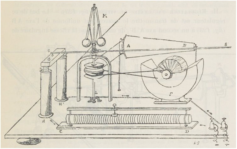
Guilleminot's cardiac gating systemGuilleminot's curiosity was not satisfied by his respiratory gating system, and shortly afterwards he devised another to synchronise the heartbeat with X-ray shots, and thus dissociate its different phases. The new system was presented by Bouchard on 17 July 1899 at the Academy of Sciences of France, reading from a note drafted by Guilleminot.30 The basic design of this device was similar to the previous one, although in this case Guilleminot used a new medical tool: Etienne-Jules Marey's portable sphygmograph, originally designed to detect the radial pulse and trace a blood pressure graph. The sphygmograph was attached to the wrist by means of strips of cloth, leaving a small ivory plate on the radial artery. The plate moved with each pulse beat, and this movement was transmitted to a needle that traced the wave on a strip of paper that was steadily advanced by means of a clockwork motor to obtain a continuous graph. For his new system, Guilleminot removed some of the pieces of the sphygmograph and adapted its mechanism so that the radial pulse moved a rod, the distal end of which indirectly opened and closed the circuit of the X-ray generator (Fig. 5), passing through an intricate electromechanical system of synchronised rheostats and dynamos (Figs. 6–8). The aim of this new device was to obtain images by firing X-rays only when the heart was in the desired cycle, and thus fix its shadow on the radiograph. In this way, the degree of displacement of the atria and ventricles in systole and diastole could be measured. This time, the results could be shown to a group of spectators, whereas previously they could only be observed in real practice on a fluoroscopic screen. In an article published at the end of 1899, in which he explained the components of the apparatus and its mode of use – albeit with important omissions that make it hard to understand – Guilleminot dubbed this new technique cineradiography.31
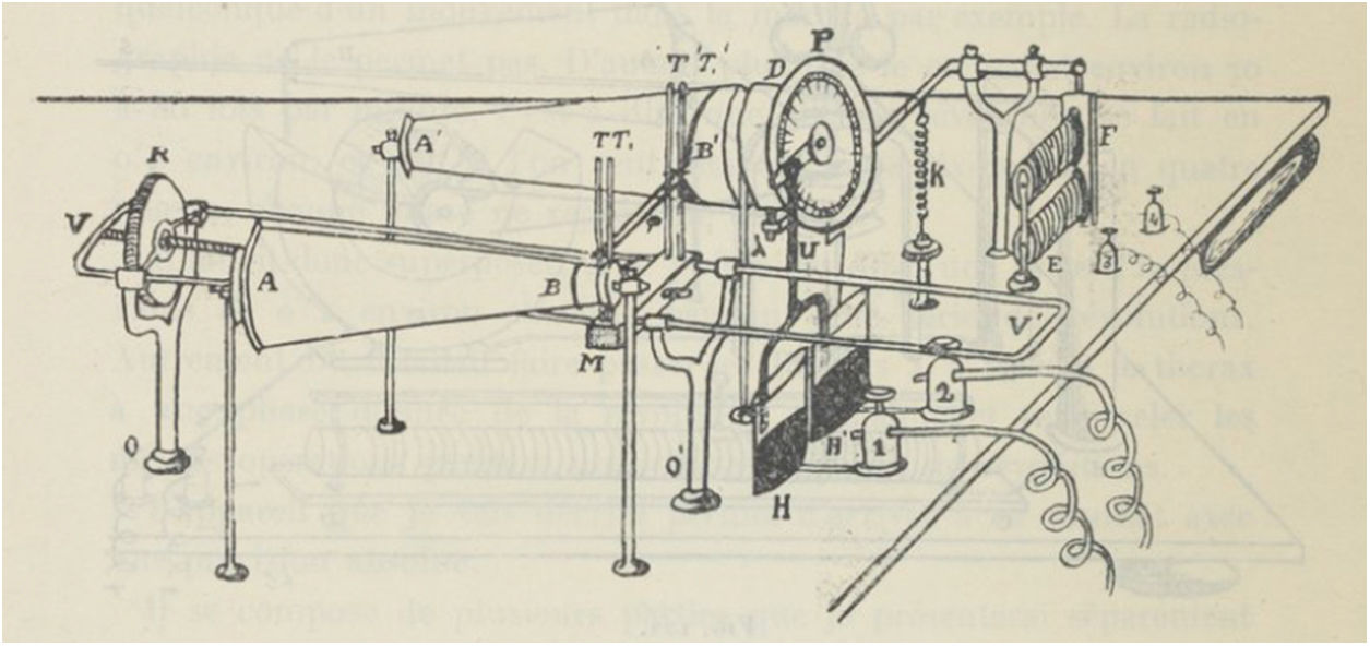
 is connected at its proximal end to the ivory plate on the wrist of the patient, which rises and falls in synchrony with the radial pulse. This pulsating movement alternately dips the rod in and out of a container filled with mercury (H). This closes and opens the electrical circuit that fired the X-rays.31")
Part of the modified sphygmograph used by Guilleminot. The rod (T) is connected at its proximal end to the ivory plate on the wrist of the patient, which rises and falls in synchrony with the radial pulse. This pulsating movement alternately dips the rod in and out of a container filled with mercury (H). This closes and opens the electrical circuit that fired the X-rays.31

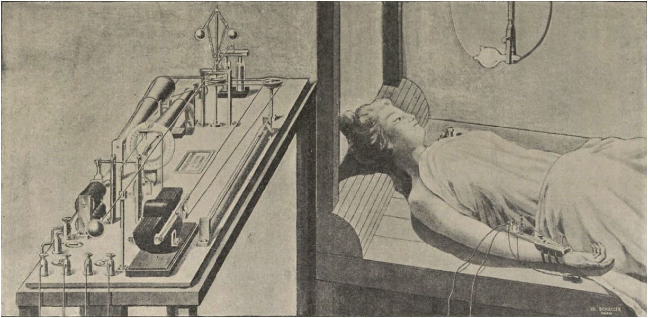
Overview of the cardiac gating system. The heart rate monitor on the patient's wrist, with the rod and mercury container acting as switches, and the complex energy generator system.37

Uniform motion generator. This device, consisting mainly of dynamos and rheostats, generated the electrical power to drive the X-ray tube. Its constant rotation generated uninterrupted energy, but this was only transmitted to the tube at the appropriate moment of the cardiac cycle, when the rod shown in Fig. 5 emerged from the mercury container.31

Optional speed regulator. This part of the apparatus could be used to regulate the rotation speed of the motion generator, which could generate more electrical energy when needed.31
We know some things about Guilleminot's personality from his contemporaries. In February 1912, Bouchard wrote in a letter: “I set aside several hours to read Guilleminot. I admire him for finding the time and strength to lead us, while also preparing for his competence”.16 After his death, Dr Paul Le Gendre described him as a “man of rare merit and even rarer modesty”, adding that he was “of medium height, frail, thin and sickly, with clear and thoughtful eyes under a broad forehead, a bony face framed by a large brown beard and large moustache. He inspired sympathy and gave the impression of energy and kindness tempered by timidity”.16 The radiologist Albert Laquerrière, meanwhile, defined him as a “modest worker and wise man, with a vast spirit and beautiful character”.16
Two of those characteristics – wise man and vast spirit – shine through in some of his books. In the four volumes of his work The new horizons of science, Guilleminot is enthusiastic about the scientific advances of the previous century. For him, these new methods and knowledge are the vehicle that will transport man away from the anguish of transcendence, and his words reveal an ideology shaped by the legacy of the French Enlightenment. The introduction of the first volume32 and the end of the fourth33 are significant insofar as they reveal his intentions in publishing the work: to disseminate and instruct in order to distance man from the philosophy that rejects the definitive nature of experimental induction. In a statement of principles that impregnates the entire work, he defends empiricism not only as a method of study, but also as a way of understanding life and distancing oneself from faith and superstition. Guilleminot upholds nature as the measure of all things, as opposed to the anthropocentric vision of the world, which he vigorously rejects. He offers succinct ideas that are at times reminiscent of the more elaborate notions put forward by Baron d’Holbach in his controversial work The System of Nature,34 which was banned for its outspoken presentation of subversive, anti-religious ideas. The book, together with the ideas of the ‘Encyclopédistes’ (Encyclopaedists) who gathered around Diderot and d’Alambert, formed the intellectual basis for the French Revolution. Guilleminot observes science, the world, and the origin of all things (the only question of interest to him) from a purely empirical, not metaphysical, perspective. He wants to know from whence, from what primary elements, life and things, even morality itself, arise; he is not interested in a speculative or metaphysical “why” or “what for” about the goals that trouble mankind. However, aware of the important role of morality in shaping civilised behaviour, in the conclusion to the fourth volume he seeks a compromise and permits morality to guide mankind, considering it a necessary subject of scholarly study, provided it is not “monopolised by a doctrine, a chapel or a party”.33 He finally reveals that positive morality and a certain ideal of finality can be extracted from his scientific proposals “and give hesitant humanity a guiding light in the night that engulfs it”.33 He echoed these thoughts in Matter and life in 1919,35 reflecting on the trauma of the Great War, an episode that altered the awareness of a large part of the European intelligentsia. In this book, Guilleminot makes a liberalist case against the collectivist ideas that had become popular at the beginning of the 20th century. He considers that the collectivists had created social laws that formally opposed natural laws that “led to the development of human intelligence, culture and individual initiative”, and appeals to the victorious nations of the war to strive to modify the mentality of society, not by force, but by the light of empirical knowledge.
Guilleminot was a true believer in science, and devoted his life to its study and dissemination. His restlessness, devotion to scientific advances and concern for the enlightenment of humanity was the driving force behind his interest in such diverse subjects as law, medicine, radiology, biology, histology, dosimetry and electricity, on which he published a large number of articles and books, some purely scientific and others more didactic.
Recognition in his time and todayCurrently, the Hyacinthe Guilleminot belt is reminiscent of the respiratory gating systems supplied by some manufacturers of MRI systems. Both systems operate basically on the same principle: they are attached to the abdomen like a belt that expands and contracts with each breath. This is a testament to the talent of the French pioneer. On 17 December 1900, his talent was further recognised when he was awarded the Montyon Prize for Medicine and Surgery by the Academy of Sciences for the invention of the two device described above.36 Although Guilleminot built the first ever gating systems, they were never popular and soon fell into disuse due to his poor descriptions of the devices25 and the rapid development of instant radiographs.37,38 Nevertheless, in a future then distant, his ideas, fading in human memory but etched in the historical treasure of specialised literature, would be rescued to a certain extent and are now used every day in MRI and CT imaging. Thus, the ingenuity of the French researcher lives on among today's radiologists. Every time a synchroniser or gating system is used in routine practice, it is a silent, though unwitting, tribute to the visionary who invented the technique.
Conflicts of interestThe author declares that there are no conflicts of interest.
Please cite this article as: Crespo Villalba FJ. El Dr. Hyacinthe Guilleminot y los primeros sistemas de sincronización respiratoria y cardíaca para obtención de imagen radiológica. Radiología. 2019;61:239–246.

