


The aim of this article is to present an appealing tool for teaching breast imaging that uses the muses of Renaissance and Baroque artists.
The examples described here show how medicine and art can be combined to arouse interest in newcomers to breast imaging.
El objetivo de este artículo es presentar una atractiva herramienta de enseñanza en la imagenología mamaria, utilizando las musas de los artistas del Renacimiento y el Barroco.
Los ejemplos aquí descritos ilustran cómo se puede conjugar la medicina y el arte para despertar el interés de quien se introduce en el aprendizaje de la lectura mamográfica.
Neuroscience has taught us that generating emotions is the key to arousing the curiosity of the learner and thus holding their attention.1
The aim of this article is to present an attractive tool for teaching mammogram interpretation skills using the muses of Renaissance and Baroque artists.
At that time, the human figure was represented in historical or biblical scenes, and drawn and sculpted with a level of detail that shows the artist's in-depth knowledge of anatomy.
In some paintings of naked torsos, we can glimpse changes in the contours of the breasts; the realism of the works leads us to assume, usually rightly, that the artists are drawing the pathology they observed in their model.
Using a series of masterpieces, we will suggest hypothetical diagnoses based on the abnormalities observed in the anatomy of the breasts and axilla, and speculate what these findings would have looked like on a modern mammogram.
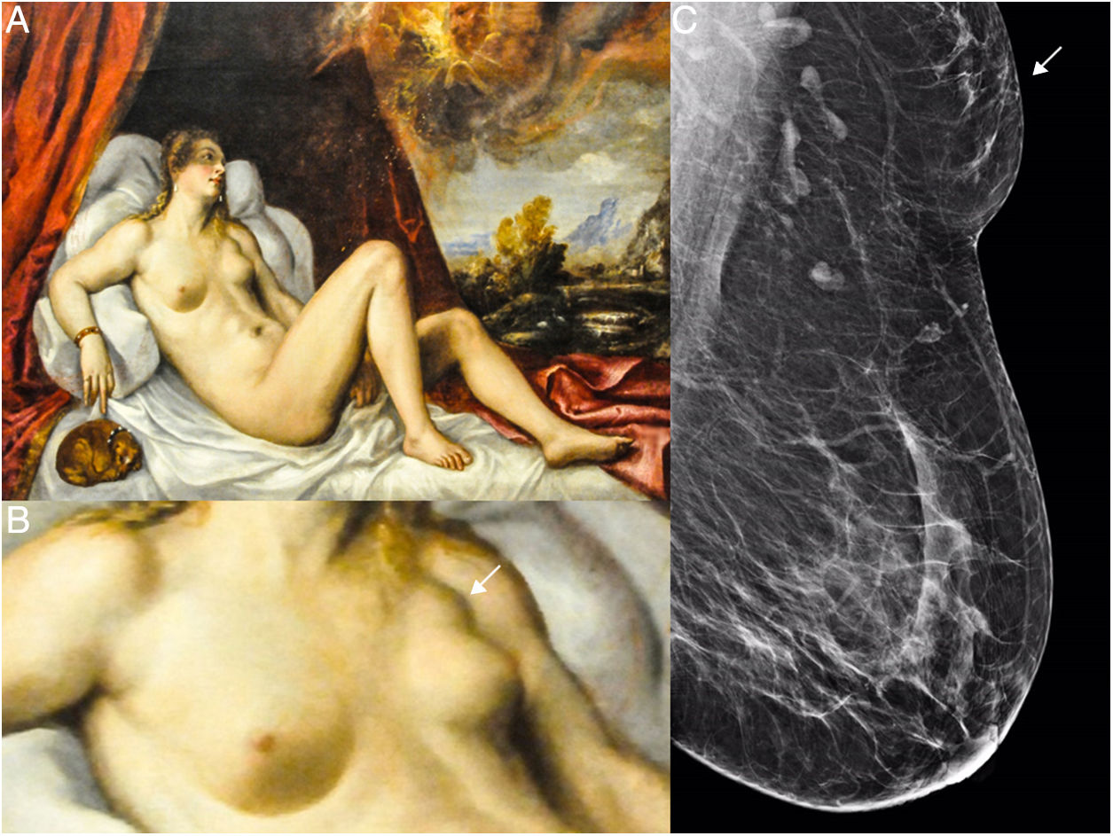
Accessory breast tissue in the axillaThe oil on canvas titled Danae, by an unknown artist from 1554, on display in the Art Institute of Chicago, represents the moment in which, according to Greek mythology, Danae conceives a son after Zeus comes to her in the form of a golden rain.
Danae was the daughter of Acrisius, king of Argos, and of Eurydice. The mythology tells that when Acrisius consulted the oracle of Delphi, he was told that his daughter would conceive a son who would eventually be the cause of his death. To prevent the prediction from being fulfilled, Acrisius locked his daughter in an underground chamber with bronze doors. But Zeus, who loved the girl, entered the prison in the form of golden rain and impregnated her. The child born of this union was Perseus.2
A careful analysis of the figure of the woman (Fig. 1) representing Danae shows a tumour in the left axillary extension. Given the young age of the model and the absence of changes in the breast, the best presumptive diagnosis would be accessory breast tissue.
 Danae (1554). Anonymous. Oil on canvas in the Art Institute of Chicago. (B) Enlargement of the left axilla of the model portraying Danae; the tumour is marked with a white arrow. (C) Left mediolateral oblique view showing ACR B glandular tissue, radiologically normal axillary and intramammary lymph nodes and accessory breast tissue in left axilla (white arrow) of a radiological density similar to breast tissue.")
(A) Danae (1554). Anonymous. Oil on canvas in the Art Institute of Chicago. (B) Enlargement of the left axilla of the model portraying Danae; the tumour is marked with a white arrow. (C) Left mediolateral oblique view showing ACR B glandular tissue, radiologically normal axillary and intramammary lymph nodes and accessory breast tissue in left axilla (white arrow) of a radiological density similar to breast tissue.
In the literature, accessory breast tissue in the anterior axillary fold or in the axillary space itself, both subcutaneous and deep, with no connection to the mammary gland, has been described in 2–6% of women, depending on the series.
On a mammogram, accessory breast tissue has the same density and morphology as breast tissue. The dimension of the tissue varies between 1.5cm and 6cm.3
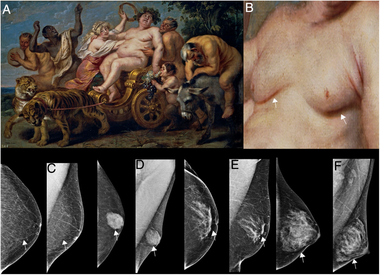
Lipomastia or pseudogynaecomastiaThe Triumph of Bacchus, an oil on canvas painting by Cornelis de Vos on display in the Prado Museum, shows the god of the grape harvest and wine (in Greek Bακχοζ or Bakkhos), an important figure in Greek and Roman mythology, son of Zeus and the mortal Semele, daughter of Cadmus, king of Thebes. Although Bacchus was part human, he gradually acquired immortality and became one of the twelve Olympians.2
Cornelis de Vos4 was a master of realism and closely followed the highly detailed Flemish style, imitating Rubens’ painting. In this painting (Fig. 2), he shows a corpulent Bacchus with a large belly, puffy face, swollen limbs and cellulitis. A close look at his torso shows enlarged breasts. This leads us to two possible diagnoses, the most likely being lipomastia, given the body's overall adiposity, or gynaecomastia, a hormonal imbalance that causes the breasts to grow.
 The triumph of Bacchus. Oil on canvas by Cornelis de Vos (1636–1638), on display in the Prado Museum, Madrid, Spain. (B) Enlarged view of the model's torso, showing enlarged breasts (white arrows). (C) Left craniocaudal oblique mediolateral view showing that the breast volume is composed of adipose tissue. Mammographic diagnosis of lipomastia (white arrows). (D) Left craniocaudal oblique mediolateral view showing the nodular mammographic pattern, consistent with the breast volume being due to gynaecomastia (white arrows). (E) Left craniocaudal oblique mediolateral view showing the dendritic mammographic pattern, consistent with enlargement of the breast due to gynaecomastia (white arrows). (F) Left craniocaudal oblique mediolateral view showing the diffuse glandular mammographic pattern, consistent with enlargement of the breast due to gynaecomastia (white arrows).")
(A) The triumph of Bacchus. Oil on canvas by Cornelis de Vos (1636–1638), on display in the Prado Museum, Madrid, Spain. (B) Enlarged view of the model's torso, showing enlarged breasts (white arrows). (C) Left craniocaudal oblique mediolateral view showing that the breast volume is composed of adipose tissue. Mammographic diagnosis of lipomastia (white arrows). (D) Left craniocaudal oblique mediolateral view showing the nodular mammographic pattern, consistent with the breast volume being due to gynaecomastia (white arrows). (E) Left craniocaudal oblique mediolateral view showing the dendritic mammographic pattern, consistent with enlargement of the breast due to gynaecomastia (white arrows). (F) Left craniocaudal oblique mediolateral view showing the diffuse glandular mammographic pattern, consistent with enlargement of the breast due to gynaecomastia (white arrows).
It is important to distinguish between gynaecomastia and true lipomastia, which is caused by an excess of adipose tissue in the breasts. In obese men in particular it can be difficult to distinguish between these conditions. Mammography, however, can show the difference and facilitate diagnosis.
On a mammogram, lipomastia is shown to be an accumulation of adipose tissue.
Gynaecomastia, meanwhile, is unilateral or bilateral breast augmentation due to ductal and stromal hyperplasia. Three types of gynaecomastia can be seen on mammography: nodular (florid), dendritic (fibrous) and diffuse glandular (similar to the female breast).5
Breast cancer in womenAndromeda is an oil painting by Rubens that shows Andromeda, the daughter of Cassiopeia and Cepheus, being offered to Poseidon to prevent the destruction of their people.
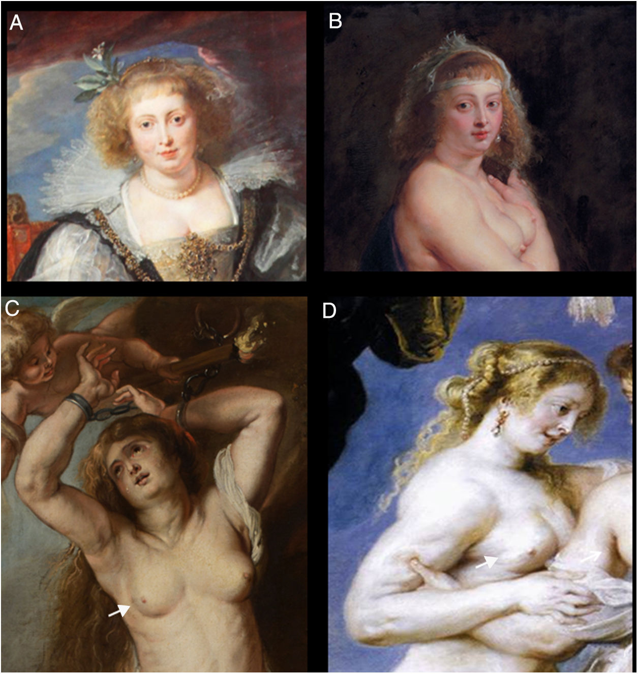
Rubens used various members of his family as his models: his first wife Isabella Brant, his second wife Hélène Fourment, and his sister-in-law Susana Fourment.6
The Andromeda of this painting is probably Hélène Fourment. I base this hypothesis on a series of physiognomic similarities between the face of Andromeda and Rubens’ portraits of his wife, among them Helena Fourment in wedding dress, in the Alte Pinakothek in Munich, which dates from 1630 to 1631, and The Fur, in which he shows a nude Hélène Fourment partially covered with a fur, dating from 1636 to 1638 and on display in the Kunsthistorisches Museum in Vienna.
Several authors describe how Rubens’ mastery of light and shadows allowed him to paint the human figure in such detail, so much so that nipple retraction and tumours can be seen in the breast and left axilla and in the right breast of his muses, as described in various medical publications.7–10
J.J. Grau, M. Prats and M. Díaz-Padrón, in their article “Cáncer de mama en los cuadros de Rubens y Rembrandt” [Breast cancer in the paintings of Rubens and Rembrandt],8 describe a tumour located in the upper outer quadrant of the left breast in a series of paintings by Rubens. Observing the figure on the left in the oil painting The Three Graces, and the figures in Orpheus and Eurydice, and Diana and her nymphs surprised by satyrs, the authors formulate the hypothesis that Rubens was documenting the evolution of a breast tumour, and that the same model might have posed for all three works.
A close examination of the breasts of the woman who posed for Andromeda shows that the right breast is smaller, retracted, with changes to the skin colouring of the lower outer quadrant at the 6 o’clock position, probably due to the presence of a malignant, locally advanced tumour.
A comparison of the right breast of the model who posed for the figure on the right in the painting of the three graces with that of Andromeda, suggests that we are observing the evolution of a breast tumour. A comparative collage (Fig. 3) shows the similarities not only in the breast, but also in the face and hair of the model.
 Detail of the portrait entitled Hélène Fourment in wedding dress, oil painting by Peter Paul Rubens (1630), on display in the Alte Pinakothek museum in Munich, Germany. (B) Detail of Het Pelsken (The Fur), portrait of Hélène Fourment by Peter Paul Rubens (1636–1638), on display in the Kunsthistorisches Museum in Vienna, Austria. (C) Enlarged view of Andromeda, by Peter Paul Rubens (1634), oil on canvas, on display in the Víctor Balaguer Museum & Library, showing retraction of the skin in the lower outer quadrant of the right breast at the 6 o’clock position (white arrow). (D) Enlarged view of the lower outer quadrant of the right breast showing retraction of the skin (white arrow) in the model on the right in The Three Graces, by Peter Paul Rubens (1639), oil on canvas. Prado Museum, Madrid, Spain.")
(A) Detail of the portrait entitled Hélène Fourment in wedding dress, oil painting by Peter Paul Rubens (1630), on display in the Alte Pinakothek museum in Munich, Germany. (B) Detail of Het Pelsken (The Fur), portrait of Hélène Fourment by Peter Paul Rubens (1636–1638), on display in the Kunsthistorisches Museum in Vienna, Austria. (C) Enlarged view of Andromeda, by Peter Paul Rubens (1634), oil on canvas, on display in the Víctor Balaguer Museum & Library, showing retraction of the skin in the lower outer quadrant of the right breast at the 6 o’clock position (white arrow). (D) Enlarged view of the lower outer quadrant of the right breast showing retraction of the skin (white arrow) in the model on the right in The Three Graces, by Peter Paul Rubens (1639), oil on canvas. Prado Museum, Madrid, Spain.
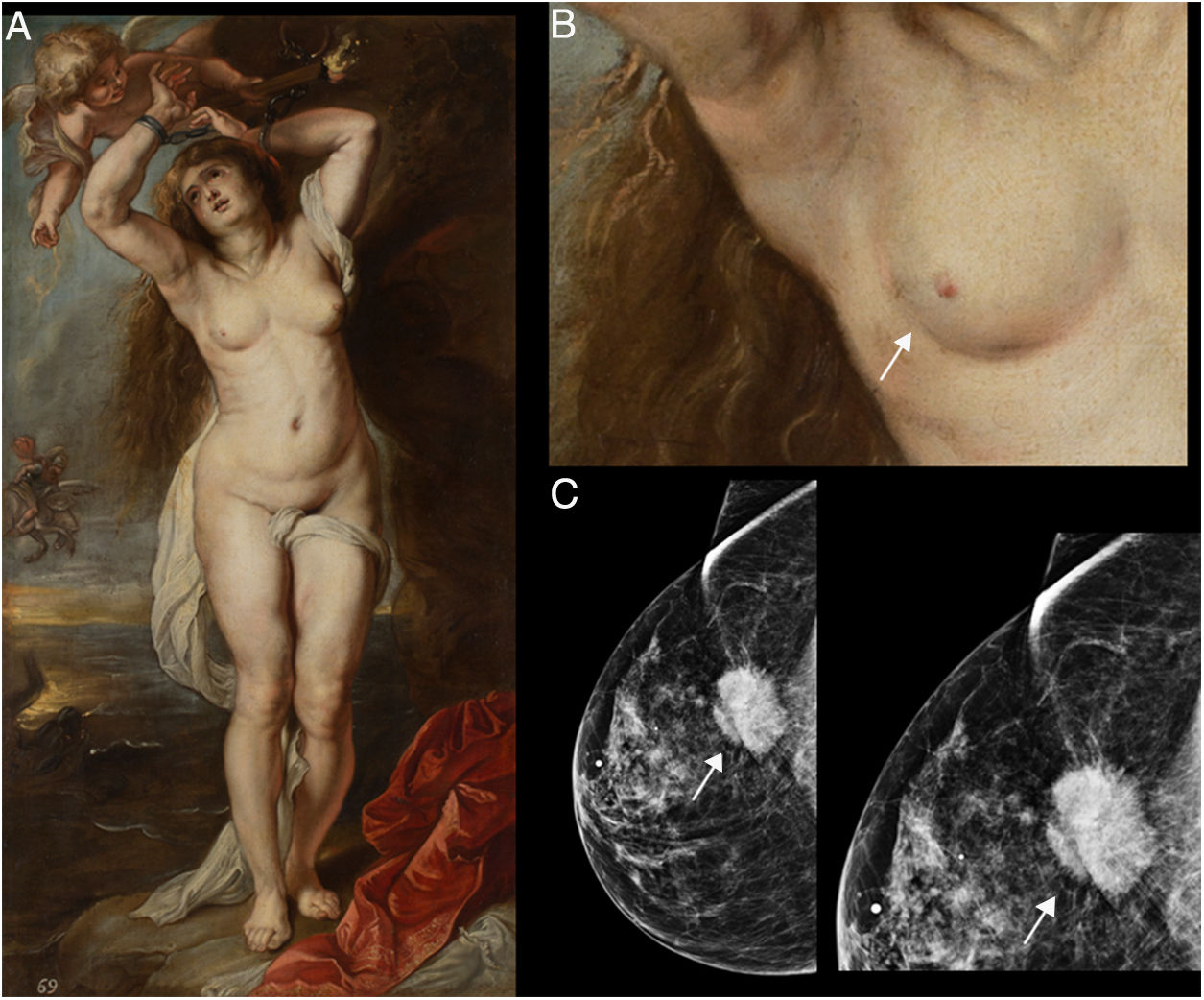
If mammography had been available in Ruben's time (Fig. 4), the image could be that of a dense, irregular, spiculated nodule with retraction of the anterior and posterior plane, as shown in the mammogram.11
 Andromeda, by Peter Paul Rubens (1634), oil on canvas, on display in the Víctor Balaguer Museum & Library. (B) Enlarged view of the right breast in the painting, where retraction of the skin (white arrow) can be seen in the lower outer quadrant. (C) Mammography. Craniocaudal view, ACR B glandular pattern in the outer quadrant, posterior plane, showing a dense, irregular spiculated nodule retracting the pectoral muscle and skin (white arrow), and enlarged view of the nodular lesion marked with a white arrow.")
(A) Andromeda, by Peter Paul Rubens (1634), oil on canvas, on display in the Víctor Balaguer Museum & Library. (B) Enlarged view of the right breast in the painting, where retraction of the skin (white arrow) can be seen in the lower outer quadrant. (C) Mammography. Craniocaudal view, ACR B glandular pattern in the outer quadrant, posterior plane, showing a dense, irregular spiculated nodule retracting the pectoral muscle and skin (white arrow), and enlarged view of the nodular lesion marked with a white arrow.
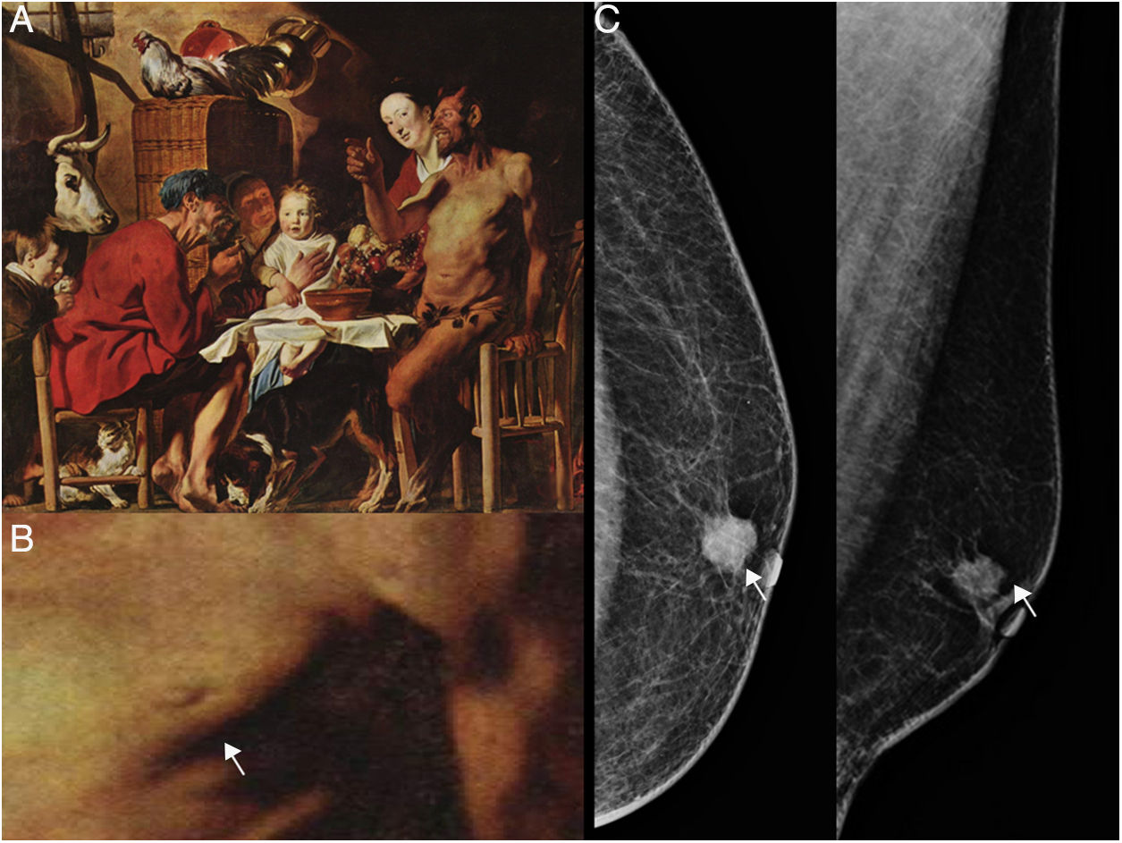
The Satyr and the Peasant is an oil on canvas painting by the Flemish artist Jacob Jordaens. This particular scene illustrates one of fables of Aesop, who lived in Greece in the 6th century BC.
Jacob Jordaens, a disciple of Rubens, was the last great master of the Flemish baroque. His paintings depict scenes from mythology and are faithful to the style of Rubens, although Jordaens lacked the creative capacity of his teacher.4
Looking at the naked torso of the figure of the satyr (Fig. 5), we see skin retraction adjacent to the nipple on the left breast, which suggests a presumptive diagnosis of male breast cancer.
 The satyr and the peasant, by Jacob Jordaens (1620–1621). Oil on oak, on display in the Alte Pinakothek museum in Munich, Germany. (B) Enlargement of the satyr's thorax showing skin retraction in the outer quadrant of the left breast next to the areola, marked with a white arrow. (C) Left craniocaudal oblique mediolateral view showing a dense, irregular, spiculated nodular lesion in the retroareolar quadrant, with associated microcalcifications and retraction of the skin (white arrow).")
(A) The satyr and the peasant, by Jacob Jordaens (1620–1621). Oil on oak, on display in the Alte Pinakothek museum in Munich, Germany. (B) Enlargement of the satyr's thorax showing skin retraction in the outer quadrant of the left breast next to the areola, marked with a white arrow. (C) Left craniocaudal oblique mediolateral view showing a dense, irregular, spiculated nodular lesion in the retroareolar quadrant, with associated microcalcifications and retraction of the skin (white arrow).
If the model had undergone mammography in 1616, the following image might have emerged: skin retraction and a dense, irregular, nodular lesion, with spiculated or microlobulated margins.12
DiscussionOne of the main contributions of neuroscience to teaching has been the discovery that it is easier to retain things in the memory if they are accompanied by an emotion. In other words, learning is not a mechanical activity; it does not only rely on repetition, but on the capacity to astonish or arouse our curiosity.1
Art supplies the astonishment and the thrill, and stirs the curiosity of the beholder.
The in-depth analysis of art and presumptive medical diagnosis is a fascinating field of medical-artistic diagnosis.
A closer look at the details of these paintings suggests the presence of abnormalities in the normal anatomy of the breasts of the models portrayed. It is highly likely that the artists intentionally reproduced the disease that changed the normal anatomy of the model's breast or axilla.
This is why paintings from the Renaissance and Baroque periods, when the human figure was painted with great realism, can be useful and innovative teaching tools to refine mammography diagnosis skills.
ConclusionsWe suggest that art can be combined with mammographic diagnosis through speculation to create a teaching tool for this field of medicine. The incorporation of humanities in medical education has been shown to increase empathy, awareness and sensitivity in the art of medicine.
Conflicts of interestThe author has no conflicts of interest to declare.
Please cite this article as: Pesce K. Diagnosticar en el arte. Radiología. 2019;61:60–65.

