


Opioid intoxication can cause potentially severe neurotoxic effects, with acute toxic leukoencephalopathy (ATL) being one of the presentations associated with the poorest prognosis. The clinical presentation is variable, ranging from mild neuropsychiatric symptoms to coma or even death.1 We report the case of a patient with ATL secondary to transdermal fentanyl and oral tapentadol intoxication.
Our patient was a 48-year-old man with history of anxiety-depressive disorder and radicular pain, under analgesic treatment with tapentadol 150mg every 12hours and transdermal fentanyl 50μg/h, who was found unconscious at home, and unresponsive to physical stimuli; the duration of this condition was unknown. Opioid treatment had begun 2 years earlier and the dosage had been progressively increased, with no previous episodes of overdose reported. No other medical or psychiatric history was documented. Five fentanyl transdermal patches were found adhered to his skin, and several empty blister packs of oral tapentadol were found nearby. Three doses of naloxone were administered, achieving no clinical response; orotracheal intubation was performed to secure the airway and the patient was transferred to our hospital. The urine toxicology screen yielded positive results for opioids and benzodiazepines, with the latter having been administered prior to orotracheal intubation. A brain CT scan revealed subtle, diffuse white matter hypodensity predominantly affecting both frontal lobes (Fig. 1). Given the patient’s critical condition and the low clinical suspicion of infection, lumbar puncture for cerebrospinal fluid analysis was not performed.

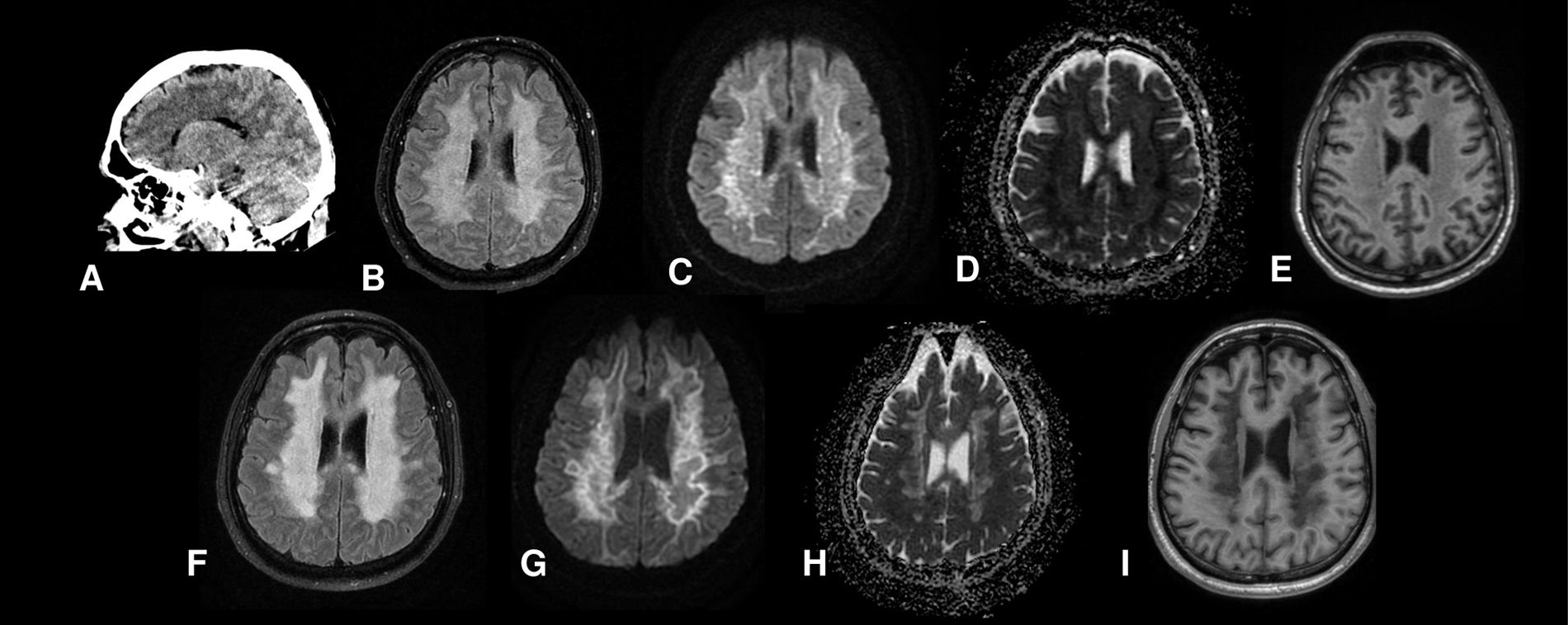
Head CT scan performed at admission showing diffuse white matter hypodensity (A). Brain MRI scan performed on day 7 revealing diffuse hyperintensity in the subcortical and periventricular white matter on T2-FLAIR sequences (B), increased signal on diffusion-weighted sequences (C), hypointensity at the periphery of the white matter on the apparent diffusion coefficient map (D), and subtle white matter hypointensity on T1-weighted sequences (E). Brain MRI scan performed on day 21 displaying progression of white matter hyperintensity on FLAIR sequences (F) and progression of cytotoxic oedema associated with deep white matter cavitations on diffusion-weighted sequences (G) and the apparent diffusion coefficient map (H), with more marked hypointensity on T1-weighted sequences, suggesting necrotic changes (I).
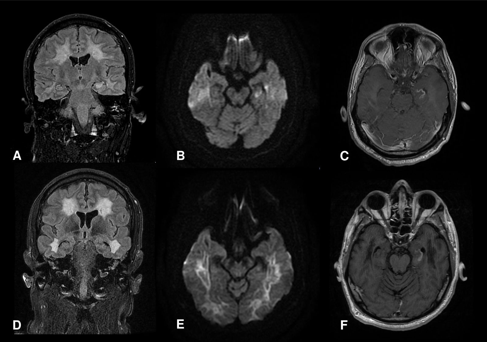
Sedation and analgesia were withdrawn 72hours after admission, but the patient remained unresponsive to stimuli. On examination, he remained with his eyes open and a fixed, centred gaze; pupils were isochoric and reactive, and brainstem reflexes were preserved. No spontaneous movement or response to painful stimuli were observed. The limbs were spastic, with brisk deep tendon reflexes and bilateral extensor plantar reflexes. Electroencephalography (EEG) revealed diffuse slowing at 2 to 5Hz, and brain MRI showed diffuse white matter hyperintensity on T2 and FLAIR sequences, sparing the U-fibres but involving the splenium of the corpus callosum and the left hippocampus. Diffusion-weighted imaging (DWI) showed areas of restricted diffusion (Fig. 1 and 2).

(A-C) Brain MRI scan performed on day 7 showing signal alterations in the left hippocampus, with restricted diffusion on DWI sequences and contrast uptake. (D-F) Brain MRI scan performed on day 21 showing mild reduction in hippocampal size with persistent contrast uptake, suggesting blood-brain barrier disruption.
The patient presented no neurological improvement over the following 3 weeks. A follow-up MRI scan revealed progression of leukoencephalopathy and the appearance of areas of necrosis and malacia at the centre of the lesion (Fig. 1 and 2). A follow-up EEG study revealed further slowing of brain electrical activity, with no response to alerting or painful stimuli, consistent with severe diffuse cerebral dysfunction. Based on these findings and the lack of clinical improvement, we considered the patient’s neurological prognosis to be poor. According to the family’s wishes, life-sustaining treatment was withdrawn, and the patient died one month after admission.
Neuroimaging findings were compatible with ATL secondary to opioid intoxication.1,2 ATL has been described in patients treated with chemotherapy or immunosuppressants, or with liver failure, among other causes,1 and is considered an extremely rare consequence of opioid intoxication. However, this condition should be included in the differential diagnosis of acute encephalopathy of unknown aetiology,1,2 particularly given that its prevalence may increase in the coming years due to the ongoing opioid overdose epidemic in Europe and North America.3,4
From a radiological viewpoint, ATL is characterised by symmetrical, confluent changes in the deep white matter corresponding to cytotoxic oedema. Alterations may be difficult to detect on head CT, making MRI the technique of choice. MRI typically shows signal alterations in the cerebral white matter, with sparing of the U-fibres,1,2,5 as observed in our patient. The radiological features of ATL differ from those seen in acute hypoxic-ischaemic injury, which frequently involves grey matter structures, suggesting different pathophysiological mechanisms. Magnetic resonance spectroscopy findings in ATL suggest a metabolic deficit, possibly secondary to mitochondrial toxicity, leading to selective white matter damage.6,7 Furthermore, another early radiological finding of acute opioid intoxication is isolated hippocampal involvement, associated with amnestic syndrome, which in some cases may progress to diffuse leukoencephalopathy.8,9
ATL must also be distinguished from delayed post-hypoxic leukoencephalopathy, which can also manifest as a result of opioid intoxication. Although the latter presents similar radiological characteristics to those of ATL, it is characterised by a biphasic clinical course, with initial improvement following the hypoxic event, followed by potentially severe neurological deterioration, characterised by cognitive impairment, parkinsonism, and even akinetic mutism.10,11
Outcomes of ATL secondary to toxic substance abuse are typically poor, with a mortality rate of up to 40%; complete recovery is infrequent.12 An association has been reported between ATL aetiology and prognosis, with cases secondary to chemotherapy or opioid misuse presenting poorer outcomes. Worse functional prognosis has also been associated with greater lesion extension on DWI and FLAIR sequences.1 Furthermore, distinguishing acute from subacute or chronic lesions may provide valuable prognostic information; therefore, MRI studies should include DWI sequences, as the extent of restricted diffusion is associated with acute lesions.12
In milder cases, early diagnosis is crucial to identifying the causal agent and preventing further exposure. In severe cases, given the poor prognosis and the lack of specific therapeutic options, supportive or palliative care is the most frequent approach.1,12
We report the case of a patient presenting the typical radiological findings of ATL secondary to opioid intoxication, as well as hippocampal involvement, contributing further evidence on the clinical and radiological picture of this entity. Recognising the characteristics of ATL is essential for early diagnosis, enabling timely supportive care and providing families with an accurate prognosis.
Author contributionsAll authors made significant contributions to this manuscript and meet the ICMJE criteria.
Patient consentThe patient’s family gave consent to the publication of this case report.
Ethics declarationThe authors have no ethical conflicts to disclose.
FundingThis study has received no funding from any public, private, or non-profit organisation.
The authors have no conflicts of interest to declare.


