


Emerging evidence increasingly suggests that poor sleep quality is associated with depressive symptoms. The hippocampus might play a crucial role in the interplay between sleep disturbance and depressive symptomatology, e.g., hippocampal atrophy is typically seen in both insomnia disorder and depression. Thus, examining the role of hippocampal volume in the interplay between poor sleep quality and depressive symptoms in large healthy populations is vital.
MethodsWe investigated the association between self-reported sleep quality, depressive symptoms, and hippocampal total and subfields’ volumes in 1603 healthy young adults from the Behavioral Brain Research Project. Mediation analysis explored the mediating role of hippocampal volumes between sleep quality and depressive symptoms.
ResultsSelf-reported sleep quality and depressive symptoms were positively correlated. In addition, it negatively related to three hippocampal subfields but not total hippocampal volume. In particular, hippocampal subfield DG and CA4 volumes mediated the interrelationship between poor sleep quality and depressive symptoms.
ConclusionsOur findings improved the current understanding of the relationship between sleep disturbance, depressive symptomatology, and hippocampal subfields in healthy populations. Considering the crucial role of DG in hippocampal neurogenesis, our results suggest that poor sleep quality may contribute to depression through a reduction of DG volume leading to impaired neurogenesis which is crucial for the regulation of mood.
A number of studies have reported bi-directions between sleep problems and mood disorders (Taylor et al., 2005), with some suggesting that poor sleep quality can increase the risk of serious mood disorders, particularly depression (Baglioni et al., 2011; Cheng et al., 2018; Hertenstein et al., 2019). Poor sleep quality has been linked to the development and exacerbation of depression by recent studies (Chai et al., 2023a, 2023b), challenging the traditional view of sleep disturbance as a symptom of mood disorders, including depression (Riemann et al., 2020). Studies have shown that sleep problems often precede depression (Boland et al., 2023; Li et al., 2016; Olfati et al., 2023; Zhang et al., 2022) and that treating insomnia can improve depressive symptoms (Boland et al., 2023) as well as prevent its relapse (Asarnow & Manber, 2019; Inada et al., 2021). Despite the well-established evidence regarding the strong link between sleep disturbance and depressive symptomatology, many researchers and clinicians ignore the role of sleep disturbance such as insomnia as a risk factor for depression. Accordingly, the assessment of neurobiological interaction between sleep disturbance and depressive symptomatology has been neglected for many years and remains poorly understood.
One potential target through which poor sleep quality may increase the severity of depressive symptoms is hippocampal structure (Meerlo et al., 2015; Novati et al., 2011), as it plays a crucial role in various important functions related to both conditions, such as emotional regulation (Walker & van der Helm, 2009), neurogenesis (Anacker & Hen, 2017), and emotional memory processing (Meerlo et al., 2015). Prior structural brain studies reported reduced hippocampal volume in sleep disorders such as insomnia disorder (Riemann et al., 2007; Joo et al., 2014; Chen et al., 2022), obstructive sleep apnea (Morrell et al., 2003; Tahmasian et al., 2016), as well as narcolepsy (Joo et al., 2012). The hippocampal volume reduction is also observed in major depressive disorder (MDD) (Tahmasian et al., 2013; Treadway et al., 2015; Anacker & Hen, 2017; Bagherzadeh-Azbari et al., 2019) and subjects with higher depressive symptom severity (Brown et al., 2014). The common reduction of hippocampal volume further suggest that depression may in part result from an alteration in hippocampal structure that can be linked to sleep disruptions.
The hippocampus is composed of distinct subfields, namely cornu ammonis fields (CA1–CA4), the dentate gyrus (DG), and the subiculum (Duvernoy et al., 2013; Sahakyan et al., 2021; Sun et al., 2023). The latest advancements in MRI acquisition have piqued a keen interest in investigating hippocampal subfields in vivo through histological examination (Iglesias et al., 2015; McHugo et al., 2018). Previous studies have explored the relationship between sleep disturbance and hippocampal subfield volumes in patients with PTSD (Neylan et al., 2010) and insomnia (Joo et al., 2014; Chen et al., 2022), and their findings are inconsistent. In healthy populations, several studies indicated no correlation between sleep quality and overall hippocampal volume atrophy in community-dwelling adults (Sexton et al., 2014; De Looze et al., 2022). However, studies that take into account hippocampal subfield volumes have revealed a significant link between poor sleep quality and hippocampal volume atrophy in healthy individuals, albeit in older adults (De Looze et al., 2022; Liu et al., 2021). Moreover, patients with MDD showed reduced volumes in the dentate gyrus (Treadway et al., 2015), which is involved in mood regulation and neurogenesis (Sun et al., 2023). Given the impact of both sleep disturbance and depressive symptomatology on hippocampal atrophy and the robust association between poor sleep quality and reduced hippocampal subfield volumes, further research is needed to understand the relationship between sleep disturbance, depressive symptomatology, and hippocampal subfields, especially in the healthy populations (Meers et al., 2020; Tahmasian et al., 2020).
Based on the abovementioned research evidence, the hippocampus may be pivotal in the interplay between sleep disturbance and depressive symptomatology, considering the typical occurrence of accelerated hippocampal atrophy in both insomnia disorder and depression. Thus, examining the role of hippocampal volumes in the interplay between poor sleep quality and depressive symptoms in large healthy populations is vital. In this context, we investigated whether measures of self-reported sleep quality, self-report depressive symptoms, and their interplay are related to individual differences in total hippocampal volume and hippocampal subfields volumes on a large sample (N = 1603) of young adults from the Behavioral Brain Research Project to Chinese Personality (BBP). The hypotheses posit that poor subjective sleep quality measured by the total score of the Pittsburgh Sleep Quality Index (PSQI) is linked to lower hippocampal total and subfield volumes and higher self-reported depression scores (hypothesis 1). Additionally, it is hypothesized that hippocampal subfield volumes could mediate the influence of poor sleep quality on self-reported depressive symptoms (hypothesis 2). Finally, we also explored whether the depressive symptoms measured by the total score of the Self-rating Depression Scale (SDS) will be linked to hippocampal total and subfield volumes.
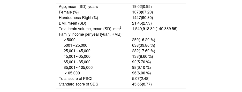
MethodsSampleThe dataset comprises individuals from the BBP (Wang et al., 2023), which was launched from September 2019 to March 2023 to recruit participants from that year's freshmen at Southwest University (SWU), Chongqing, China. To ensure the eligibility of participants for MRI scanning and minimize confounding factors, a set of inclusion and exclusion criteria were implemented. Firstly, all participants underwent standardized physical examinations at the affiliated hospital of the SWU, ensuring the absence of major physical illnesses. Furthermore, prior to the brain scanning and measurement of behavioral variables, participants provided written informed consent, which included self-reported medical and psychiatric disorder information. Only individuals who reported no neurological or psychiatric disorders (such as depression or sleep disorders), no usage of psychoactive medications, and no other chronic diseases were considered for inclusion in this research. Additionally, participants had to meet safety requirements for MRI scanning, including not having claustrophobia, metallic implants, pregnancy, or a history of head trauma and fainting. These meticulous criteria were applied to create a homogeneous and reliable participant group, thus enhancing the validity of the study's findings. Specifically, a total of 1640 undergraduates who met the inclusion criterion were recruited via mobile telephone text message to complete the cross-sectional neuroimaging protocol, demographic information, as well as subjective measures of sleep quality and depression. Thirty-seven participants were excluded from the quality control procedure of the hippocampal subfield segmentation (for the details please refer to section 2.7), resulting in a final sample of 1603 participants (mean age=19. 02 years old, SD= 0.95; 525 males and 1078 females, Table 1). Full informed consent from each participant was obtained by BBP Consortium, and research procedures and ethical guidelines were followed in compliance with SWU institutional review board approval.
Demographic characteristics of participants in the present study (N = 1603).
Note. SD: Standard Deviation. PSQI: Pittsburgh Sleep Quality Index. SDS: Self-rating Depression Scale. BMI: Body Mass Index. RMB: Ren Min Bi.
The collection of demographic information consisted of participants’ age, sex, handedness, body mass index (BMI) and self-reported family income per year. The total brain volume (including gray matter, white matter, and cerebrospinal fluid) of the participants was estimated by FreeSurfer (https://surfer.nmr.mgh.harvard.edu/). See Table 1 for detailed demographics of the dataset.
Sleep quality assessmentSleep quality was self-assessed using the Pittsburgh Sleep Quality Index (Buysse et al., 1989; Tsai et al., 2005), which is a comprehensive 19-item questionnaire that evaluates an individual's self-reported sleep quality over the last month. It consists of seven distinct components, which include subjective sleep quality, latency, duration, efficiency, disturbance, sleeping medication, and daytime fatigue. The disturbance component includes subcomponents such as the inability to sleep within 30 min, nocturnal awakening due to insomnia, and breathing comfortably, coughing/snoring related to sleep apnea. To determine the global score of the PSQI, the scores of all seven components are summed up, which ranges from 0 to 21. Higher scores indicate poorer sleep quality. A global PSQI score of ≥5 indicates subjective poor sleep quality, according to Buysse et al. (1989). The Chinese version of PSQI had an overall reliability coefficient of 0.82–0.83 for all subjects (Tsai et al., 2005). The included sample has a mean total PSQI score of 5.07 with a standard deviation of 2.48 (Table 1). Notably, 610 out of 1603 participants or 38 % of the present sample, have a total PSQI score higher than 5.
Depression assessmentThe severity of depressive symptoms in individuals was assessed using the Self-rating Depression Scale (SDS), which was first developed by Zung in 1965. Since its development, the SDS has been widely used in both clinical and research settings (Zung, 1965). The SDS consists of 20 items that assess a range of symptoms commonly associated with depression, including sadness, guilt, insomnia, fatigue, and loss of appetite. Each item is rated on a 4-point Likert scale ranging from 1 (rarely or none of the time) to 4 (most or all of the time). The standard score of SDS was computed for further analysis. The total standard score can range from 25 to 100, with higher scores indicating more severe depression. A standard score of SDS >53 indicates the presence of depressive symptoms (Duan & Sheng, 2012). The included sample has a mean standard SDS score of 45.65 with a standard deviation of 8.77 (Table 1). Also, 273 out of 1603 participants, or 17 % of the present sample, have a standard SDS score higher than 53.
MRI data acquisitionAll MR Images were acquired using a 3T Siemens Prisma-fit scanner with a standard 32 channel head coil located at Southwest University. High-resolution T1-weighted (T1w) structural image was obtained using a three-dimensional gradient sequence in order to facilitate alignment of individual subject images into a common space: repetition time (TR) = 2530 ms, time of echo (TE) = 2.98 ms, field of view (FOV) = 256 × 256 mm2, thickness = 1 mm, voxel size = 0.5 × 0.5 × 1 mm3, flip angle = 7°, resolution matrix = 256 × 256, slices = 192, slice oversampling = 33.3 %, phase encoding direction = AC » PC.
Imaging data processing and hippocampal subfield segmentationThe FreeSurfer software package version 7.3.2 (https://surfer.nmr.mgh.harvard.edu/) was used to process the remaining structural T1-weighted images to extract volume estimates for left and right hippocampal total and subfield volumes as well as intracranial volume (ICV). For preprocessing, the main reconstruction pipeline (“recon- all”) was used for volumetric segmentation. This processing includes motion correction, removal of non-brain tissue, automated Talairach transformation, tessellation of the gray matter/white matter boundary, and segmentation of hippocampus (Fischl, 2012; Fischl et al., 2004). Hippocampal structures were further parcellated using the Hippocampal Subfields protocol (Iglesias et al., 2015), automatically segmenting the hippocampal formation into 12 subfields (cornu ammonis (CA)−1, CA-3, CA-4, fimbria, granule cell layer of dentate gyrus (GC-ML-DG), hippocampus- amygdala-transition-area (HATA), hippocampal tail, hippocampal fissure, molecular layer, parasubiculum, presubiculum and subiculum, also see Fig. 1) for each hemisphere and calculating their volumes, using a probabilistic brain atlas (Leemput et al., 2008). The validity and reliability of this procedure have been demonstrated in previous studies (Iglesias et al., 2015; Sahakyan et al., 2021). To account for differences in brain size, we used the estimated total intracranial volume (eTIV) to adjust each subfield volume. Specifically, raw each subfield volume was corrected for the eTIV using the following formula: Voladj= (Volraw ×eTIVmean)/eTIVparticipant, where Voladj is the adjusted regional volume, Volraw is the raw volume for each participant, and eTIVmean is the mean value of the eTIV from all included participants, which is a common practice from the previous studies (Jiang et al., 2019; La Joie et al., 2010; Yeung et al., 2017). The head size correction was conducted separately for each subfield in each hemisphere. It should be noted that the total volume of hippocampus was also adjusted using the same procedure.
; the bottom row shows the relation of these original FreeSurfer 7.3.2 hippocampal subfields to functional models separating the hippocampus into a model grouping cornu ammonis (CA) and dentate gyrus (DG) within the head-body-tail separation.")
FreeSurfer7.3.2 output showing hippocampal substructure. Top row shows 3D model of the different segmented hippocampal subfields with bottom and top views on the segmented hippocampus (color legend of individual subfield segments on bottom right); the bottom row shows the relation of these original FreeSurfer 7.3.2 hippocampal subfields to functional models separating the hippocampus into a model grouping cornu ammonis (CA) and dentate gyrus (DG) within the head-body-tail separation.
We followed the proposed quality control procedure guidelines for the FreeSurfer-based segmentation of the hippocampal subregions designed for the Enhancing Neuro Imaging Genetics through Meta-Analysis (ENIGMA) consortium (Sämann et al., 2022). Briefly, the proposed procedures for quality control included the following steps: (1) The original T1-weighted images were visually checked to exclude images with visible artifacts, i.e., subject motion and ghosting. (2) The general (sub-)cortical segmentation accuracy were snapshot-based visually checked to exclude images with a noticeable error; (3) we then conducted HTML-snapshot-based visual QC of hippocampus segmentation of all participants, paying attention to the hippocampus mask as a whole and hippocampus fissure within the hippocampal mask; (4) we further used interactive FreeSurfer viewing tool to conduct dynamic visual inspection of suspicious hippocampus segmentation with statistical outliers (based on group-wise outlier detection (2 standard deviations from group mean) and rank-order rules of subfield volumes. Based on this QC procedure, thirty-seven participants were excluded due to gross errors in the hippocampal subfield segmentation (atypical segmentation results). We also checked the processed data quality using the rank-order rules of subfield volumes, as detailed in the same paper (Sämann et al., 2022).
Statistical analysisStatistical analysis was performed using SPSS® (version 29). All analyses included combined hippocampal volumes across both hemispheres (left + right volume) as dependent variables. Results from the main analyses for the left and right hippocampal subfields are reported in supplementary materials Table S1. To test our a priori hypothesis 1, partial correlational analyses were run between total scores of PSQI and hippocampal and hippocampal subfields volume, between total scores of PSQI and SDS by adding age, sex, handedness, BMI, and family income as covariates to control for their potential effects. Multiple comparison correction was performed across hippocampal subfields with the Bonferroni correction. A a level of 0.05/12 was used to denote statistical significance in these analyses.
To test hypothesis 2 whether the volume of hippocampal subfields that are significantly correlated with sleep quality can mediate the influence of poor sleep quality on self-reported depressive symptoms, mediation analysis was conducted with an SPSS-based package named Process version 3.4.1 (Hayes, 2009). Mediation analysis investigates how much of the covariance between two variables can be explained by the mediator variable(s). The dependent variable was chosen to be the SDS total scores. PSQI total score was selected as the independent variable. The mediator variable was chosen to be the volume of hippocampal subfields that are significantly correlated with overall sleep quality (i.e., DG, CA-3, and CA-4). Age, sex, handedness, BMI, and family income were also added as covariates for the mediation analysis. The bootstrap test was performed 5000 times to determine the significance. The significance level was set at 0.05, 2-tailed. In a confirmatory analysis, we further repeated the analysis based on the seven components of PSQI to identify which component of sleep quality affects depressive symptoms via hippocampal subfields. As an exploratory analysis, partial correlational analyses were also run between total scores of SDS and hippocampal and hippocampal subfields volume by adding age, sex, handedness, BMI, and family income as covariates. Bonferroni correction was applied for the multiple comparison issues.
Confirmatory analysisGiven item #4 (“I have trouble sleeping at night”) in the SDS reflects a sleep problem, we removed it to calculate the total score of SDS and repeated the above-mentioned analysis as a confirmatory analysis (Leerssen et al., 2020; Olfati et al., 2023). We also explored a different option for our mediation analysis as a further confirmatory analysis, i.e., sleep quality is outcome variables, depressive symptoms are the dependent variables, and hippocampal subfield volumes as the mediators.
ResultsThe included sample has a mean hippocampal total volume of 5513 mm3 with a standard deviation of 596 mm3. Our partial correlational analysis revealed that sleep quality measured by the total score of PSQI was positively correlated with the SDS scores (r = 0.354, p<0.001, Fig. 2a). Notably, sleep quality measured by the total score of PSQI was still correlated with the SDS scores removing the sleep item (r = 0.312, p<0.001).
")
a, the total score of PSQI was positively correlated with self-reported depressive symptoms measured by SDS. b, the total score of PSQI was negatively correlated with the volume of hippocampal subfield DG. c, the total score of PSQI was negatively correlated with the volume of hippocampal subfield CA-3. d, the total score of PSQI was negatively correlated with the volume of hippocampal subfield CA-4. DG, dentate gyrus; CA, cornu ammonis; PSQI, Pittsburgh Sleep Quality Index; SDS, Self-rating Depression Scale. (For interpretation of the references to colour in this figure legend, the reader is referred to the web version of this article.)
Sleep quality measured by the total score of PSQI did not correlate with total hippocampal volume (r= −0.047, p = 0.063), while the total score of PSQI was negatively correlated with the volume of hippocampal subfields including DG (r= −0.088, p<0.001, Fig. 2b), CA-3 (r= −0.089, p<0.001, Fig. 2c) and CA-4 (r= −0.088, p<0.001, Fig. 2d). For the rest of the hippocampal subfields, we did not find a significant correlation with sleep quality (all ps>0.05/12, also see supplementary Table S2).
Our exploratory analysis indicated that total score of SDS was negatively correlated with the volume of hippocampal subfields including DG (r= −0.088, p<0.001), CA-1 (r= −0.076, p = 0.002), CA-3 (r= −0.073, p = 0.004), HATA (r= −0.072, p = 0.004) and CA-4 (r= −0.083, p<0.001), as well as the total hippocampal volume (r= −0.073, p = 0.004). For the rest of the hippocampal subfields, we did not find a significant correlation with depressive symptoms (all ps>0.05/12, also see supplementary Table S2). These significant correlations were again evident when repeated with the SDS scores removing the sleep item. Specifically, the SDS scores removing the sleep item was negatively correlated with the volume of hippocampal subfields including DG (r= −0.087, p<0.001), CA-1 (r= −0.076, p = 0.002), CA-3 (r= −0.070, p = 0.005), HATA (r= −0.072, p = 0.004) and CA-4 (r= −0.081, p = 0.001), as well as the total hippocampal volume (r= −0.072, p = 0.004).
Our mediation analysis revealed that volume of hippocampal subfield DG (β = 0.018, 95 % CI: [0.003,0.038]) and CA-4 (β = 0.017, 95 % CI: [0.002,0.036]) significantly mediated the influence of poor sleep quality on self-reported depressive symptoms. The hippocampal subfield CA-3 (β = 0.013, 95 % CI: [−0.001,0.032]) did not have a mediating role in the interplay between sleep quality and self-reported depression (see supplementary figure 1 for more details). These mediation effects were repeated with the SDS scores removing the sleep item. Specifically, volume of hippocampal subfield DG(β = 0.018, 95 % CI: [0.004,0.037]) and CA-4 (β = 0.016, 95 % CI: [0.002,0.035]) significantly mediated the influence of poor sleep quality on self-reported depressive symptoms without sleep issue. The hippocampal subfield CA-3 (β = 0.013, 95 % CI: [−0.002,0.031]) did not have a mediating role in the interplay between sleep quality and self-reported depression not involving sleep issue.
Moreover, it turned out that the first component of PSQI, namely subjective sleep quality was significantly correlated with the hippocampal subfield CA-3 (r=−0.092, p<0.001), CA-4 (r=−0.10, p<0.001) and DG (r=−0.10, p<0.001). Furthermore, the first component of PSQI also affects depressive symptoms via hippocampal subfield DG (β = 0.06, 95 % CI: [0.01,0.13], Fig. 3a) and CA-4 (β = 0.06, 95 % CI: [0.01,0.12], Fig. 3b). Again, these mediation effects were repeated with the SDS scores removing the sleep item. Specifically, the first component of PSQI affects depressive symptoms not involving sleep issue via hippocampal subfield DG (β = 0.07, 95 % CI: [0.01,0.13]) and CA-4 (β = 0.06, 95 % CI: [0.009,0.12]). Furthermore, the optional mediatory analysis indicated that the volume of hippocampal subfields DG, CA4 and CA3 did not mediate the influence of depressive symptoms on self-reported sleep quality. Please refer to Supplementary Results for more details.

a, volume of hippocampal subfield DG was found to mediate the association between poor sleep quality and self-reported depression. The volume of hippocampal subfield DG was selected as mediation variable. The total score of SDS was used as the dependent variable and the score of the first component of PSQI, namely subjective sleep quality was determined as the independent variable. b, volume of hippocampal subfield CA-4 was also found to mediate the association between poor sleep quality and self-reported depression. The volume of hippocampal subfield CA4 was selected as mediation variable. The total score of SDS was used as the dependent variable and the score of the first component of PSQI, namely subjective sleep quality was determined as the independent variable. Age, sex, handedness, BMI, and family income were included as covariates to rule out confounding when performing mediation analysis. DG, dentate gyrus; CA, cornu ammonis; PSQI, Pittsburgh Sleep Quality Index; SDS, Self-rating Depression Scale; CI, confidant interval.
The present study directly tested the role of hippocampus and hippocampal subfields volume in the interplay between poor sleep quality and depressive symptoms in healthy young populations. In coincide with recent studies investigating the association between self-reported sleep quality and hippocampal subfield volumes in healthy elderly group (Sexton et al., 2014; De Looze et al., 2022), the present study also revealed sleep quality did not correlate with total hippocampal volume in healthy populations. However, self-reported sleep quality was found to be negatively correlated with the volume of hippocampal subfields, including DG, CA-3 and CA-4. Notably, the CA-4 is frequently considered as a part of the DG (Duvernoy et al., 2013; Liu et al., 2021). Moreover, self-reported depressive symptoms were found to be negatively correlated with several hippocampal subfields including DG, CA-1, CA-3, CA-4, HATA as well as the total hippocampal volume. In addition, the present study revealed that the volume of hippocampal subfields DG (including CA-4) significantly mediated the influence of poor sleep quality on self-reported depressive symptoms. Because DG is considered to play a crucial role in hippocampal neurogenesis (Meerloa et al., 2009), poor sleep quality may contribute to depression through a reduction of DG volume leading to impaired neurogenesis, which is crucial for the regulation of mood ( Treadway et al., 2015; Sun et al., 2023).
With a large sample size (n = 1603), this study indicated an association between poor sleep quality and volume loss of the CA-3/CA-4/DG hippocampal subfields in healthy young adults. Notably, the correlations are weak but statistically significant (i.e., unlikely to have occurred by chance). The significance mainly comes from our large sample size, similar to the small effect size but significant correlations in previous studies with large sample sizes (Cheng et al., 2018; He et al., 2021; Li et al., 2022a). The significant correlations between hippocampal subfields and sleep quality are consistent with previous research by Neylan et al. (2010), which showed that subjective insomnia severity is associated with volume loss of the CA-3/dentate subfields (Neylan et al., 2010) in PTSD patients. The association of self-reported sleep quality with the CA-3/CA-4/DG subfield volume is also consistent with animal models that have shown that chronic sleep disruption is associated with decreased neurogenesis and dendritic branching in these structures (Novati et al., 2011; Meerlo et al., 2015). Moreover, this association between sleep quality and hippocampal subfield volume is in alignment with both previous studies that have investigated the association between sleep disorders and hippocampal subfield volumes ( Neylan et al., 2010; Joo et al., 2014) and previous studies that have indicated that reduced hippocampal subfield volume was associated with short sleep and sleep disturbance in the elderly group (Fjell et al., 2020; Liu et al., 2021; De Looze et al., 2022) . Animal studies involving sleep and sleep-dependent manipulations demonstrated that the CA-3/CA-4/DG hippocampal subfields are crucial in neural plasticity, reduction of hippocampal cell proliferation and neurogenesis (Kreutzmann et al., 2015). Our findings further indicated that certain biological factors, such as experimental sleep restriction or disrupted sleep may have significant effects on neurogenesis in the dentate gyrus, causing changes in the volume of the CA-3/CA-4/dentate region and possibly affecting other subfields, potentially impacting the hippocampal subfield volume. Given the weak correlations between hippocampal subfields and sleep quality, our study merely proposes a potential intervention pathway, and its effectiveness needs to be tested through clinical practice.
It is important to note that the present study revealed that poor sleep quality was not associated with the total hippocampal volume. In the literature, the evidence of a relationship between poor sleep and total hippocampal volume is divergent. On one side, a negative association between hippocampal volume and sleep deprivation severity or duration has been reported in disorders characterized by sleep disruption (Joo et al., 2012, 2014; Morrell et al., 2003), in healthy older adults (Noh et al., 2012; Liu et al., 2018) and in animal studies (Novati et al., 2011; Raven et al., 2018). Cumulative evidence, therefore, suggests that sleep may be an important risk factor for hippocampal atrophy (Polsek et al., 2018; Levenstein, 2019; Li et al., 2022b). On the other side, other human studies report no association between poor sleep and overall hippocampal volume (Spiegelhalder et al., 2013; Sexton et al., 2014; Fjell et al., 2020). One reason for these inconsistent findings may be due to the low sensitivity of a global hippocampal volumetric approach,which may obscure hippocampal subfield distinct vulnerability to poor sleep quality. Indeed, our study revealed that poor sleep quality was associated with volume loss of the CA-3/CA-4/DG subfields. This rationale is consistent with a recent study which also did not find associations between sleep and total hippocampal volume but found associations between sleep duration and specific hippocampal subfield volumes (De Looze et al., 2022).
The present study has revealed that the volume of hippocampal subfields DG (including CA-4) plays a significant role in mediating the impact of poor sleep quality on self-reported depressive symptoms. The DG, a crucial component of the hippocampal trisynaptic circuit, serves as a gate that controls the information flow from cortical regions into the hippocampus (Abrous et al., 2005; Ehninger & Kempermann, 2008), and plays a critical role in the hippocampal neurogenesis (Meerloa et al., 2009). Additionally, the DG has been implicated in mood regulation (Sun et al., 2023), in addition to its critical role in learning and memory (Hainmueller & Bartos, 2020). To this end, poor sleep quality may contribute to depressive symptoms through a reduction of DG volume leading to impaired neurogenesis which is crucial for the regulation of mood. Moreover, the observed mediating effects of DG in the interplay between sleep quality and depressive symptoms are supported by several lines of evidence in humans with psychiatric disorders and in animal models of depression. For example, patients with MDD have decreased hippocampal volume, including DG (Kronmüller et al., 2008; Gerritsen et al., 2011), and manipulation of DG circuits or neurogenesis in mice can induce depression-like behaviors (Hill et al., 2015; Yun et al., 2018). This alteration in hippocampal subfield volume in depression indicates that the depression may, in part, result from a suppression of hippocampal neurogenesis (Anacker & Hen, 2017). Considering the observed negative correlation between poor sleep quality and volume size of the CA-3/CA-4/DG hippocampal subfields mentioned above, sleep deficiency can have a significant role in the reduction of hippocampal neurogenesis, which further contributes to the development of depression. Taken together, these observations suggest that interventions aimed at altering the size of DG (Fotuhi et al., 2012; Boland et al., 2023) could be a promising therapeutic strategy for depressive symptoms.
Leveraging a large sample size, the present study investigated hippocampal subfield volumes in association with self-reported sleep quality and its mediating role in the determination of how poor sleep quality influences depressive symptoms in healthy young adults. Although a few studies have examined the associations between sleep, hippocampus, and depressive symptoms, they assessed them separately using univariate analysis (e.g., only correlation), using global hippocampal volumetric measurements only, relatively small sample sizes, and/or based on patient populations (e.g., chronic insomnia, depression, and PTSD) and healthy elderly groups. Nonetheless, the present study has several limitations. First, a subjective measure of sleep quality was used. Subjective sleep measures are readily available and can be used conveniently to screen and track sleep quality including sleep duration and sleep disorders. Future studies examining the influence of sleep quality on depressive symptoms can incorporate more objective sleep measures such as the duration, efficiency, and regularity of sleep obtained by actigraphy and polysomnography. Secondly, causality between sleep quality and depressive symptoms cannot be established in this cross-sectional study. Future studies can use a longitudinal design to test whether poor sleep leads to depressive symptoms via accelerating hippocampal subfield volume loss. Thirdly, our investigation centered on examining the hippocampal structure; nonetheless, it is worth considering that there may be connections with additional regions, including the amygdala, thalamus, and prefrontal cortex. These associations hold potential and should be explored in future research to better understand the impact of poor sleep quality on depressive symptoms. Finally, the study participants were all college students with a limited age range, limiting the generalizability of our results to other age groups.
ConclusionThis study demonstrated that self-reported sleep quality was negatively associated with the volume of hippocampal subfields, such as DG, CA-3, and CA-4, in healthy young adults. However, no significant correlation was observed between self-reported sleep quality and total hippocampal volume. For the first time, the present study revealed that the volume of hippocampal subfields DG (including CA-4) significantly mediated the influence of poor sleep quality on self-reported depressive symptoms, thus improving our understanding of the relationship between sleep disturbance, depressive symptomatology, and hippocampal subfields in the healthy populations. Given DG's crucial role in hippocampal neurogenesis, poor sleep quality may contribute to depression through a reduction of DG volume leading to impaired neurogenesis, which is crucial for the regulation of mood. These findings could open opportunities for considering sleep intervention (e.g., behavioral therapy or physical exercise) to minimize volume loss in the affected hippocampal subfield DG, and to delay the onset/severity of depression.
FundingThis work has been funded by the National Key Research and Development Program of China (2021YFC2501500), the National Natural Science Foundation of China (31971028; 82202247; 32300861), and 2021 International Exchange Program and Introduction Project Funding.
Author contributionsY.L.W. conceived and implemented the study, carried out the data analysis and wrote the manuscript. X.L. supervised the work. M.T. supported the data analysis, reviewed and edited the manuscript. Y.T. helped with the acquiring of the BBP dataset, as well as the calculation of the total scores of SDS removing the sleep item. Both Z.L.L. and D.B.D. helped with the hippocampal subfield segmentation and commented on the manuscript. Q.H.H., J.Q., T.Y.F., H.C., and X.L. funded, designed, and implemented the Behavioral Brain Research Project to Chinese Personality (BBP) project.
The authors declare that they have no known competing financial interests or personal relationships that could have appeared to influence the work reported in this paper.
