


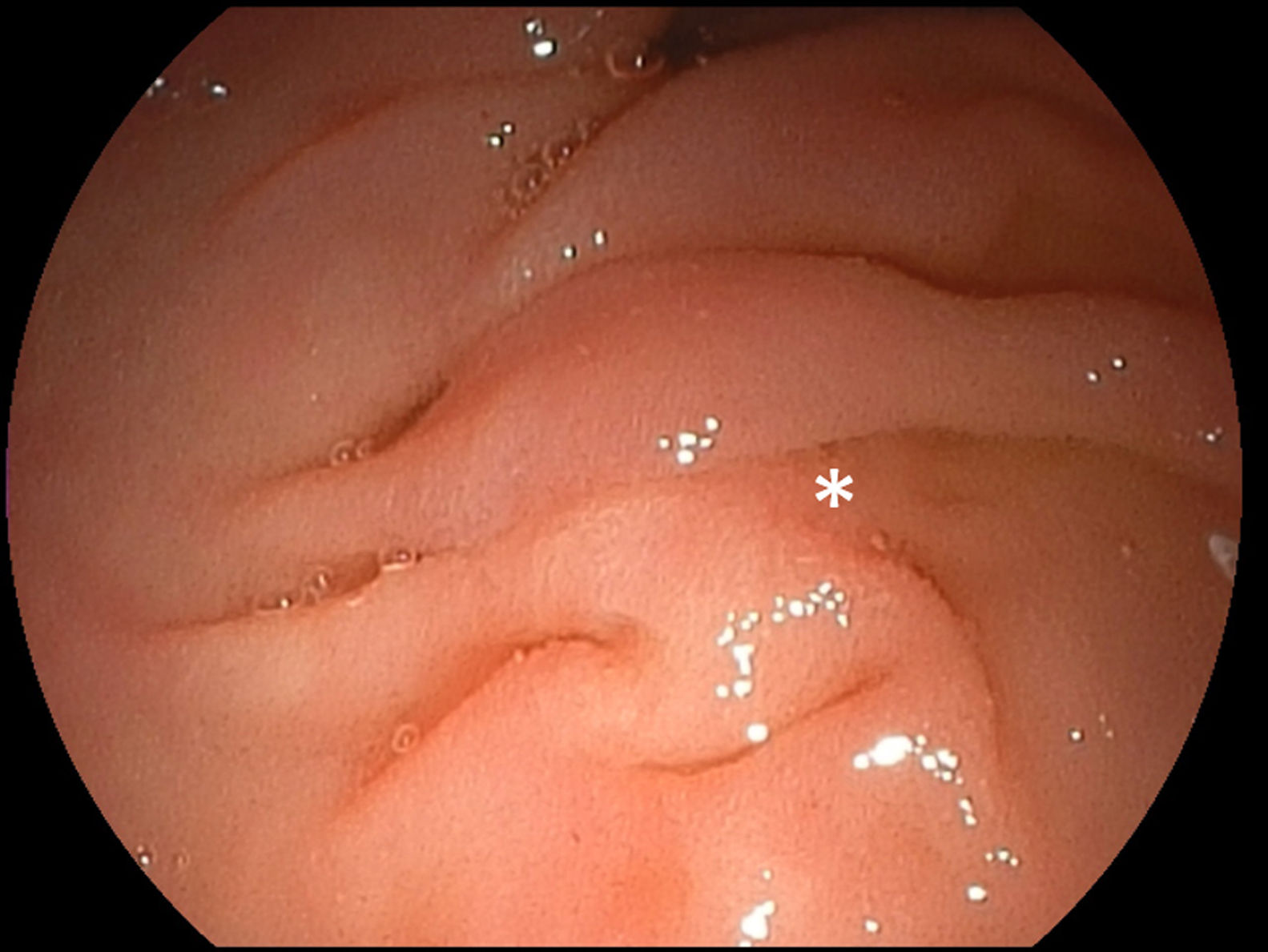
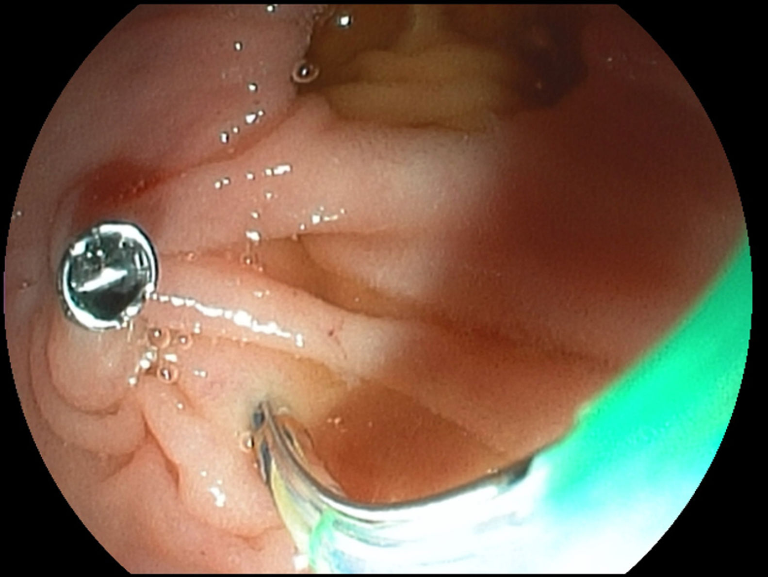
A 71-year-old female patient was referred for endoscopic retrograde cholangiopancreatography (ERCP) for pre-papillary bile duct stone disease detected by outpatient magnetic resonance cholangiopancreatography (MRCP). After insertion of a therapeutic duodenoscope, redundant duodenal folds became visible in the descending duodenum (D2), initially giving rise to a “hiddenpapilla” situation. In fact, the papilla only became appreciable after ERCP catheter manipulation in between the stack of folds, confirming a regular localization at the medial aspect of D2, however, obscured by an overriding fold. The papilla itself was classified as a type 2 configuration (small <3mm diameter and flat) as suggested by the Scandinavian Association for Digestive Endoscopy (SADE) (Fig. 1, asterisk).1 Administration of butylscopolamine for duodenal antimotility and repeated lifting of the fold by a guidwire-loaded papillotome as our first-line standard cannulation technique failed to provide sustained visualization of the papilla. Therefore, we switched to a clip-assisted ERCP approach. To this end, the overriding fold was lifted by one branch of an 11-mm rotable clip and attached to the more cranially and laterally neighboring mucosa, providing optimal ERCP settings for successful papillotome cannulation and safe papillotomy with stone extraction (Fig. 2).
While mostly reported in papilla-related pathology, such as peripapillary lipoma and/or diverticulum, this is the first report of clip-assisted ERCP in the not uncommon setting of redundant duodenal folds initially precluding instrumentation.2 Therefore, while clip-assisted cannulation is well established maneuver to externalize and fix intradiverticular papillae, the presented alternative application for redundant duodenal folds, obscuring visualization and manipulation of the papilla, may represent an indispensible rescue approach of special value to remind less experienced endoscopists of this basically old technique.
