Buscar en
Gastroenterología y Hepatología
Toda la web
Inicio Gastroenterología y Hepatología Bile duct stone formation around migrated surgical clip 17 years after laparosco...
Información de la revista
Vol. 44. Núm. 9.
Páginas 662-663 (Noviembre 2021)
Compartir
Compartir
Descargar PDF
Más opciones de artículo
Vol. 44. Núm. 9.
Páginas 662-663 (Noviembre 2021)
Image of the month
Acceso a texto completo
Bile duct stone formation around migrated surgical clip 17 years after laparoscopic cholecystectomy
Formación de cálculos en el conducto biliar alrededor de una grapa quirúrgica migrada 17 años después de una colecistectomía laparoscópica
Visitas
183
Aleksandar Gavrica,b, Sebastian Stefanovica,b, Samo Pluta,b, Luka Strnisaa,b,
Autor para correspondencia
luka.strnisa@gmail.com

Corresponding author.
a University Medical Center Ljubljana, Department of Gastroenterology and Hepatology, Ljubljana, Slovenia
b Ljubljana Digestive Endoscopy Research Group (LuDERG), Department of Gastrointestinal Endoscopy, Ljubljana, Slovenia
Este artículo ha recibido
Información del artículo
Texto completo
Bibliografía
Descargar PDF
Estadísticas
Figuras (2)
Texto completo

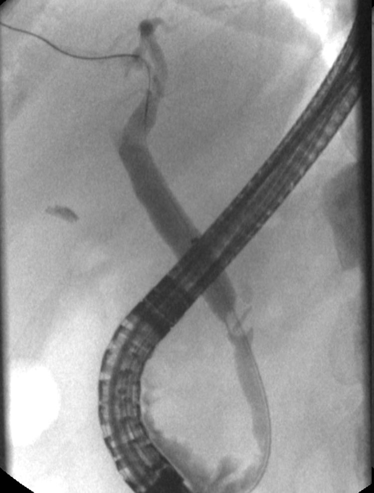
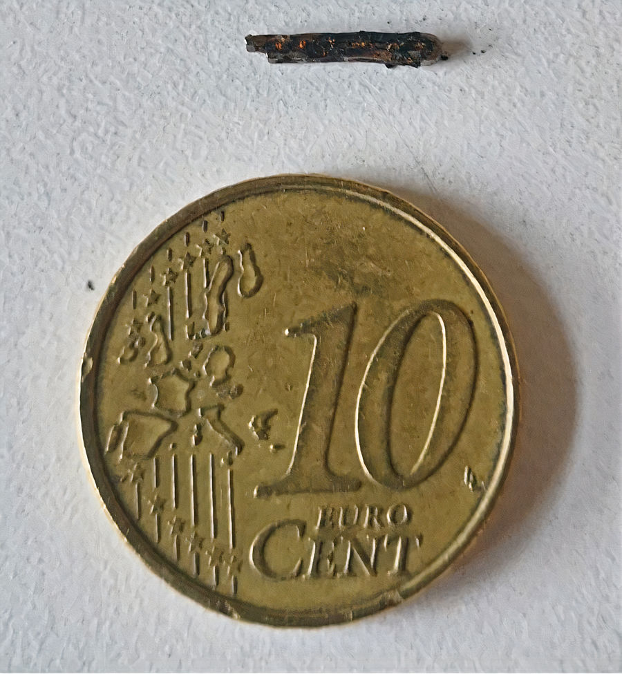
69-year old female presented with a monthlong abdominal pain and nausea with vomiting. Initial workup showed mild increase of aminotransferases (AST, 64.8U/L; ALT, 99.6U/L). Ultrasound was unremarkable. Few days later aminotransferases increased (AST, 1652U/L; ALT, 1785U/L), with elevated total bilirubin (1.7mg/dL), CRP (47mg/L) and leukocytosis. Her medical history was notable for laparoscopic cholecystectomy 17 years earlier during which cystic duct and artery were ligated. During current work up patient underwent endoscopic ultrasound that showed 11mm gallstone in the bile duct. At subsequent ERCP a 10mm×6mm suspected stone with a central oval radiopaque defect (Fig. 1) in the common hepatic duct at the level of cystic duct was successfully extracted. Surgical clip within the stone was identified (Fig. 2).

Figure 1.

A 10mm×6mm suspected stone with a central oval radiopaque defect in the common hepatic duct.

(0,06MB).
Figure 2.

Surgical clip within the stone was identified.

(0,15MB).

Surgical clip migration is an uncommon complication of laparoscopic cholecystectomy, which may occur days to years following surgery and can lead to clip-related biliary stones.1 The exact mechanism is not fully understood. Some authors hypothesize that clip migration is consequence of the pressure induced by surrounding structures such as liver on the clipped cystic duct, which is inverted into the lumen of the common bile duct.2 Clip migration with gallstone formation should be considered in the differential diagnosis of postcholecystectomy biliary colic and cholangitis. Management with ERCP is the treatment of choice although some cases require surgical therapy.

Author contributions

AG, SS drafted the manuscript; LS, SP reviewed the manuscript. LS performed ERCP. AG is the article guarantor.

Patient consent

Written informed consent was obtained from the patient.

References
[1]
A.M. Hussameddin, I.I. AlFawaz, R.F. AlOtaibi.
Common bile duct stone formed around a migrated clip: an unexpected complication of laparoscopic cholecystectomy.
Case Rep Gastrointest Med, 2018 (2018), pp. 5892143
[2]
V.H. Chong, C.F. Chong.
Biliary complications secondary to post-cholecystectomy clip migration: a review of 69 cases.
J Gastrointest Surg, 14 (2010), pp. 688-696
Copyright © 2020. Elsevier España, S.L.U.. All rights reserved
Opciones de artículo
Herramientas
es en pt

¿Es usted profesional sanitario apto para prescribir o dispensar medicamentos?

Are you a health professional able to prescribe or dispense drugs?

Você é um profissional de saúde habilitado a prescrever ou dispensar medicamentos

Quizás le interese:
10.1016/j.gastrohep.2023.03.004
No mostrar más