Journal Information
Vol. 72. Issue 3.
Pages 428 (May - June 2006)
Share
Share
Download PDF
More article options
Visits
6034
Vol. 72. Issue 3.
Pages 428 (May - June 2006)
Case Report
Open Access
Bilateral frontal sinus mucocele
Visits
6034
Flavio Akira Sakaea,*, Bernardo Cunha Araújo Filhob, Marcus Lessac, Richard Lois Voegelsd, Ossamu Butugane
a Post-graduate student and physician of the Otorhinolaryngological Clinical Division of the USP Medical College
b Post-graduate student and physician of the USP Medical College
c Collaborating physician of the Otorhinolaryngology Department of the Federal Universityof Bahia
d Physician, professor of the Otorhinolaryngological Clinical Division of the USP Medical College, associate professor of the Otorhinolaryngological Clinical Division of the USP Medical College
e Professor of the Otorhinolaryngological Clinical Division of the USP Medical College, associate professor of the Otorhinolaryngological Clinical Division of the USP Medical College. Clinical Hospital of the Sao Paulo University Medical College
This item has received

Under a Creative Commons license
Article information
Full Text
Download PDF
Statistics
Figures (1)
Full Text
INTRODUCTION

Mucoceles are benign lesions, covered by pseudostratified epithelium, that affect paranasal sinuses. Most of them occur in the frontal sinus (60%). Bilateral involvement is extremely rare1.
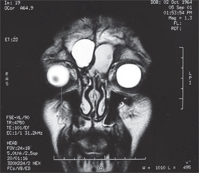
CASE REPORT

A male patient aged 37 years with a history of cranioencephalic trauma 21 years ago, reports a left frontal tumor beginning 4 months ago. Magnetic resonance imaging revealed a lesion suggesting mucocele (Figure 1). The patient underwent endoscopic bilateral frontal ethmoid sinusectomy. The option was made for external left frontal ethmoidectomy due to intense bone sclerosis in the frontal recess. The intersinusal septum was intact and was opened during the procedure. At 5 months follow-up there is no evidence of mucocele recurrence.

Figure 1.

Magnetic resonance image of paranasal sinuses, coronal section, T2. Bilateral Frontal Mucocele.

(0.06MB).
DISCUSSION

Mucoceles develop following obstruction of the drainage ostia, growing slowly within the sinuses, eventually eroding adjacent bone structures1. The etiology includes inflammatory processes, neoplasms, post-operative complications and post-traumatic sequelae2.

Mucoceles are mostly located in the frontal sinus, although bilateral involvement is extremely rare1,3. In literature only two such cases have been reported2,4.

The diagnosis is made based on the clinical history, physical examination and radiologic imaging. Symptoms vary in frontal ethmoid mucocele from absence of symptoms to incapacitating pain, headache and visual disturbances3.

Computed tomography shows an isodense homogeneous image, with loss of the normal sinus contour that is not enhanced with contrast if not infected3.

Magnetic resonance imaging is indicated if there is any doubt in diagnosis. The typical finding is a T1 hyposignal area T1 and a T2 hypersignal area, however any combination of signal intensities may be found depending on the presence of blood particles or the degree of hydration of the content5.

Currently ample marsupialization of the mucocele and simple drainage of the sinus by endoscope has been done with excellent surgical results. The advantages include low morbidity, low risk of complication and a rare recurrence rate6. The surgeon should have deep knowledge of frontal recess anatomy to perform endoscopic treatment of this condition.

FINAL COMMENTS

Endoscopic treatment seems to be the best treatment option. However, for technical reasons anatomical variants may preclude ample marsupialization done only endoscopically.

REFERENCES
[1]
Arrue P , Kany MT , Serrano E , Lacroix F , Percodani J , Yardeni E , Pessey JJ , Manelfe C .
Mucoceles of the paranasal sinuses: uncommon location.
J Laryngol Otol, 112 (1998), pp. 840-844
[2]
Santoro PP , Medeiros IRT , Queiroz E , Voegels RL , Butugan O .
Mucocele Frontal Bilateral.
Arquivos da Fundação Otorrinolaringologia, 3 (1999), pp. 14-18
[3]
Voegels RL , Balbani AP , Santos Junior RC , Butugan O .
Frontoethmoidal mucocele with intracranial extension: a case report.
Ear Nose Throat J, 77 (1998), pp. 117-120
[4]
Rao ABN .
A Case of Bilateral Complicated Frontal Mucocele.
Pract. Oto-Rhino-Laryngol, 28 (1966), pp. 117-120
[5]
Lloyd G , Lund VJ , Savy L , Howard D .
Optimum imaging for mucoceles.
J Laryngol Otol, 114 (2000), pp. 223-226
[6]
Har-El G .
Transnasal endoscopic management of frontal mucoceles.
Otolaryngol Clin North Am, 34 (2001), pp. 243-251

Paper submitted to the ABORL-CCF SGP (Management Publications System) on March 20th, 2005 and accepted for publication on May 1st, 2006.

Copyright © 2006. Associação Brasileira de Otorrinolaringologia e Cirurgia Cérvico-Facial
Idiomas
Brazilian Journal of Otorhinolaryngology (English Edition)
Article options
Tools
en pt
Announcement Nota importante
Articles submitted as of May 1, 2022, which are accepted for publication will be subject to a fee (Article Publishing Charge, APC) payment by the author or research funder to cover the costs associated with publication. By submitting the manuscript to this journal, the authors agree to these terms. All manuscripts must be submitted in English.. Os artigos submetidos a partir de 1º de maio de 2022, que forem aceitos para publicação estarão sujeitos a uma taxa (Article Publishing Charge, APC) a ser paga pelo autor para cobrir os custos associados à publicação. Ao submeterem o manuscrito a esta revista, os autores concordam com esses termos. Todos os manuscritos devem ser submetidos em inglês.