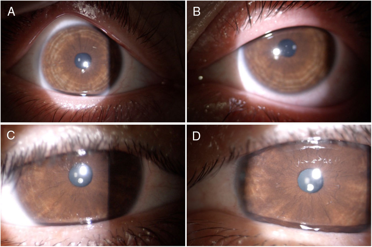
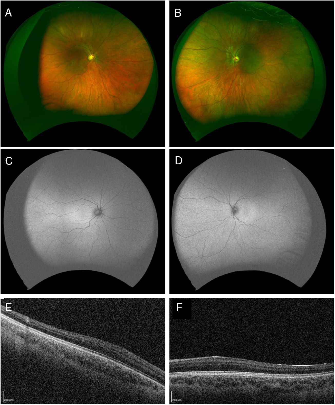
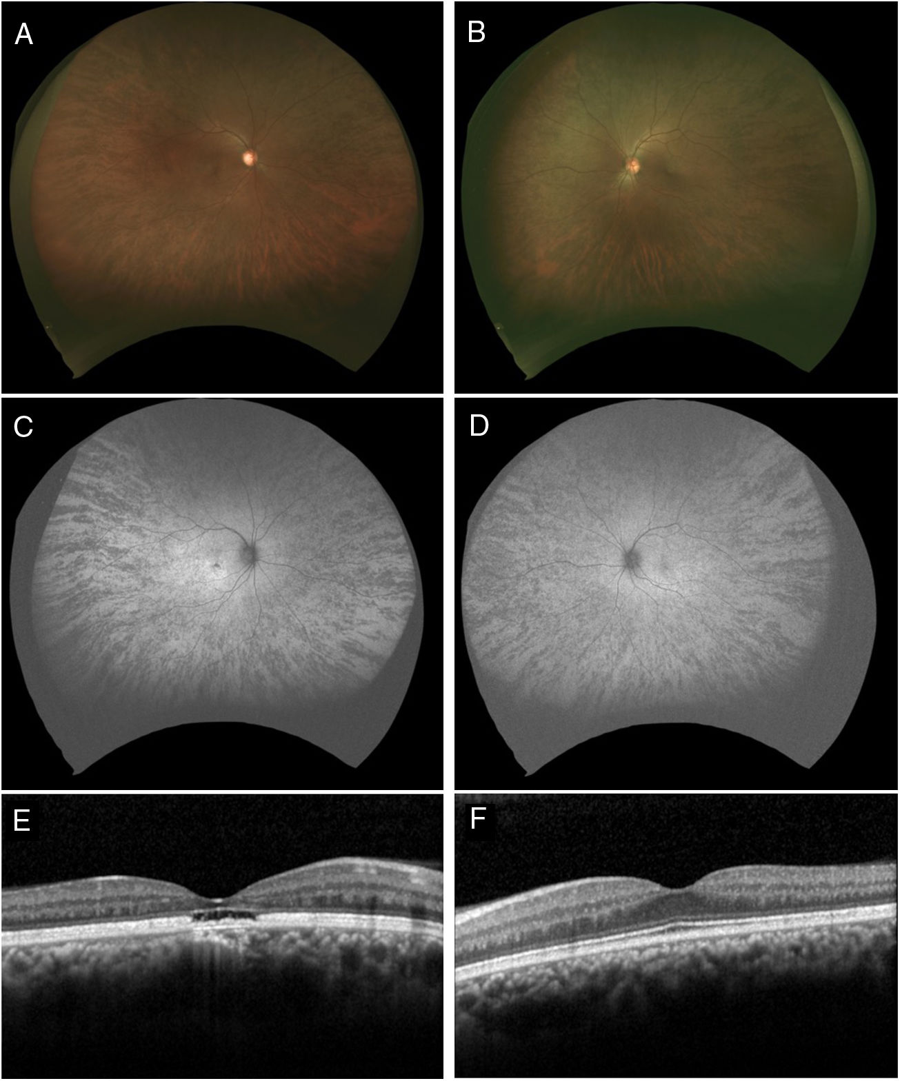
A 12 year-old boy who consulted due to nystagmus and low vision from birth. His mother also consulted for low vision of the right eye since she was a child, which worsened recently. The physical examination revealed no alterations in skin and hair pigmentation. In the examination of the anterior segment of the child, areas of slight circumferential hypopigmentation were observed in the iris in both eyes. The fundus examination revealed a choroidal fundus due to the absence of melanin in the retinal pigment epithelium. In the autofluorescence, an absence of physiological macular hypo-autofluorescence was observed and, in optical coherence tomography, foveal hypoplasia was observed in both eyes. In the ocular fundus examination of the mother, slight macular pigmentary changes were observed in the right eye, with hyperpigmented radiated spots in the retinal periphery of both eyes, which were hypo-autofluorescent in the wide-field autofluorescence. In the optical coherence tomography of the right eye, a cavitation of the outer retinal layers was observed in the fovea. The genetic study by nucleotide sequencing was performed on the mother and the child. In the mutation found in the GPR143 gene, the son was hemizygous and the mother was heterozygous. X-linked ocular albinism was diagnosed and the genetic counselling was carried out.
Ocular albinism linked to X is the most frequent genetic variant of this disease. Peripheral pigment alterations in heterozygous mothers have been previously described in the literature, but there are no reports of cavitations in the external retinal layers using optical coherence tomography.
Varón de 12 años que consulta por nistagmo y visión baja desde el nacimiento. Su madre también consulta por visión baja de ojo derecho desde pequeña que empeoró últimamente. El examen físico no reveló alteraciones en la pigmentación de piel y cabello. En el examen del segmento anterior del niño se observaron áreas de hipopigmentación circunferencial leve de iris en ambos ojos. El examen de fondo de ojo reveló un fondo coroideo por ausencia de melanina en el epitelio pigmentario retiniano. En la autofluorescencia se observa una ausencia de la hipoautofluorescencia macular fisiológica y en la tomografía de coherencia óptica se objetivó hipoplasia foveal en ambos ojos. En el examen de fondo de ojo de la madre se observaron cambios pigmentarios maculares tenues en el ojo derecho y unas manchas radiadas hiperpigmentadas en la periferia retiniana de ambos ojos que en la autofluorescencia de campo amplio se vieron hipoautofluorescentes. En la tomografía de coherencia óptica del ojo derecho se objetivó una cavitación de las capas externas retinianas en la fóvea. Se hace el estudio genético por secuenciación nucleotídica en madre e hijo y se encuentra una mutación en el gen GPR143: el niño fue hemicigote y la madre heterocigote. Se diagnostica albinismo ocular ligado a X y se realiza el aconsejamiento genético respectivo.
El albinismo ocular ligado a X es la variante genética más frecuente de esta enfermedad. Si bien las alteraciones pigmentarias periféricas en las madres heterocigotas fueron descritas en la literatura previamente, no existen reportes de cavitaciones en las capas retinianas externas por tomografía de coherencia óptica.