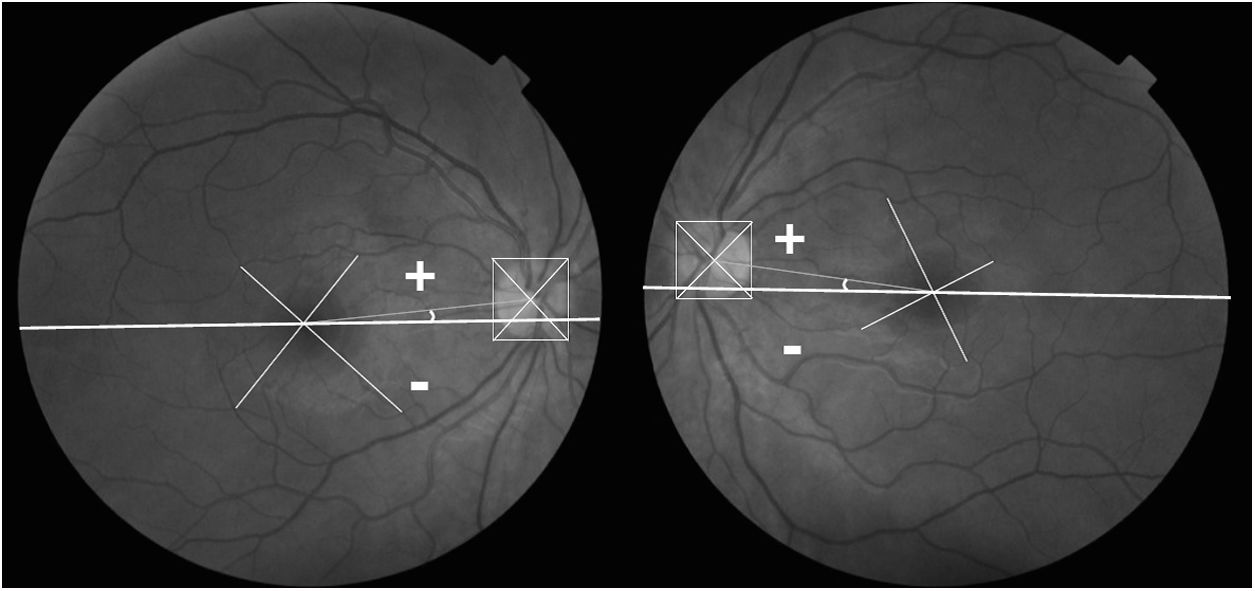
Describe a time-sparing technique to measure disc-foveal angle (DFA), determine normal values and its role when analyzing paired fundus photographs.
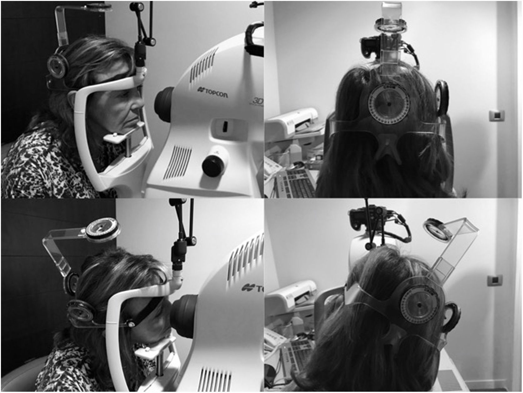
MethodsDFA was analysed using the software program Keynote V 6.2.2 on 440 fundus photographs (3D OCT 2000, Topcon) of 20 individuals. The 11 different head positions were determined with the cervical range of motion device (CROM, Performance Attainment Associates). A reproducibility and correlation study between two fundus cameras (OCT 3D-2000 and TRC-50EX, Topcon) was performed.
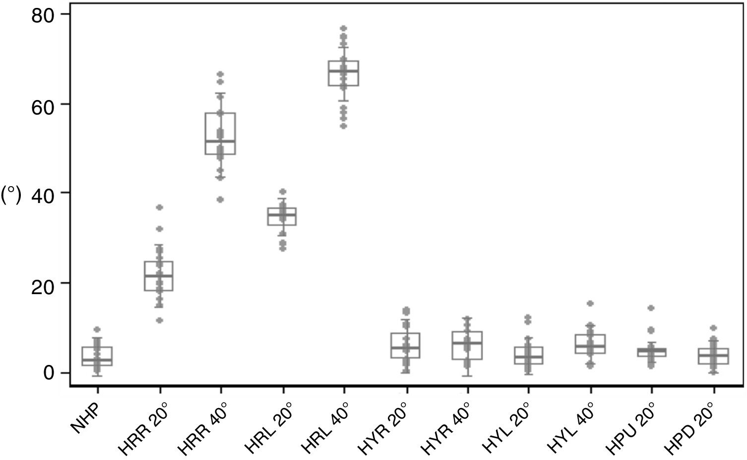
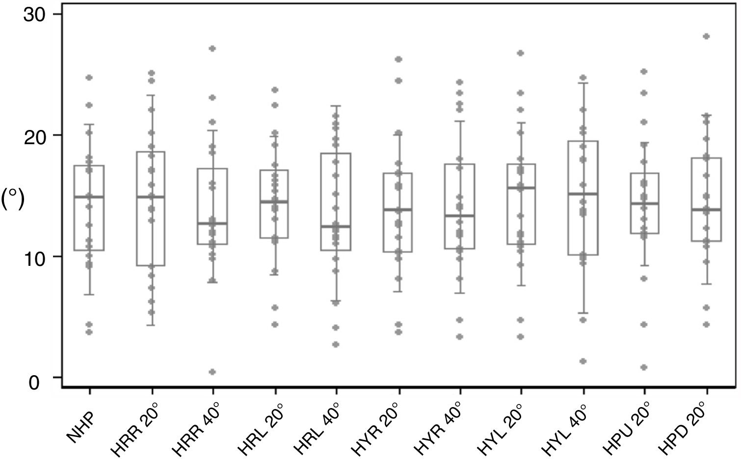
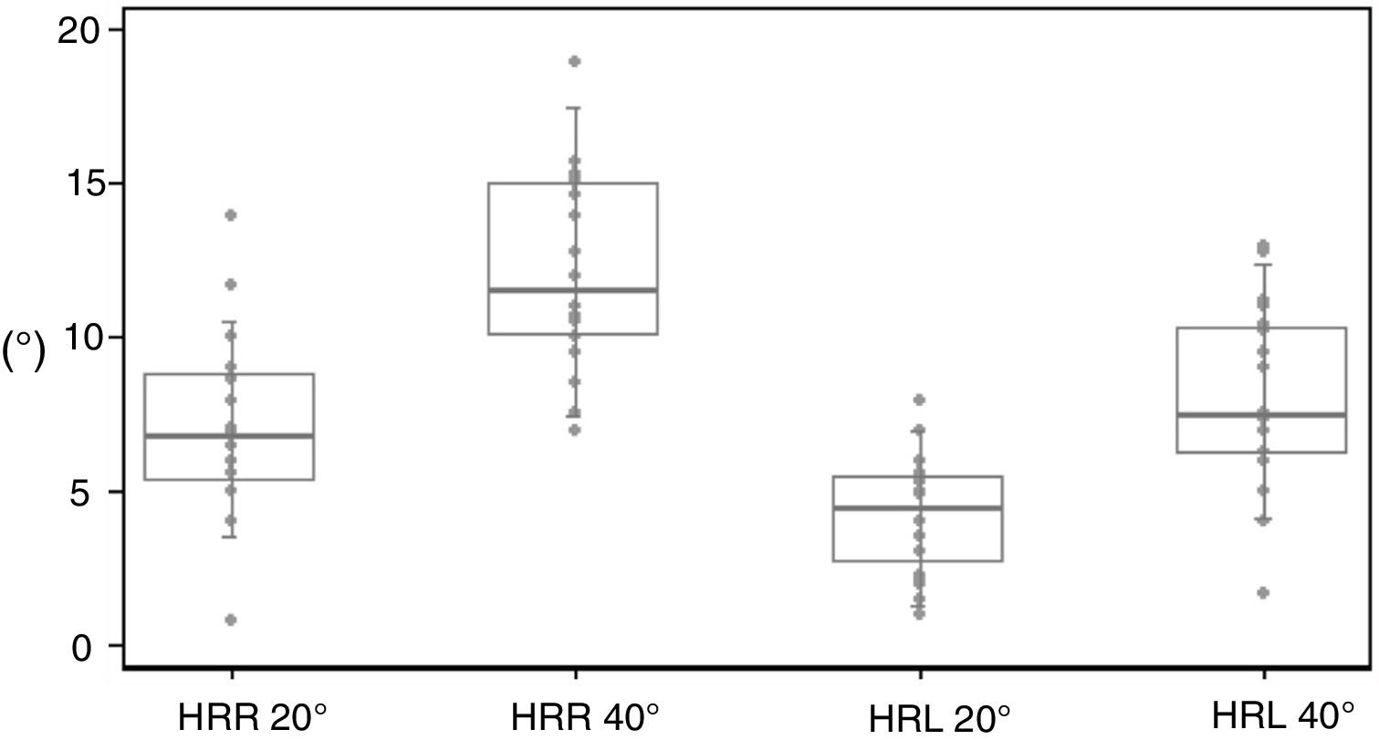
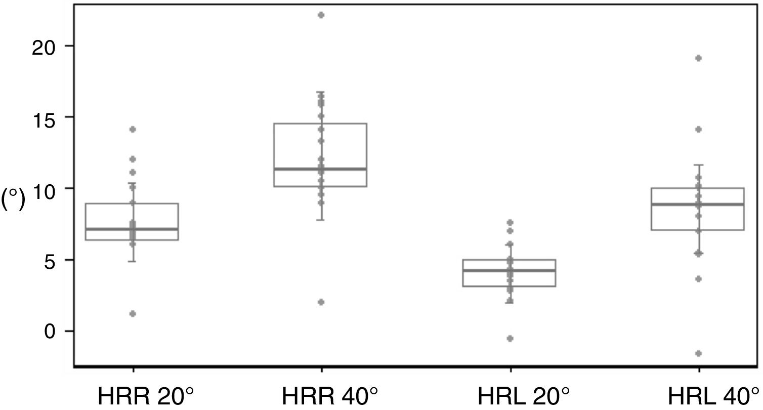
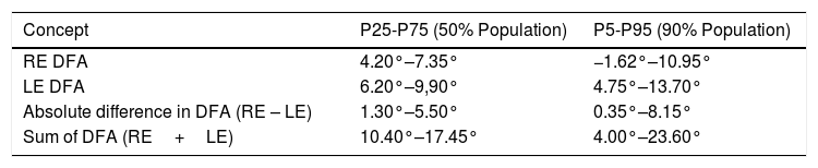
ResultsMean DFA of the right and left eye was 5.5±3.4° and 8.6±2.9°, with a difference of 3.1° (p=0.001 Wilcoxon signed-rank test) in the upright head position. Mean absolute difference in DFA between eyes was 3.5±2.6°; an increase was seen with increasing head tilt (p=0.000 Wilcoxon signed-rank test). Mean sum of DFA in both eyes was 14.1±5.4°. On head-tilt of 20° and 40° to the right, mean ocular counterrolling (OCR) was 7.1° and 12.2° in right eye and 7.7° and 12.1° in left eye. On head-tilt of 20° and 40° to the left, OCR was 4.4° and 8° in right eye and 4.2° and 8.7° in left eye (p=0.000 Wilcoxon signed-rank test). The two cameras showed strong correlation and high reproducibility.
ConclusionsOur DFA measurement technique is time-sparing and reproducible. Left eye shows higher disc-foveal angle than right eye. OCR occurs only in the roll plane. Absolute difference in disc-foveal angle between eyes changes according to head tilt, this information is of value when analyzing paired fundus photographs.
Describir una técnica rápida para medir el ángulo papila-fóvea (APF), determinar sus valores normales y su relevancia a la hora de analizar retinografías apareadas.
Métodosen 20 sujetos se realizaron 440 retinografías (3D OCT 2000, Topcon) en 11 posiciones diferentes de la cabeza (CROM, Performance Attainment Associates). Se analizó el APF mediante el software Keynote V 6.2.2 y se estudió la reproducibilidad y correlación entre los retinógrafos OCT 3D-2000 y TRC-50EX (Topcon).
ResultadosLa media del APF en el ojo derecho (OD) y en el izquierdo (OI) fue de 5.5±3.4° y de 8.6±2.9°, con una diferencia de 3.1° (p=0.001 test del signo-rango de Wilcoxon). La media de la diferencia absoluta del APF entre ambos ojos fue 3.5±2.6°; aumentando con la inclinación cefálica en el plano frontal (p=0.000 test del signo-rango de Wilcoxon). La media de la suma del APF de ambos ojos fue 14.1±5.4°. La media de la torsión ocular compensatoria (TOC) con la cabeza inclinada 20° y 40° a la derecha fue 7.1° y 12.2° en OD y 7.7° y 12.1° en OI. Con la cabeza inclinada 20° y 40° a la izquierda, la media fue 4.4° y 8° en OD y 4.2° y 8.7° en OI (p=0.000 test del signo-rango de Wilcoxon). Los retinógrafos mostraron alta correlación y reproducibilidad.
ConclusiónNuestra técnica es rápida y reproducible. El OI muestra mayor APF. La TOC sólo ocurre en el plano frontal. La diferencia absoluta de APF varía con la inclinación de la cabeza, aspecto relevante al analizar retinografías apareadas.