To compare corneal epithelial thickness (CET) between patients who underwent LASIK surgery for the correction of myopia at least one year ago and healthy subjects.
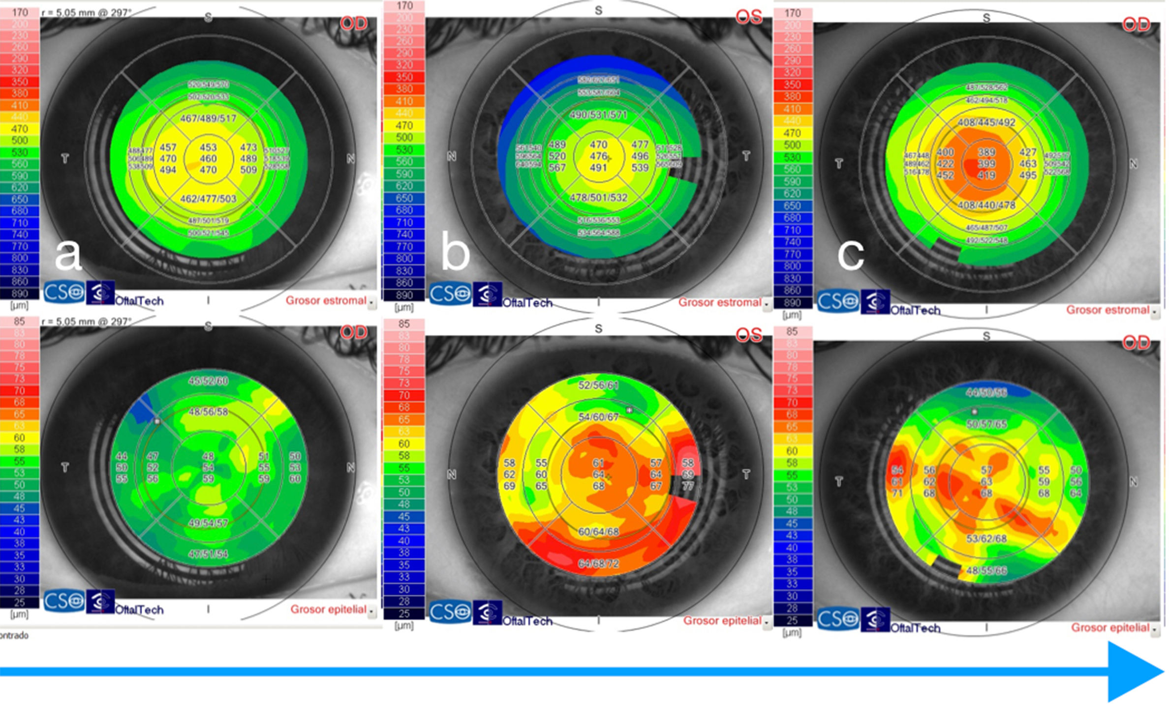
MethodsA retrospective observational study was conducted that included 93 healthy subjects (186 eyes) and 26 subjects (52 eyes) that underwent myopic LASIK surgery. OCT-SA, combined with Placido disk, was performed on all subjects, and CET maps were measured. Statistical analysis was performed to analyze differences between groups. Multivariate analysis was also performed to look for possible predictors of final CET.
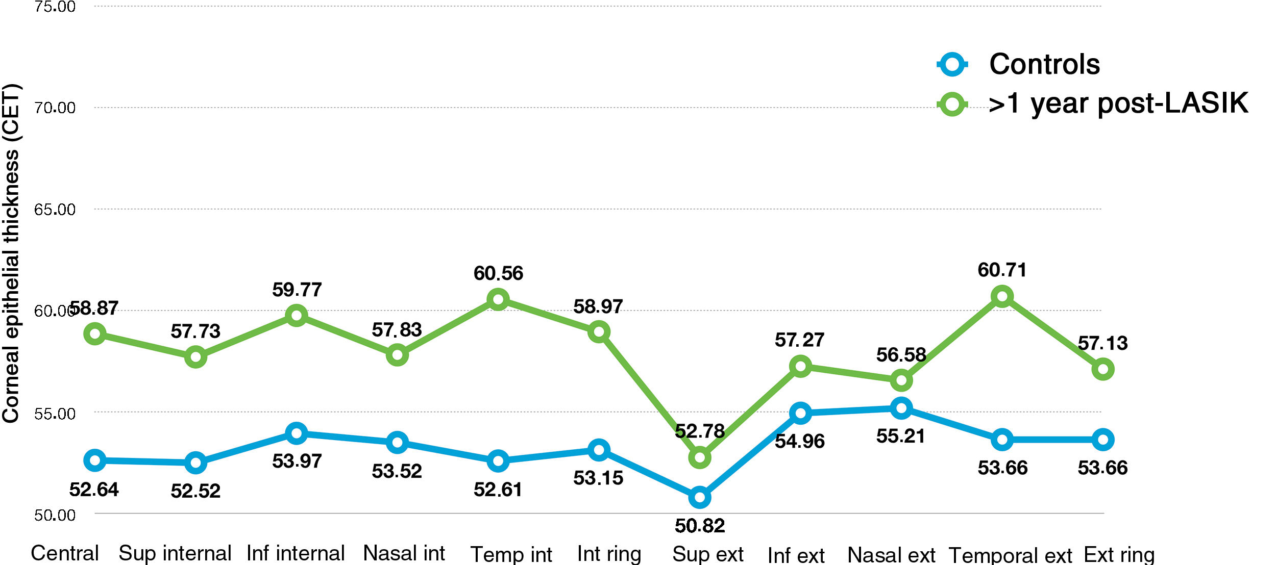
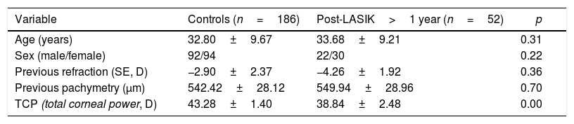
ResultsThere was no statistically significant differences between the groups in the demographic (age, sex) or anterior segment parameters (spherical equivalent, pachymetry) (all p>0.05). Statistically significant differences (p<0.05) were obtained between both groups when comparing CET, including central, internal, and external rings (higher in patients that underwent LASIK surgery ≥1 year). With the exception of the time elapsed since surgery (p=0.00), no correlation was found between the CET and age, sex, ablated dioptres, or other variables studied (p<0.05).
ConclusionsCET values measured by the OCT-SA were higher in patients that underwent LASIK surgery ≥1 year. The only variable that correlated with the CET after LASIK was the time elapsed since surgery. CET changes should be taken into consideration when planning refractive surgery due to its implications on the final outcome.
Comparar el grosor epitelial corneal (GEC) en pacientes intervenidos de LASIK miópico de más de un año respecto a sujetos no operados.
MétodosEn este estudio retrospectivo observacional se incluyeron 93 sujetos no operados (186 ojos) y 26 sujetos (52 ojos) operados de LASIK miópico. Se realizó una tomografía óptica de segmento anterior (OCT-SA) combinada con anillo de Plácido en todos los sujetos y se midió el GEC por sectores. Se hizo análisis estadístico para determinar diferencias entre las variables medidas en ambos grupos, así como análisis multivariante para buscar predictores de GEC.
ResultadosNo hubo diferencias significativas entre los grupos en términos demográficos (edad, sexo) ni de segmento anterior (equivalente esférico, paquimetría) (todas las p>0,05). Se obtuvieron diferencias estadísticamente significativas (p<0,05) entre los dos grupos en todos los sectores estudiados, central, anillos interno y externo, siendo mayores todos los valores de GEC en los pacientes intervenidos de LASIK ≥1 año. A excepción del tiempo transcurrido desde la cirugía (p=0,00), no se encontró correlación entre el GEC y la edad, el sexo, las dioptrías ablacionadas ni otra variable estudiada (p>0,05).
ConclusionesEl GEC medio y por sectores medido mediante la OCT-SA es mayor en pacientes intervenidos de LASIK hace más de un año. La única variable correlacionada con el GEC tras el LASIK es el tiempo desde la cirugía. Las modificaciones del GEC deben ser tenidas en cuenta al planear la cirugía refractiva por sus implicaciones en el resultado final.