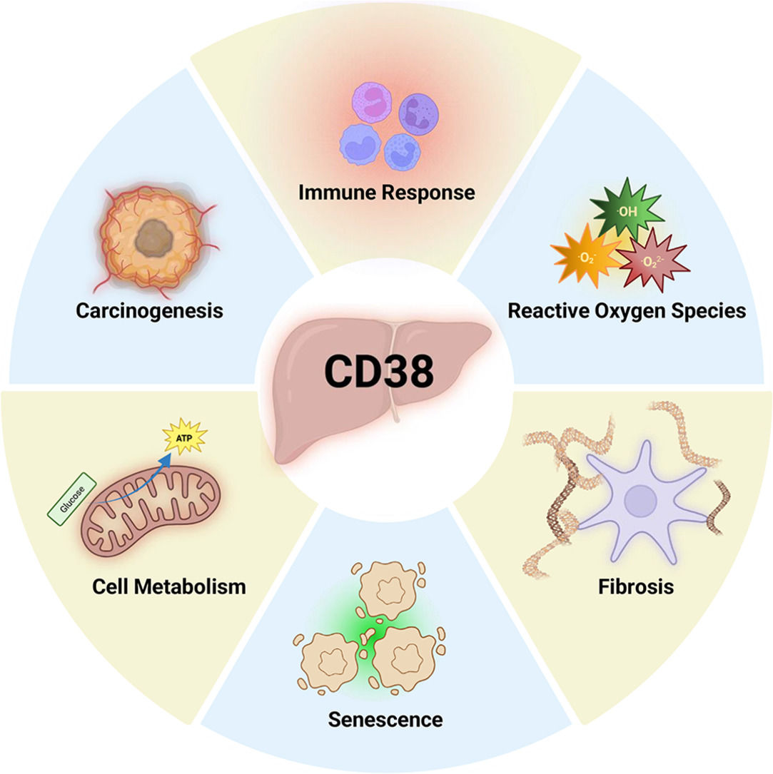

CD38 is a ubiquitously expressed 46 kDa transmembrane protein with both enzymatic and receptor functions [1,2]. This protein has been extensively studied for its roles in NAD⁺ metabolism and immune cell regulation. Originally identified as an immune activation marker, it is now recognized as a regulator of numerous physiological and pathological processes. CD38 enzymatically degrades NAD⁺ and generates calcium-mobilizing second messengers including cADPR and NAADP [3,4]. NAD⁺ itself is a critical cofactor for enzymes such as sirtuins and PARPs involved in DNA repair, cell cycle, and apoptosis. By modulating NAD⁺ availability, CD38 can impact cellular replication, aging, and senescence across multiple tissues.
In recent years, the role of CD38 in non-immune tissues has attracted increased attention. In the liver, both hepatocytes and non-parenchymal cells express detectable CD38. It is involved in a broad range of biological functions, including energy metabolism, oxidative stress, inflammation, and tissue recovery. Importantly, dysregulated CD38 activity has been implicated in the pathogenesis of several liver diseases, including metabolic dysfunction-associated steatotic liver disease (MASLD), cirrhosis, and HCC. Given its dependence on tightly regulated NAD⁺ levels for metabolic and regenerative processes, hepatic CD38 has emerged as a potential driver of disease progression. Indeed, its expression and activity are upregulated in chronic liver injury, indicating a link between CD38 and mechanisms of hepatic dysfunction and fibrosis [5]. Furthermore, age-related increases in CD38 expression across multiple tissues directly impact DNA and telomeric damage; however, the exact mechanisms by which CD38 contributes to this process remain unclear [6]. Recently, the orientation of CD38, which can adopt extracellular (type II) or intracellular (type III) topology with its enzymatic subunit, has emerged as an area of significant interest. While the mechanisms which dictate the differing conformations remain unknown, they have implications for cellular function since NAD+ is greatly concentrated within the cytosol, and the type III conformation more directly impacts intracellular signaling activity by catabolizing NAD+ into Ca2+ second messengers. These studies highlight a central role of CD38 in liver disease development and provide support for further investigation into its various functions.
This review aims to provide a consolidated overview of CD38 in liver biology, including its expression across hepatic cell types, its contributions to physiological liver function, and its involvement in the progression of acute and chronic liver diseases. We also explore emerging therapeutic strategies targeting CD38 which could restore NAD⁺ homeostasis, as well as mitigate liver injury. Lastly, we will highlight several outstanding questions and discuss avenues for future investigation which could lead to a better understanding of its mechanistic role in hepatic health and disease.
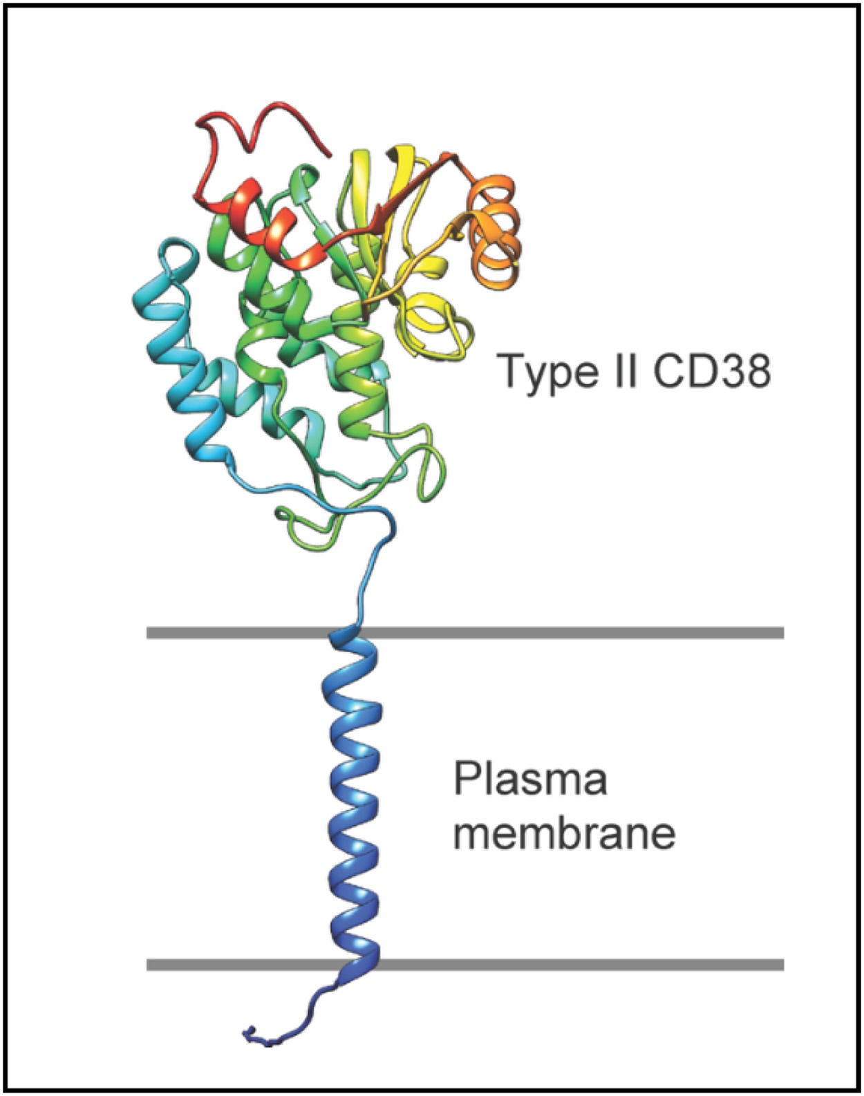
1.1Basic molecular biology of CD38 and its role in the liverInitially identified as a leukocyte cell surface receptor, CD38 is now recognized as a broadly expressed molecule found across different tissue types. Structurally, a 20-amino acid N-terminal domain, a short transmembrane region, and a larger 245-amino acid C-terminal domain comprise the main portions of CD38. Although the N-terminus lacks catalytic function, it participates in stabilizing the protein and orienting it within the membrane [7,8] (Fig. 1). It also acts as a cytoplasmic scaffold, interacting with other proteins such as Lck, to propagate intracellular signaling cascades that induce inflammatory cytokine transcription including IL-2 and IL-6 [9]. The C-terminal domain, by contrast, exhibits both enzymatic and receptor functions. The latter includes interactions with CD31, a molecule involved in immune cell activation and trans-endothelial migration. This process relies on mechanical adhesion and tethering by leukocytes to arrest on the luminal surface of vessels before entering injured or infected tissues. Multiple immune cells, including neutrophils, DCs, and lymphocytes, utilize the CD38-CD31 interactions to facilitate endothelial translocation [10]. Pathological conditions such as multiple myeloma also exploit this interaction to maintain malignant cells within the bone marrow niche [11]. Anti-CD38 therapies, like daratumumab, disrupt these interactions, displacing myeloma cells from the stromal environment and enhancing their sensitivity to agents like bortezomib [12].

A model of full-length CD38. The classic type II orientation of CD38 is oriented with the principal C-terminal, catalytic domain towards the extracellular space. Several residues including Glu226, Trp125, and Trp189 form the enzymatic subunit, while Glu146 regulates the hydrolase and cyclase activities [108]. The transmembrane and N-terminal domains have no known enzymatic function. Generated using AlphaFold.
While calcium signaling is required for CD38-mediated extravasation, its enzymatic activity may not be essential for this process. Frasca et al. [13] showed that DC migration persisted despite treatment with inhibitors of downstream CD38 s messenger signals. In neutrophils, intracellular Ca2+ influx continued despite blockade, implying that CD38 can influence calcium signaling through multiple mechanisms. Additional evidence from lamina propria T cells, which lack PLC and do not induce calcium-dependent pathways, suggests alternative signaling pathways operate in tandem [14]. These cells employ poorly defined adapter proteins and scaffolding complexes to initiate intracellular responses, further underscoring the cell-type specificity of CD38 signaling.
Despite decades of study, novel functions for CD38 continue to emerge. These varied responses are shaped by its subcellular localization, its capacity to interact with membrane-associated proteins, and its topological orientation. Two principal membrane bound topologies have been identified for CD38: type II and type III. The more well-studied type II topology projects its catalytic site extracellularly, allowing it to function as a receptor in addition to an NAD+glycohydrolase [15]. In this orientation, CD38 serves as a scavenger of extracellular NAD+, likely released from nearby dying cells, to produce precursor substrates and act as a danger sensor for injured or damaged tissues [16]. This conformation also facilitates lateral interactions with other proteins, including T and B cell receptors, CD203/CD73, and CD16, enabling the formation of highly specific signaling complexes [17]. Non-enzymatic ligand binding, inflammatory stimuli and cellular stress, such as ROS, can trigger CD38 internalization via clathrin-mediated endocytosis [18]. Within the acidic milieu of endolysosomes, CD38 generates NAADP from NADP and nicotinic acid. NAADP, a potent calcium-mobilizing metabolite, activates two-pore channels to release endolysosomal calcium, which initiates calcium-induced calcium release from the endoplasmic reticulum (ER) [19,20]. Endosomal calcium further directs cell migration through autophagy, in particular the formation of autophagolysosomes [21]. Inflammatory chemokines such as IL-8 also participate by stimulating CD38 internalization and increasing endolysosomal uptake of NADP into vesicles for NAADP synthesis. Disruption of this axis impairs cell motility [22]. Moreover, inhibition of CD38 and its adapter protein LRRK2 suppresses LPS-induced inflammation, reinforcing the importance of CD38 in endosomal activity [23].
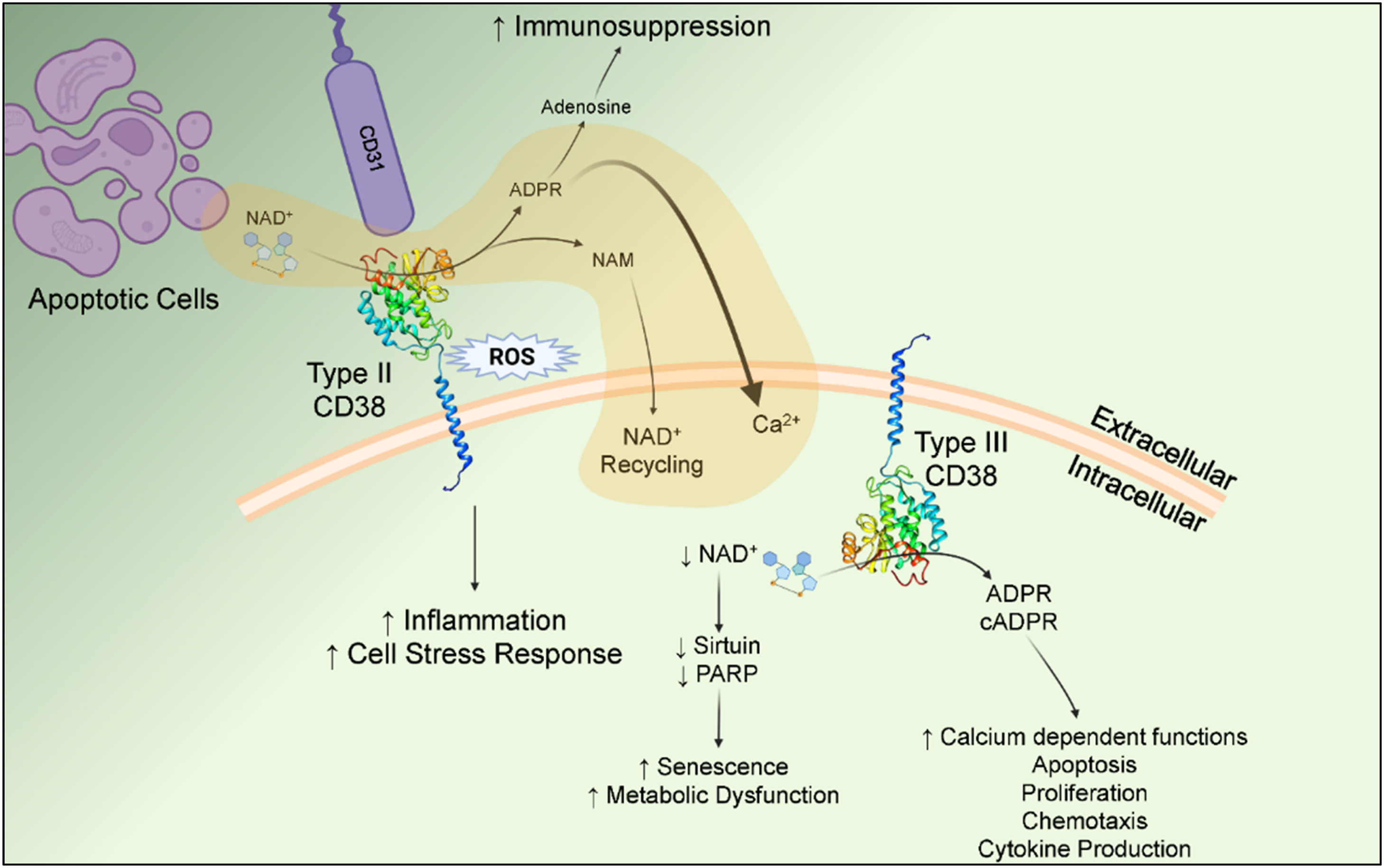
While type II CD38 is involved in extracellular interactions, the less abundant type III arrangement orients the catalytic domain into the cytoplasm, granting access to intracellular NAD⁺. Type III CD38 is favored when the N-terminal domain adopts a negative charge, which can be achieved experimentally through mutation of positive residues to negative residues in the N-terminal tail, or endogenously in cells by posttranslational phosphorylation of serine residues [7]. Interactions with chaperones, including heat shock proteins (HSPs), have been proposed as critical regulatory steps in establishing and maintaining the type III membrane topology of CD38. Consistent with this model, siRNA-mediated knockdown of Hsp90 and Hsp40 results in decreased type III CD38 protein stability and steady-state expression. As it stands, how these complexes direct type II versus type III conformations and their insertion into the membrane remains speculative [24,25]. The type III conformation resolves longstanding questions about how interactions with cytoplasmic NAD+, where concentrations of ∼400 µM are observed, occur when the predominant form of CD38 projects its catalytic domain into the extracellular space [26]. NAD⁺ does not cross the plasma membrane and low concentrations (0.1–0.5 µM) are observed extracellularly [27]. In this orientation, CD38 catalyzes adenosine diphosphate ribose (ADPR) formation and, through its cyclase activity, cADPR, which modulate intracellular calcium dependent functions including apoptosis (Fig. 2). These effects are fine-tuned through interactions with cytosolic partners such as CIB1, which stabilizes the enzyme’s active conformation to enhance NAD⁺ hydrolysis [28]. Although ADPR is the dominant product produced by CD38, cADPR is also a potent signaling molecule which activates ryanodine receptors on the ER to mobilize Ca2+. ADPR, on the other hand, targets TRPM2 channels on the plasma membrane, facilitating extracellular Ca2+ influx. Though smaller in magnitude, extracellular influx synergizes with cADPR-driven ER calcium release to amplify signaling [29]. TRPM2 channels also exhibit slower activation rates compared with the rapidly activating ryanodine receptor channels, suggesting that they contribute to more sustained signaling [30]. Oxidative stress can likewise activate type III CD38 [31,32]. Intracellularly, other membrane associated proteins including Nox4 and phosphorylated-p22phox interact with CD38. The Nox4-CD38 network promotes several functions including immune cell activation and endothelial inflammation. In fact, inhibition of CD38 has been shown to improve endothelial nitric oxide synthase activity and restore vascular integrity in the brain [33]. As illustrated in Fig. 2, these nuanced activities together allow for tightly controlled and robust intracellular responses.

Cellular responses regulated by type II and type III CD38. Oriented toward the extracellular space, type II CD38 possesses both enzymatic and receptor-ligand functions. Its enzymatic function includes scavenging NAD+(and NADPH) released from surrounding apoptotic cells to facilitate transport and recycling of nicotinamide substrates and promote Ca2+ dependent signaling. As a receptor, type II CD38 can interact with CD31 or internalize in response to ROS, leading to upregulation of inflammatory mediators and cytokines. Conversely, type III CD38 regulates intracellular NAD+pools and directs cellular processes involved in senescence, cell metabolism, proliferation and survival. Generated with BioRender.
Soluble forms of CD38 are also found in different settings [34]. These forms retain enzymatic activity, as evidenced by their ability to catalyze cADPR synthesis. Circulating, soluble CD38, generated by proteolysis of the type II form at Arg-58, contributes to immune regulation including long-term memory cell maintenance [35,36]. Although its pathophysiological significance remains unclear, circulating CD38 may also paracrine signal to other cell types to regulate processes such as cellular senescence. Additional speculation hints that soluble CD38 helps recycle extracellular NAD⁺ back into cells in the form of NAM via transporters such as SLC proteins [37]. Overall, the presence of CD38 in membrane-bound, cytosolic, and extracellular forms highlights its diverse biological roles [34] and its potential as a biomarker for inflammation and tissue damage.
The amount of CD38 is dynamically regulated in response to tissue injury and repair. Inflammatory signals serve as the primary stimulus that enhances CD38 production. For instance, angiotensin II upregulates CD38 via ROS-mediated secondary signaling pathways, such as STAT1 activation, which enhances gene expression in vascular smooth muscle cells [38,39]. The angiotensin II receptor also generates additional ROS through NADPH oxidase, thus promoting a positive feedback loop to sustain CD38 transcription [40]. In cardiovascular tissues, studies demonstrate that hypoxia further induces CD38, such as in response to ischemia-reperfusion injury (IRI) in endothelial cells (ECs) [31]. Additional regulatory checkpoints further influence protein stability, function, and turnover, including post-translational modifications and miRNA interactions [41,42].
In the liver, CD38 is expressed across multiple cell types including hepatic stellate cells, sinusoidal endothelial cells, hepatocytes, Kupffer cells, and infiltrating lymphocytes, but is notably absent in cholangiocytes [43]. Although some studies report low hepatocyte transcription, transcriptomic and proteomic analyses confirm the functional presence in these parenchymal cells [44,45]. Like other tissues, CD38 governs hepatic calcium signaling and NAD⁺ metabolism. Studies show that it modulates glucose metabolism in the liver by regulating gluconeogenesis. CD38-activated calcium signaling results in translocation of CREB to the nucleus, which promotes transcription of gluconeogenic regulators including phosphoenolpyruvate kinase. Further investigation revealed that CD38 also localized to the inner nuclear membrane where it participated in hepatic gluconeogenesis regulation by activating calmodulin kinase II and IV [46,47]. NAD+ shuttling into the nucleus is required for this process and can be abolished by preventing TRPM2-mediated calcium influx. As a result, CD38-deficient mice show improved glucose tolerance, suggesting that increased CD38 impairs metabolic regulation [48]. Treatment with CD38 inhibitors restores glucose sensitivity leading to decreased insulin secretion and lower serum glucose levels [49]. Other strategies for targeting CD38, including vaccination models, have shown efficacy in inhibiting age-related NAD+ decline and diminishing glycolysis in the liver [50]. Other metabolic processes, including hepatic thermogenesis, may also rely on CD38 activity. In turn, these pathways can impact lipogenesis in an organism leading to alterations in fat storage and obesity [6]. These studies underscore the influence of CD38 on multiple processes involved in both hepatic and systemic metabolism.
2CD38 in liver pathobiology2.1Inflammatory conditionsThe role of CD38 is perhaps most clearly defined within the context of inflammation [51]. CD38 not only coordinates broader responses through intercellular interactions in the surrounding microenvironment but also contributes to immune responses by working with other signaling molecules to activate myeloid cells and lymphocytes. Ligand binding and internalization of the receptor into lysosomes further enhance inflammatory cascades by generating NAADP and releasing calcium depots. The acidic calcium stores are essential for the activation and propagation of inflammasomes mediated by NLRP3 upregulation [52]. In addition to direct signaling, CD38 modulates the immunological environment by influencing adenosinergic pathways. Its enzymatic byproducts, and particularly ADPR, can be further metabolized by extracellular CD203 and CD73 to generate adenosine, a potent immunosuppressive molecule [53]. This function is particularly relevant in the tumor microenvironment, where accumulated adenosine inhibits anti-tumor immune responses.
Despite these interactions, the extent to which CD38 directly contributes to liver inflammation remains unclear. Autoimmune hepatitis (AIH), primary biliary cholangitis (PBC), and primary sclerosing cholangitis (PSC) represent inflammatory liver diseases initiated by immune recognition of hepatic antigens, either broadly, as in AIH, or specifically within the biliary epithelium, as in PBC and PSC. Other liver conditions such as alcoholic hepatitis or drug-induced liver injury also depend heavily on immune-mediated damage [54]. Transcriptomic analysis of the immune infiltrate in several autoimmune liver conditions demonstrates enrichment of CD38+ T cell populations [55]. In AIH, elevated hepatic CD38 expression correlates with the presence of cirrhosis, suggesting its potential as a biomarker for advanced fibrosis [56]. Beyond hepatocytes, CD38 activity plays a crucial role in the recruited immune cells within the liver. Notably, CD38+ IL-15–producing T lymphocytes have been observed infiltrating liver tissue across multiple diseases, though predominantly in viral hepatitis. Secreted IL-15 from these cells further enhances tissue damage by expanding cytotoxic T cell pools and leads to fibrosis progression [57]. Similarly, PBC is characterized by the presence of CD38-high plasma cells localized around bile ducts in patient biopsies [58]. In vitro studies have shown that these plasma cells suppress regulatory T cell function and promote Th1 polarization, thereby intensifying inflammatory damage to the biliary epithelium [59]. Collectively, these observations underscore the role of CD38-expressing immune cells in liver inflammation and fibrosis across a spectrum of autoimmune conditions.
2.2CirrhosisCirrhosis is characterized by prominent fibrosis and hepatic dysfunction, and represents the final stages of chronic liver injury [60]. It arises from a variety of etiologies, including chronic alcohol consumption, viral hepatitis, steatohepatitis, or inherited metabolic disorders such as hemochromatosis. The development of fibrosis in end stage liver disease involves the persistent activation of immune cells and stellate cells, leading to extracellular matrix deposition, hepatocyte turnover, and the formation of fibrotic nodules. While the role of CD38 in hepatic fibrosis remains unclear, evidence from other organ systems demonstrates that CD38 can promote fibrogenesis [61,62]. CD38-mediated NAD⁺ depletion downregulates sirtuins and other senescence-related molecules such as p16 [63], and in turn, sensitizes fibroblasts to profibrotic cues like TGF-β [64].
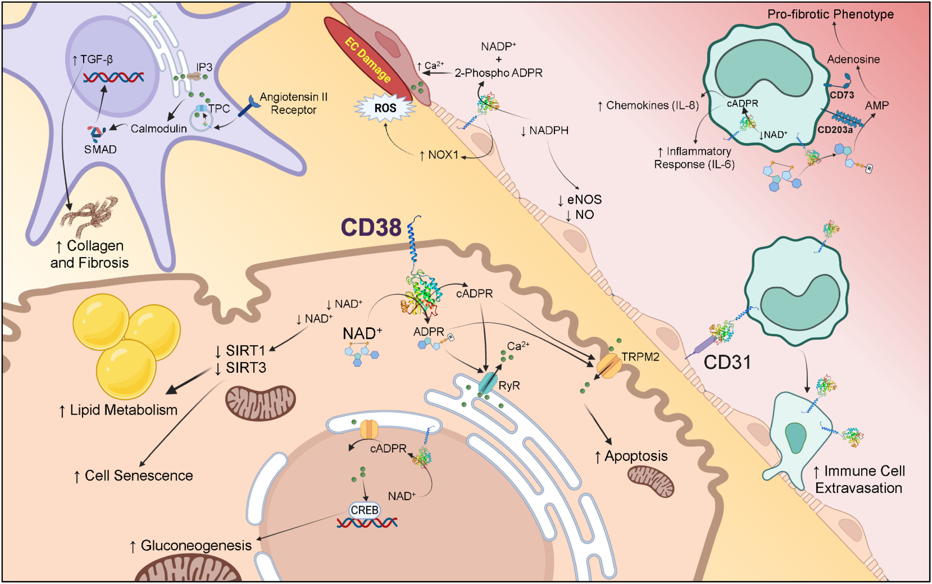
In vivo, CD38 expression is increased in liver fibrosis models. For instance, elevated CD38 transcripts were observed in cirrhotic rats induced by thioacetamide [65]. Although the producing cells were initially unknown, subsequent studies deduced that hepatic stellate cells were responsible for the upregulated CD38 which paralleled fibrosis severity. Similar findings have been observed in liver biopsies from patients with HCV. Mechanistic experiments further revealed that CD38 activation triggers intracellular calcium release, leading to the production of inflammatory mediators such as IL-6, VCAM1, and ICAM-1, which support immune cell recruitment, activate stellate cells, and induce fibroblast differentiation to collagen-depositing myofibroblasts. These findings suggest that CD38 not only contributes to inflammatory damage, but also directly regulates fibrotic remodeling leading to end-stage liver disease (Fig. 3).

CD38-regulated pathways in liver injury and fibrosis. CD38 is present on nearly all cells within the liver. In hepatocytes, it can promote cell senescence, increased lipid storage, gluconeogenesis and cellular apoptosis through Ca2+ mediated signaling pathways. Another hallmark of chronic liver disease, EC dysfunction, induced by CD38, can downregulate nitric oxide production and increase ROS leading to cellular damage. Inflammation triggered by CD38 engagement on infiltrating immune cells further facilitates liver injury and fibrosis, whereas hepatic stellate cells can differentiate into myofibroblasts leading to increased collagen and extracellular matrix deposition that enhances liver cirrhosis. Generated with BioRender.
A leading cause of cirrhosis, MASH has been closely associated with CD38 activity. CD38-knockout mice exhibit resistance to obesity and display reduced severity in steatohepatitis, indicating a protective role against MASH development [66]. Systemic metabolic disorders, including diabetes, dyslipidemia, and obesity are known drivers of lipid accumulation in the liver and MASH progression. Cardiovascular disease, in particular, is a closely linked comorbidity and in recent studies, myocardial infarctions also accelerated MASH through the systemic release of inflammatory chemokines that promoted myeloid cell recruitment to the liver, an effect that may be mediated in part by CD38-dependent pathways [67,68]. In metabolic liver disease, CD38 contributes to both immune cell activation and the depletion of NAD⁺ which participates in lipid metabolism. The consequences of decreased NAD+ are the impairment of other enzymes, such as sirtuins, which utilize it as a catalytic substrate. In one report, knockout of the NAD+ dependent deacetylase, Sirt1, resulted in increased hepatic steatosis in high fat diet mice due to impaired fatty acid oxidation resulting from diminished PPARγ [69]. Likewise, decreased NAD+ impacts the function of PARP-1, an enzyme involved in DNA repair. Recent work from Guo et al. interrogated the effects of NAD+loss in lipotoxicity. The authors revealed that NAD+ dependent PARP-1 activity was crucial for protecting hepatocytes from apoptosis induced by palmitate-mediated TLR4 signaling [70].
Beyond the liver, CD38-driven NAD⁺ depletion affects other metabolic tissues that participate in MASH pathogenesis. The conversion of white adipose tissue to energy-burning brown fat is diminished when CD38 activity is increased in adipocytes, further exacerbating any underlying metabolic syndrome [71]. These combined effects highlight the potential ways in which CD38 links metabolic and inflammatory processes that drive MASH-related fibrosis and cirrhosis. Similar mechanisms have been observed in alcohol-induced liver injury, where alcohol binging in aged mice increased hepatic CD38 expression, promoted fibrosis, and induced CD38+T cell infiltration [54]. In these experiments, pharmacologic inhibition of CD38 with the small molecule 78c reduces liver injury, partly by suppressing fatty acid synthase and restoring Sirt1 activity [49]. Though further research is needed to define its cell-specific functions and full therapeutic potential in cirrhosis, these data highlight the contributions of CD38 to chronic liver injury and fibrosis.
2.3Acute liver failure and acute-on-chronic liver failureThe role of CD38 in ALF and ACLF remains poorly understood. Both conditions are characterized by rapid deterioration of hepatic function, often progressing to multi-organ failure with high mortality rates [72]. ALF refers to a condition associated with irreparable organ dysfunction due to severe, acute injury to the liver such as APAP toxicity, exposure to hepatotoxic agents, or vascular compromise from thrombosis in individuals with no underlying liver disease. On the other hand, ACLF broadly involves patients with underlying liver conditions and advanced tissue fibrosis. Decompensation occurs in the context of cirrhosis and is commonly precipitated by acute insults such as infection, hemorrhage, or toxic exposures, including alcohol and APAP. Due to clinical severity, affected patients frequently require urgent liver transplantation to survive. Although liver dysfunction triggers the initial decompensation, ALF and ACLF patients evolve extrahepatic organ complications which can lead to profound renal, pulmonary and cardiovascular compromise.
Over the past decade, accumulating evidence has implicated CD38 in the pathogenesis of sepsis in both preclinical models and critically ill patients. For example, Peng et al. [73] found that pharmacological inhibition of CD38 attenuated organ damage in experimental sepsis. Since infection is the most common precipitant of ACLF, accounting for nearly half of all inciting events, CD38 may contribute mechanistically to disease progression by amplifying immune dysfunction and promoting systemic inflammation in septic liver patients [74]. Surprisingly, Rah et al. [75] found that liver protection in response to lipopolysaccharide (LPS) exposure was mediated by autophagosome formation preventing apoptosis, which required NAADP-driven calcium signaling. Since CD38 has a role in regulating autophagosome function, these findings appear counter to much of the literature that suggests CD38 activity exacerbates liver injury. One possibility for the discrepancies may lie in experimental model differences; specifically, the D-galactosamine model employed by Rah et al. is dependent on intracellular Ca2+ levels [76]. Given the importance of CD38 in regulating calcium, its activity may affect the degree of cellular damage and cytotoxicity following D-galactosamine treatment, whereas cecal ligation and puncture models a polymicrobial infection with broader systemic inflammatory mediators. These divergent stimuli likely activate distinct immune and metabolic pathways, which may explain the conflicting roles of CD38 observed in each system.
NAD+ is critical for oxidative stress responses, mitochondrial function, and cellular repair in liver cells [6,77,78]. While the precise role of CD38 in ACLF remains poorly defined, its function as the primary NAD⁺-consuming enzyme in mammalian cells suggests that it influences disease trajectory through NAD⁺ depletion. Elevated NADH levels promote reductive stress and impaired cellular function in the liver such as during steatosis development [79,80], whereas elevated NAD⁺ availability provides protective and buffering capabilities against genotoxic insults. Although CD38 has not been directly linked to ALF, the pathological consequences of NAD⁺ depletion are well established in this condition. In APAP-induced ALF models, NAD⁺ decreases precipitously in tandem with worsening hepatic function. Experimental approaches aimed at restoring NAD⁺, by supplementation with precursors or inhibition of catabolic enzymes such as NAD kinase, have resulted in marked improvement [81]. Similarly, in murine models of partial hepatectomy, NAD⁺ augmentation enhances liver regeneration by supporting DNA synthesis and repair enzyme activity [82].
These observations underscore the potential for targeting CD38 as a means of modulating NAD⁺ and its availability in acute liver damage. In Zhao et al., blocking CD38 on myeloid cells improved bile duct damage and fibrosis in ischemic cholangiopathy by suppressing NAD⁺/Sirt1 signaling and attenuating pro-inflammatory polarization of liver-infiltrating monocytes and macrophages. In the study, CD38 ablation led to improvement in bile ducts and cholangiocyte recovery following IRI both in vitro and in vivo. Interestingly, only the myeloid cells appeared to have any impact on biliary injury progression despite multiple other cell types expressing CD38. These results suggest that the timing of the depletion, the cellular localization of CD38 (i.e. plasma membrane vs nuclear localized), or the type II or type III conformation contributes to the effects [83]. Collectively, these findings emphasize the multifaceted nature of CD38 in liver injury and underscore the urgent need to elucidate new therapeutic tools in both acute and chronic liver failure.
2.4Hepatocellular carcinomaCD38 is increasingly recognized as a contributor to cancer development. In the liver, HCC is the most common malignancy and will be the focus of this section. Among other liver cancers such as cholangiocarcinoma, the role of CD38 is relatively unexplored and its contribution to disease progression is unclear. Conversely, several reports suggest that CD38 promotes HCC tumorigenesis through both tumor cell intrinsic and extrinsic mechanisms. Notably, CD38 expression is elevated in tumor-infiltrating immune populations such as immunosuppressive regulatory T cells and myeloid-derived suppressor cells. On effector T cells, increased CD38 levels correlate with immune exhaustion markers PD-1 and CTLA-4 and are associated with higher-grade HCC [84]. Similar patterns are observed in NK cells, where CD38 expression is linked to functional impairment. Clinically, HCC development in hepatitis C (HCV) patients strongly correlates with an expansion of CD38⁺ Tim-3⁺ NK cells compared with those lacking this subset. These CD38⁺ NK cells display reduced cytotoxicity, consistent with a diminished anti-tumor immune response [85]. Additionally, CD38 contributes to the production of adenosine which can dampen the tumoricidal functions of innate immune cells. Recent work demonstrated that silencing CD38 promotes the polarization of macrophages toward the cytotoxic M1 phenotype and fosters greater tumor control in HCC [86]. In fact, some reports suggest that CD38 expression on immune cells carries favorable prognostic implications. In one study, increased infiltration of CD38-positive immune cells was associated with improved responses to immune checkpoint blockade therapy. In this context, CD38 may serve as a therapeutic biomarker rather than a direct regulator of tumor growth [87]. Much like PD-1 expression or mismatch repair deficiencies, the presence of CD38 in tumor infiltrating immune cells may offer an avenue for therapeutic intervention that reshapes the inflammatory milieu. Collectively, these findings underscore the complex and context-dependent roles of CD38 in the immunologic and metabolic landscape of HCC.
Although the function of CD38 within HCC cells remains poorly characterized, the broader NAD⁺ network has been examined in liver cancer. NAD⁺ is a vital coenzyme required for DNA repair and the maintenance of genomic stability. One study demonstrated that NAD⁺ depletion, driven by hepatocyte expression of the unconventional prefoldin RPB5 interactor (URI), promotes HCC development. Supplementation with vitamin B3, a precursor in NAD⁺ biosynthesis, restored DNA repair capacity and prevented diethylnitrosamine-induced carcinogenesis in this model by enhancing L-tryptophan/kynurenine catabolism [88]. Similarly, NAD⁺-dependent enzymes such as Sirt7 were found to suppress HCC by deacetylating critical gene targets for preserving genomic integrity [89]. Restoration of NAD⁺ pools with lenvatinib also promoted apoptosis in HCC cells and synergistically re-polarized surrounding M2 macrophages, contributing to tumor control [90]. Together, these studies highlight another possible mechanism by which CD38 could regulate liver tumorigenesis through direct modulation of NAD⁺ homeostasis.
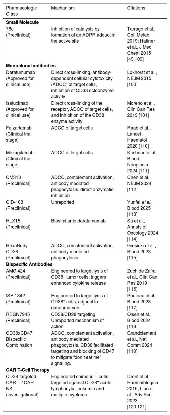
CD38 has attracted interest as a therapeutic target in HCC. Although the use of CD38-specific agents in liver cancer remains underexplored, preclinical studies using CD38-blocking antibodies or small-molecule inhibitors in other malignancies, such as hematologic cancers and breast cancer, show promising results [91,92]. Moreover, in vivo work has shown that daratumumab can bind HCC cells with high affinity [93] and could be worth exploring clinically. Currently, several compounds including small molecule inhibitors and monoclonal antibodies have entered varying stages of development and clinical use (Table 1). Exploratory studies with daratumumab and isatuximab have shown promise in liver cancer [94,95]. Tumors with high CD38 expression exhibit features of immune exhaustion but show enhanced responsiveness to immune checkpoint blockade. Several challenges remain in this arena, including our poor understanding of the heterogeneity in CD38 expression across HCC subtypes and our lack of insight into the role of CD38 in non-tumor cell populations such as fibroblasts, ECs or nerve cells. Continued investigation is needed to better develop CD38 as a therapeutic target in HCC.
Therapeutic agents targeting CD38. Multiple agents have been identified or generated that target different facets of the CD38 molecule. 78c remains one of the only available small molecules that inhibit CD38 enzymatic function in the presence of NAD+. Biologic agents and cell-based therapies principally target cells expressing CD38 for elimination.
| Pharmacologic Class | Mechanism | Citations |
|---|---|---|
| Small Molecule | ||
| 78c (Preclinical) | Inhibition of catalysis by formation of an ADPR adduct in the active site | Tarrago et al., Cell Metab 2019; Haffner et al., J Med Chem 2015 [49,109] |
| Monoclonal antibodies | ||
| Daratumumab (Approved for clinical use) | Direct cross-linking, antibody-dependent cellular cytotoxicity (ADCC) of target cells, inhibition of CD38 ectoenzyme activity | Lokhorst et al., NEJM 2015 [100] |
| Isatuximab (Approved for clinical use) | Direct cross-linking of the receptor, ADCC of target cells, and inhibition of the CD38 enzyme activity | Moreno et al., Clin Can Res 2019 [101] |
| Felzartamab (Clinical trial stage) | ADCC of target cells | Raab et al., Lancet Haematol 2020 [110] |
| Mezagitamab (Clinical trial stage) | ADCC of target cells | Krishnan et al., Blood Neoplasia 2024 [111] |
| CM313 (Preclinical) | ADCC, complement activation, antibody mediated phagocytosis, direct enzymatic inhibition | Chen et al., NEJM 2024 [112] |
| CID-103 (Preclinical) | Unreported | Yunfei et al., Blood 2025 [113] |
| HLX15 (Preclinical) | Biosimilar to daratumumab | Su et al., Annals of Oncology 2024 [114] |
| HexaBody-CD38 (Preclinical) | ADCC, complement activation, antibody mediated phagocytosis | Grosicki et al., Blood 2023 [115] |
| Bispecific Antibodies | ||
| AMG 424 (Preclinical) | Engineered to target lysis of CD38+ tumor cells; triggers enhanced cytokine release | Zuch de Zafra et al., Clin Can Res 2019 [116] |
| ISB 1342 (Preclinical) | Engineered to target lysis of CD38+ cells; adjunct to daratumumab | Pouleau et al., Blood 2023 [117] |
| REGN7945 (Preclinical) | CD38/CD28 targeting. Unreported mechanism of action | Olsen et al., Blood 2024 [118] |
| CD38xCD47 Bispecific Combination | ADCC, complement activation, antibody mediated phagocytosis. CD38 facilitated targeting and blocking of CD47 to mitigate “don’t eat me” signaling. | Grandclement et al., Nat Comm 2024 [119] |
| CAR T-Cell Therapy | ||
| CD38-targeted CAR-T / CAR-NK (Investigational) | Engineered chimeric T-cells targeted against CD38+ acute lymphocytic leukemia and multiple myeloma | Drent et al., Haematologica 2016; Liao et al., Adv Sci 2023 [120,121] |
The field of transplantation may represent the most compelling area of exploration for CD38 in hepatology. Its relevance in transplant immunology has already been explored in other organs, including the heart and kidney, where CD38 has emerged as a potential target for anti-rejection therapy. In kidney transplantation, seminal work by Mayer et al. [96] showed that targeting CD38 led to resolution of antibody mediated rejection (AMR). Follow-up molecular analyses confirmed that CD38 inhibition downregulated interferon and NK cell signatures [97]. Additional therapeutic benefit was also obtained through depletion of plasma cells, which promotes humoral rejection. Compared with other forms of anti-rejection therapy, CD38 targeting agents showed the strongest response in patients with moderate AMR scores but were less effective in severe rejection, indicating a potential therapeutic limitation. Similar outcomes have been reported in heart transplantation, where a patient with refractory AMR achieved resolution following daratumumab therapy without adverse effects [98]. Comparable responses have been described in lung transplantation [99]. Improved graft survival was noted with no significant increase in treatment-associated morbidity. Both daratumumab and isatuximab can inhibit CD38 enzymatic activity, likely in response to conformational changes in the folded structure from antibody binding [100,101]. The contribution of CD38 enzyme inhibition or cell-type depletion remains unclear as there are no studies which explore CD38 enzyme inhibition alone as a treatment strategy for rejection. Due to the lipid bilayer preventing passage of large molecules, targeting intracellular type III CD38 with these agents is further limited. Collectively, these studies indicate that CD38 blockade may be a viable addition to the therapeutic regimen against solid organ rejection.
Beyond rejection, CD38 potentially influences IRI, an ongoing challenge in liver transplantation. With growing use of extended criteria donors and donation-after-circulatory-death organs, IRI remains a pertinent issue despite increased use of normothermic perfusion which has reduced ischemic cholangiopathy risks [102]. Preclinical work in heart transplant first suggested a protective role for CD38 deficiency in IRI by reducing oxidative stress and attenuating Ca²⁺ overload resulting in smaller infarct sizes. The improvement in organ injury was secondary to enhanced SOD2 activity that prevents oxidative stress damage to the myocardial tissue [103]. Additionally, NADPH levels, which are directly regulated by CD38, diminished in ECs leading to dysfunction. NADPH depletion led to further ROS and vascular injury which impaired blood flow to the transplanted organ and perpetuated hypoxic insult [104]. Recent studies implicated myeloid CD38 as another vital contributor to IRI. Increased CD38 activity enhanced macrophage polarization towards an inflammatory phenotype. In this state, the innate cells produced cytokines such as TNF-α and IL-β which led to organ injury [83]. Further, SIRT1, whose activity is modulated by CD38, regulated myeloid pyroptosis during experimental orthotopic liver transplantation, whereas hepatocyte apoptosis during cold ischemia was driven by IL-8–dependent inflammatory signaling. Interestingly, IL-8 neutralization protected hepatocytes, though effects on immune recruitment were not assessed [105]. However, ongoing debate persists as several groups propose that elevated NAD+ in the liver instead exacerbates IRI progression. A recent report demonstrated that inhibition of NAMPT, an enzyme responsible for recycling NAD+ stores, in myeloid cells attenuated IRI in the liver [106]. The discrepancy in these findings may be related to alternative NAMPT functions beyond NAD+ recovery. When released extracellularly, NAMPT can induce systemic inflammatory responses along with TLR4 and function as a danger-associated molecular pattern leading to tissue damage [107]. Thus, a deeper dissection of these molecules is needed to understand their impact on disease states. Altogether, these findings highlight the complex post-transplant involvement of CD38 and NAD⁺ metabolism. Moving forward, the key challenge lies in developing therapies that not only deplete CD38-expressing immune populations but also modulate the CD38 enzymatic function within liver cells.
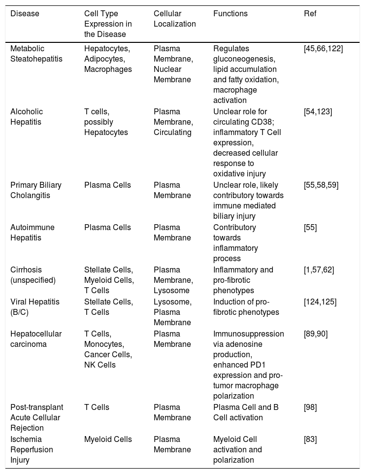
4ConclusionCD38 is a multifaceted protein and its role in liver disease continues to evolve as new mechanistic data emerge. It is now understood to be a key modulator of cellular NAD⁺ metabolism, calcium signaling, and inflammatory responses across various hepatic cell types. In addition, the connection between CD38, cellular senescence and aging likely plays a critical role in the liver, yet this aspect remains largely unexplored. Table 2 summarizes a list of diseases which involve CD38. Looking to the near future, targeted therapies hold the most promise in liver transplantation. By influencing cellular senescence and the immune landscape, strategies that alter CD38 signaling may prevent liver failure or preserve organ function. These multifaceted pathways create opportunities to improve patient outcomes at numerous stages including enhancing graft quality, preventing graft loss and decreasing post-operative complications. Given its role in jumpstarting regeneration in pre-clinical studies, CD38 directed therapies could be useful tools in repairing traumatic liver injuries or even enhancing organ growth following resection. Although work in kidney and heart transplantation has begun to establish therapeutic possibilities, studies focused specifically on liver transplantation remain scarce. The ability to selectively target the type III versus type II conformation of CD38 remains an area of ongoing investigation. While agents interacting with the extracellular catalytic ectodomain are more readily available, targeting the cytosolically-oriented type III CD38 conformation is complicated by multiple barriers, including membrane impermeability, active efflux via ABC transporters, endosomal/lysosomal entrapment, and cytosolic metabolic inactivation. Overcoming these barriers will require pharmacologic optimization of existing agents such as 78c or carrier-based strategies such as liposomal formulations to facilitate cytosolic access. Realizing the therapeutic potential of CD38 will require further mechanistic investigations and targeted translational studies across the spectrum of liver disorders.
Summarization of liver disease states associated with CD38 expression. Several liver conditions have been associated with CD38 activity. CD38 expression is present on multiple cell types and localization of the receptor within the cell structure dictates functional responses. Published studies have identified its role in these various disease states.
| Disease | Cell Type Expression in the Disease | Cellular Localization | Functions | Ref |
|---|---|---|---|---|
| Metabolic Steatohepatitis | Hepatocytes, Adipocytes, Macrophages | Plasma Membrane, Nuclear Membrane | Regulates gluconeogenesis, lipid accumulation and fatty oxidation, macrophage activation | [45,66,122] |
| Alcoholic Hepatitis | T cells, possibly Hepatocytes | Plasma Membrane, Circulating | Unclear role for circulating CD38; inflammatory T Cell expression, decreased cellular response to oxidative injury | [54,123] |
| Primary Biliary Cholangitis | Plasma Cells | Plasma Membrane | Unclear role, likely contributory towards immune mediated biliary injury | [55,58,59] |
| Autoimmune Hepatitis | Plasma Cells | Plasma Membrane | Contributory towards inflammatory process | [55] |
| Cirrhosis (unspecified) | Stellate Cells, Myeloid Cells, T Cells | Plasma Membrane, Lysosome | Inflammatory and pro-fibrotic phenotypes | [1,57,62] |
| Viral Hepatitis (B/C) | Stellate Cells, T Cells | Lysosome, Plasma Membrane | Induction of pro-fibrotic phenotypes | [124,125] |
| Hepatocellular carcinoma | T Cells, Monocytes, Cancer Cells, NK Cells | Plasma Membrane | Immunosuppression via adenosine production, enhanced PD1 expression and pro-tumor macrophage polarization | [89,90] |
| Post-transplant Acute Cellular Rejection | T Cells | Plasma Membrane | Plasma Cell and B Cell activation | [98] |
| Ischemia Reperfusion Injury | Myeloid Cells | Plasma Membrane | Myeloid Cell activation and polarization | [83] |
JE wrote, illustrated, arranged and edited the manuscript. BP wrote, illustrated, arranged and edited the manuscript. SB conceptualized, wrote, arranged and edited the manuscript.
FundingThis project did not receive any grant funding from public, non-profit or commercial agencies.
The authors have no declaration of interests associated with this work.