Buscar en
Allergologia et Immunopathologia
Toda la web
Inicio Allergologia et Immunopathologia Elevated levels of activated CD4 T cells in common variable immunodeficiency: as...
Journal Information
Vol. 34. Issue 4.
Pages 131-135 (July 2006)
Share
Share
Download PDF
More article options
Vol. 34. Issue 4.
Pages 131-135 (July 2006)
Full text access
Elevated levels of activated CD4 T cells in common variable immunodeficiency: association with clinical findings
Valores aumentados de células T CD4+ activadas en pacientes con inmunodeficiencia variable común: asociación con características clínicas
Visits
5579
J. Carbonea, E. Sarmientoa, D. Michelouda, J. Rodríguez-Molinaa, E. Fernández-Cruza
a Immunology Department. Hospital General Universitario Gregorio Marañón. Madrid. Spain.
This item has received
Article information
Abstract
Full Text
Bibliography
Download PDF
Statistics
Tables (2)
Table 1.
Table. II
Show moreShow less
Background. Common variable immunodeficiency (CVID) is a very heterogeneous syndrome defined by impaired immunoglobulin production. The primary defect remains unknown, but many reports describe peripheral blood T and B lymphocyte dysfunctions in a substantial proportion of CVID patients. Immunophenotypic alterations on memory B lymphocytes correlate with clinical findings. A B-cell-oriented classification principle of the patients has been proposed. Methods and results. We investigated the expression of activation surface molecules on CD4 and CD8 T-cells from 14 patients with CVID, 6 non-CVID hypogammaglobulinemic patients with recurrent infections, 47 asymptomatic HIV-positive patients without AIDS defining conditions and 23 healthy subjects. Lymphocyte subsets were analysed by three-colour flow cytometry. Monoclonal panel: CD38-FITC/HLADR-PE/CD4 or CD8-PerCP. In CVID patients serum levels of CD4 T-cells co-expressing the activation marker HLA-DR [CD4+DR+ (34 %), CD4+CD38+DR+ (18 %)] were significantly elevated compared with controls. Significant increases in CD8+DR+ (54 %), CD8+ CD38+ (43 %) and CD8+CD38+DR+ (29 %) T-cells were observed in comparison with healthy controls. CVID patients with splenomegaly, lower pre-infusion IgG levels (< 600 mg/dl), autoimmune or lymphoproliferative conditions demonstrated even higher levels of CD4+CD38+DR+T cells (22, 22, 21 and 21 % respectively) compared with other CVID patients (13, 13, 15 and 15 % respectively). Conclusion. These findings indicate a state of ongoing T lymphocyte activation which is associated with clinical findings frequently observed in CVID.
Keywords:
Immunodeficiency
Common variable immunodeficiency
CD4
Lymphocyte activation
Classification
Fundamento. La inmunodeficiencia variable común (IDVC) es una enfermedad caracterizada por una deficiencia en la formación de anticuerpos. El defecto primario sigue sin conocerse, pero los pacientes exhiben alteraciones funcionales de los linfocitos T y B. Recientemente se ha demostrado que las alteraciones inmunofenotípicas de los linfocitos B se correlacionan con características clínicas de la enfermedad, habiéndose propuesto clasificaciones de los pacientes sobre la base de las alteraciones inmunofenotípicas. Métodos y resultados. Mediante un estudio descriptivo transversal, comparamos la expresión de marcadores de activación linfocitaria sobre células T CD4+ y CD8+ de 14 pacientes con IDVC, 6 pacientes con hipogammaglobulinemia (sin criterio de IDVC), 47 pacientes con infección VIH sin criterio clínico de SIDA y 23 controles sanos. Las subpoblaciones linfocitarias se estudiaron mediante citometría de flujo de tres colores. Panel de anticuerpos monoclonales: CD38-FITC/HLADR-PE/CD4 o CD8-PerCP. El porcentaje medio de linfocitos T CD4+DR+ (34%), CD4+CD38+DR+ (18%) en los pacientes con IDVC era mayor que en los controles. Los pacientes con IDVC tenían valores más altos de células T CD8+DR+ (54%), CD8+CD38+ (43%) y CD8+CD38+DR+ (29%) que los controles sanos. Los pacientes con IDVC que tenían esplenomegalia, niveles más bajos de IgG pre-infusión de GGIV (<600 mg/dl), enfermedad autoinmune o condiciones linfoproliferativas, mostraron valores mayores de linfocitos T CD4+CD38+DR+ (22%, 22%, 21% y 21%, respectivamente) en comparación con pacientes con IDVC sin estas condiciones clínicas (13%, 13%, 15% y 15%, respectivamente). Conclusiones. Valores mayores de activación linfocitaria de células T CD4+ se asocian con ciertas características clínicas en pacientes con IDVC.
Full Text

INTRODUCTION

Common variable immunodeficiency (CVID) is the most clinically relevant primary antibody deficiency in the adult age 1-2. Hypogammaglobulinemia and defective antibody production are the hallmark of CVID syndrome, predisposing patients to recurrent bacterial infections. An increased frequency of lymphoproliferative disease, splenomegaly, granulomatosis and or autoimmune diseases is also observed in CVID patients 1-3. Many reports describe peripheral blood T and B lymphocyte dysfunctions in a substantial proportion of CVID patients 4-5. Recent studies point toward defects at various stages of B-cell differentiation in CVID subgroups and support the value of a B-cell-oriented classification principle 6-12. A good correspondence between a low number of memory B cells and splenomegaly, lymphoid proliferation, granulomatous disease and/or autoimmune diseases has been suggested 6-12. In this study, we investigated if the alterations in peripheral T-cell activated lymphocyte subsets correlated with clinical findings in CVID patients.

MATERIAL AND METHODS

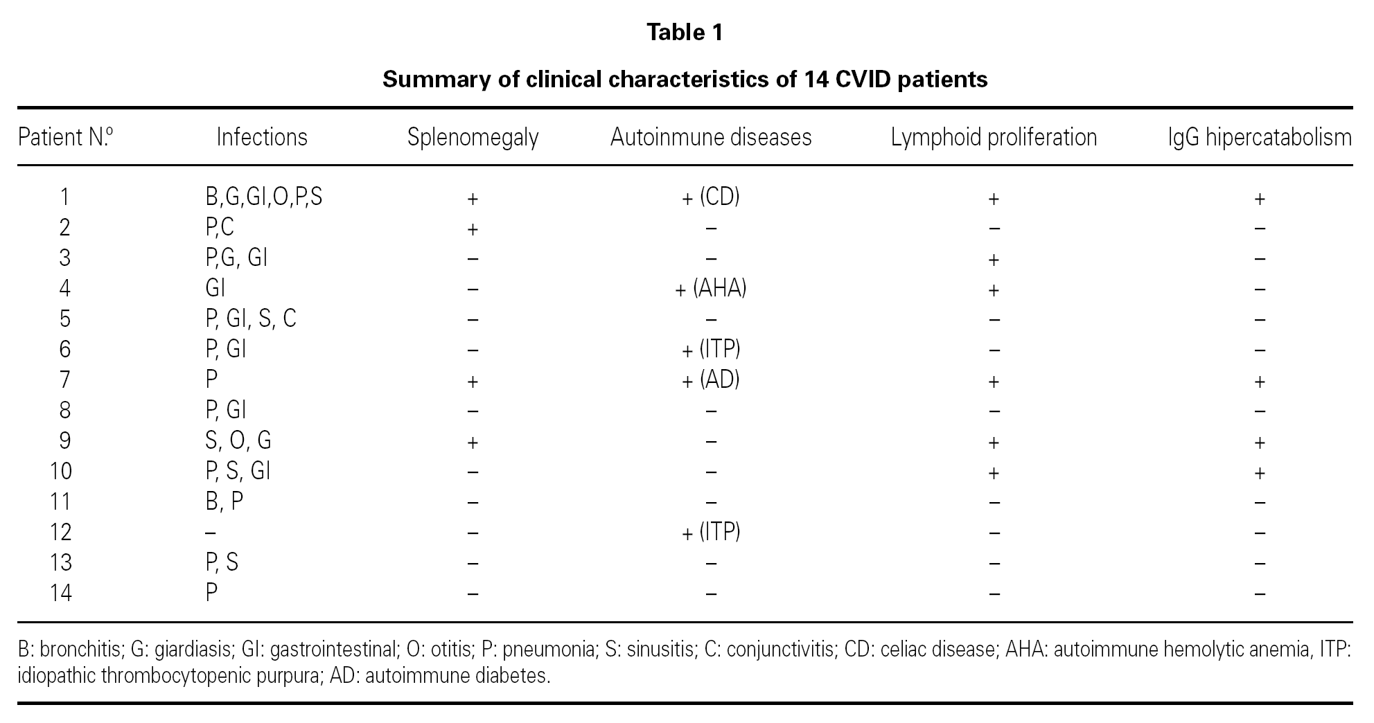
In a transversal descriptive study we analyzed activated CD4+ and CD8+ T cell-subset percentages in 14 CVID patients (21-68 years old, mean age 37.4 years, 8 men and 6 women). All patients had been diagnosed as having CVID based on diagnostic criteria for primary immunodeficiencies 13. The clinical characteristics of the patients are summarized in table I. At the time of the immunological study all the patients were receiving substitutive therapy with intravenous immunoglobulin (IVIG), at a dose of 400 mg/kg every 3 weeks. Blood samples were obtained immediately before IVIG administration. Increased levels of lymphocyte activation markers can be observed during chronic infections. For that reason, in this study we compared the percentages of lymphocyte activated subsets in CVID patients with the percentages observed in 47 asymptomatic HIV-positive patients whithout clinical criteria of AIDS; and with 6 non-CVID hypogammaglobulinemic patients who had chronic bacterial infections and who were receiving IVIG. We also compared lymphocyte subsets percentages in CVID patients against values observed in a health control group composed of 23 healthcare workers. Splenomegaly and lymphoid proliferation were confirmed by computed tomography. Increased catabolism of serum IgG antibodies was suspected in those patients that did not reached IgG levels greater than 600 mg/dl despite conventional dosis of IVIG. None of the patients had protein losing diseases. Patients with acute infections or opportunistic infections were not included in the study.

Activated CD4 and CD8 T-cell subsets were quantitated by three color flow cytometry (FACScan, Becton & Dickinson, San Jose, CA, USA). We enumerated T-cell subsets using FITC/PE/PerCP combinations of CD38/HLA-DR/CD4 or CD8 and isotype controls. Percentages of activated CD4 and CD8 T cell subsets are expressed as a percentage of total CD4 and CD8 lymphocytes. Details of adquisition and analysis of lymphocyte subsets have been reported elsewere 14. T-cell activated percentages were compared between groups using the non-parametric Mann-Whitney test.

RESULTS

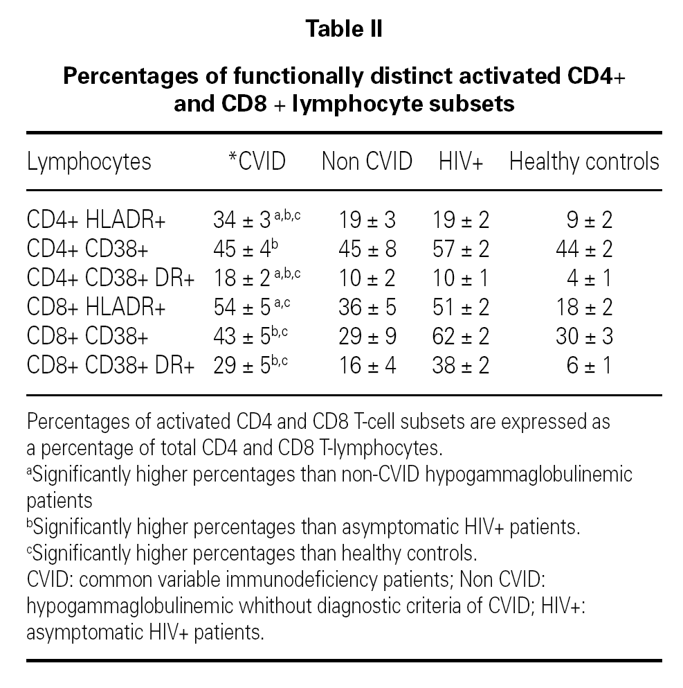
Mean concentration of serum IgG at the time of diagnosis of CVID was 127 mg/dl (15 to 441 mg/dl) and at the time of the immunological study it was 837 mg/dl (384 to 1320 mg/dl). Table II shows the mean percentages of the functionally distinct activated CD4 and CD8 T-cell subsets in the study groups. In CVID patients serum levels of CD4+ T-cells co-expressing the activation marker HLA-DR [CD4+ DR+ (34 %), CD4+ CD38+ DR+ (18 %)] were significantly elevated compared with all control groups. A tendency toward a negative correlation between the percentage of CD4+ CD38+ DR+ T-cells and the concentration of serum IgG at the time of the diagnosis of CVID was observed (Spearman Coefficient, r = ­0.48 (p = 0.08)]. CVID patients showed higher percentages of CD8 + DR + T-cells (54 %) than HIV-negative controls. Significant increases of CD8+ CD38+ (43 %) and CD8+ CD38+ DR+ (29 %) T-cells were observed in CVID patients as compared with healthy controls. CVID patients with splenomegaly, lower pre-infusion IgG levels (< 600 mg/dl), autoimmune or lymphoproliferative conditions demonstrated even higher levels of CD4 + CD38 + DR + T cells (22, 22, 21 and 21 % respectively) compared with other CVID patients (13, 13, 15 and 15 % respectively).

Interestingly enough, the group of CVID patients whithout any complication (n = 5) showed higher percentages of CD4 + DR + (27 %), CD4+ CD38+ DR+ (12 %), CD8+ DR+ (49 %) and CD8+ CD38+ DR+ (22 %) T-cells than healthy controls.

DISCUSION

Recent studies have proposed a new classification of CVID patients using class-switched memory B-cell counts determined by flow cytometry in peripheral blood 8-9,11. CD27+ (memory) B lymphocytes can be divided into two distinct subpopulations: IgD-CD27+, which produces IgG, IgM or IgA (class-switched memory phenotype), and IgD+ CD27+, which predominantly produces IgM (non-class-switched memory phenotype). A good correspondence has been observed between a low number of memory B cells and splenomegaly, lymphoid proliferation and/or autoimmune diseases which are common complications in CVID patients 1-2,8-9,11.

Several T-cell abnormalities have also been observed in CVID patients including decreased lymphocyte proliferation in response to mitogens and antigens, a tendency towards increased T-cell apoptosis and elevated levels of T-cell activation markers 12,15-20. However, few studies have correlated T-cell functional abnormalities with clinical findings 12,16,20. Studies performed in acquired states of immunodeficiency, such as in HIV infection, have demonstrated that the increase in the percentages of CD4 and CD8 T-cell activated subsets provided additional independent prognostic information on the progression of the disease 14. We hypothezised that increased levels of CD4 and CD8 T-cells might be associated with clinical findings commonly observed in CVID.

Previous studies have compared the values of the functionally distinct subsets of T-cells observed in CVID patients with healthy controls. Since it is not easy to completely exclude the presence of infections in CVID patients, in this study we have compared the percentages of CD4 and CD8 T-cell activated subsets in CVID patients with the percentages observed in patients with a viral chronic infection (HIV + asymptomatic patients) or with chronic bacterial infections (non-CVID hypogammaglobulinemic patients with bacterial infections). Interestingly, CVID patients disclosed significantly higher percentages of activated CD4 T-cells than controls with chronic infections, which suggest that CD4 lymphocyte activation does not merely reflect a chronic infection state in CVID patients. Furthermore, when we split our patients into two groups according to the presence or absence of complications (splenomegaly, autoimmune disease and/or lymphoid proliferation), we found higher percentages of CD4+ CD38+ DR+ T-cells in CVID patients with such complications. In addition to previous studies, we have observed significantly higher percentages of CD4+ CD38+ DR+ T-cells in patients with suspected IgG hypercatabolism.

CVID patients showed increased percentages of activated CD8 T-cell subsets than healthy controls. It has been recently reported that the expresión of the HLA-DR molecule on CD8 T-cells was restricted to patients with clinical complications 20. In our study we observed higher percentages of CD8+ DR+ in CVID patients with clinical complications (splenomegaly, autoimmune disease and/or lymphoid proliferation), but the difference was not significant (data not shown). Since the percentage of CD8+ DR+ T-cells in CVID patients was similar to that observed in HIV positive controls, we speculate that a subclinical or undetected infection could at least partially explain the higher percentage of activated CD8+ T cells observed.

The results presented in this study indicate a state of ongoing T lymphocyte activation which is associated with clinical findings frequently observed in CVID patients. Further studies with a greater number of patients are necessary to establish if the evaluation of CD4 and CD8 T-cell activated subsets might be useful to identify subgroups of patients for etiological studies and classification.

ACKNOWLEDGEMENTS

The authors would like to thank Dr. Jose Miguel Benito for performing lymphocyte subset analysis of HIV + and healthy controls. They also acknowledge Dr. Juana Gil (Cellular Immunity Section) and Dr Margarita Rodriguez-Mahou (Autoimmunity Section) for laboratory diagnostic evaluation of the patients.


Correspondencia:
J. Carbone
Clinical Immunology Unit.
Immunology Department
Hospital General Universitario Gregorio Marañón
Dr. Esquerdo, 46
28007 Madrid. Spain
E-mail: carbone@teleline.es

Bibliography
[1]
Iglesias J, Matamoros N..
Common variable immunodeficiency. Review..
Allergol Immunopathol, 29 (2001), pp. 113-8
[2]
Cunningham-Rundles C, Bodian C..
Common Variable Immunodeficiency: Clinical and Immunological Features of 248 Patients..
Clin Immunol, 92 (1999), pp. 34-48
[3]
Carbone J, Sarmiento E, Rodriguez-Molina JJ, Fernandez-Cruz E..
Atypical presentation of common variable immunodeficiency without infections..
Allergol Immunopathol, 32 (2004), pp. 218-22
[4]
Spickett GP, Webster AD, Farrant J..
Cellular abnormalities in common variable immunodeficiency..
Immunodefic Rev, 2 (1990), pp. 199-219
[5]
Farrant J, Spickett G, Matamoros N, Copas D, Hernandez M, North M, et al..
Study of B and T cell phenotypes in blood from patients with common variable immunodeficiency (CVID)..
Immunodeficiency, 5 (1994), pp. 159-69
[6]
Brouet JC, Chedeville A, Fermand JP, Royer B..
Study of the B cell memory compartment in common variable immunodeficiency..
[7]
Agematsu K, Futatani T, Hokibara S, Kobayashi N, Takamoto M, Tsukada S, et al..
Absence of memory B cells in patients with common variable immunodeficiency..
Clin Immunol, 103 (2002), pp. 34-42
[8]
Warnatz K, Denz A, Drager R, Braun M, Groth C, Wolff-Vorbeck G, et al..
Severe deficiency of switched memory B cells (CD27(+)IgM(­)IgD(­)) in subgroups of patients with common variable immunodeficiency: a new approach to classify a heterogeneous disease..
Blood, 99 (2002), pp. 1544-51
[9]
Ferry BL, Jones J, Bateman EA, Woodham N, Warnatz K, Schlesier M, et al..
Measurement of peripheral B cell subpopulations in common variable immunodeficiency (CVID) using a whole blood method..
Clin Exp Immunol, 140 (2005), pp. 532-9
[10]
Carsetti R, Rosado MM, Donnanno S, Guazzi V, Soresina A, Meini A..
The loss of IgM memory B cells correlates with clinical disease in common variable immunodeficiency..
J Allergy Clin Immunol, 115 (2005), pp. 412-7
[11]
Piqueras B, Lavenu-Bombled C, Galicier L, Bergeron-var der Cruysse.n, Mouthon L, Chevret S, et al..
Common variable immunodeficiency patient classification based on impaired B cell memory differentiation correlates with clinical aspects..
J Clin Immunol, 23 (2003), pp. 385-400
[12]
Vlkova M, Thon V, Sarfyova M, Blaha L, Svobodnik A, Lokaj J, Litzman J..
Age dependency and mutual relations in T and B lymphocyte abnormalities in common variable immunodeficiency patients..
Clin Exp Immunol, 143 (2006), pp. 373-9
[13]
Conley ME, Notarangelo LD, Etzioni A..
Diagnostic Criteria for Primary Immunodeficiencies..
Clin Immunol, 93 (1999), pp. 190-7
[14]
Carbone J, Gil J, Benito JM, Navarro J, Muñóz-Fernández A, Bartolomé J, et al..
Increased levels of activated subsets of CD4 + T cells add to the prognostic value of low CD4 + T cell counts in a cohort of HIV-infected drug users..
AIDS, 14 (2000), pp. 2823-9
[15]
Vukmanovic S, Vuckovic S, Stosic-Grujicic S, Ramic Z, Abinun M..
An unusual T-cell surface phenotype in vivo correlates with the failure to proliferate and produce IL-2 in vitro in a patient with common variable immunodeficiency..
Clin Immunol Immunopathol, 65 (1992), pp. 261-70
[16]
Nordoy I, Muller F, Aukrust P, Froland SS..
Adhesion molecules in common variable immunodeficiency (CVID) a decrease in L-selectin-positive T lymphocytes..
Clin Exp Immunol, 114 (1998), pp. 258-63
[17]
North ME, Spickett GP, Webster AD, Farrant J..
Raised serum levels of CD8, CD25 and beta 2-microglobulin in common variable immunodeficiency..
Clin Exp Immunol, 86 (1991), pp. 252-5
[18]
Baumert E, Wolff-Vorbeck G, Schlesier M, Peter HH..
Immunophenotypical alterations in a subset of patients with common variable immunodeficiency (CVID)..
Clin Exp Immunol, 90 (1992), pp. 25-30
[19]
Ferrer JM, Iglesias J, Hernandez M, Matamoros N..
Alterations in interleukin secretion (IL-2 and IL-4) by CD4 and CD4 CD45RO cells from common variable immunodeficiency (CVI) patients..
Clin Exp Immunol, 102 (1995), pp. 286-9
[20]
Viallard JF, Blanco P, Andre M, Etienne G, Liferman F, Neau D, et al..
CD8(+)HLA-DR(+) T lymphocytes are increased in common variable immunodeficiency patients with impaired memory B-cell differentiation..
Clin Immunol, 119 (2006), pp. 51-8
Article options
Tools
es en pt

¿Es usted profesional sanitario apto para prescribir o dispensar medicamentos?

Are you a health professional able to prescribe or dispense drugs?

Você é um profissional de saúde habilitado a prescrever ou dispensar medicamentos