


Stone characterization is becoming important before decision of treatment such as percutaneous nephrolithotomy (PCNL) and extracorporeal shock wave lithotripsy (ESWL). Some studies have reported that the twinkling artifact (color-flow ultrasonography artifact) may be useful to detect urinary stones. This study aims to determine whether the presence or absence of the twinkling artifact is correlated with the chemical composition of the stones.
Material and methodPatients with renal stones ≥0.5cm were included in a prospective study. Sixty patients were examined with X-ray film, intravenous pyelography, non-contrast computerized tomography, and color and spectral Doppler ultrasonography. The artifact was considered grade 1 when it occupied only one portion of the acoustic shadowing and when the artifact occupied the entire acoustic shadowing was considered grade 2. Patients with stones smaller than 2cm were treated with SWL and patients with stones larger than 2cm were treated with PCNL.
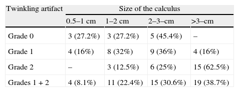
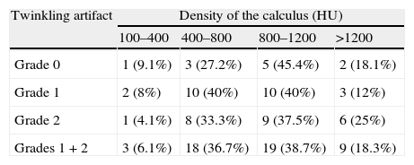
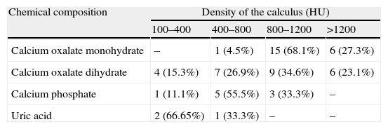
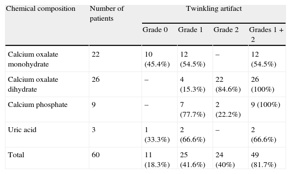
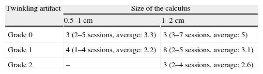
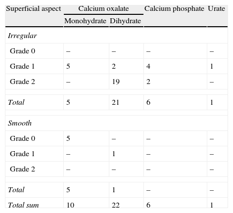
ResultsNo artifact (grade 0) was detected in 11 subjects, grade 1 in 25 and grade 2 in 24. Significant relationship was found between the increase in twinkling artifact and stone size (p<0.001). When the relation between the composition of the stones and the twinkling artifact was analyzed, artifact was detected nearly in all of the calcium oxalate dihydrate and calcium phosphate stones, whereas the artifact was detected in more than half of the calcium oxalate monohydrate and uric acid stones. In ESWL group it was observed that as the grade of the twinkling artifact increases, the number of required ESWL sessions decreases (p<0.001). In PCNL group twinkling artifact was found in all the patients (100%) with roughly surfaced stones.
ConclusionThe roughness of stone surface is the most important factor in terms of formation of the twinkling artifact in kidney stones. This artifact can be of use in anticipating the breakability of the stones of those patients to be treated with applied ESWL. One might anticipate that cases where the size of the stone is larger than 2cm but no twinkling artifact is detected are calcium oxalate monohydrate, which is one of the stones with highest level of breakability.
La caracterización de los cálculos renales está cada vez cobrando mayor importancia como paso previo a la toma de decisiones terapéuticas tales como la nefrolitotomía percutánea (NLP) y la litotricia extracorpórea por ondas de choque (LEOC). En algunos estudios se ha publicado que el artefacto de centelleo (artefacto de ecografía de flujo en color) puede ser de utilidad en la detección de piedras en el riñón. En este estudio se pretende dilucidar si la presencia o ausencia del artefacto de centelleo tendría alguna relación con la composición química de los cálculos.
Material y métodoEn un estudio prospectivo se incluyó a pacientes con cálculos renales de ≥ 0,5cm. Se examinó a 70 pacientes mediante rayos X, pielografía intravenosa, tomografía computarizada sin contraste y ecografía doppler espectral y a color. El artefacto se consideró de grado 1 si sólo ocupaba una parte de la sombra acústica, considerándose de grado 2 si ocupaba la totalidad de la sombra. Se trató a los pacientes con cálculos de menos de 2cm con LEOC, y a aquellos con piedras de mayor tamaño se les derivó a tratamiento con NLP.
ResultadosNo se detectó artefacto alguno (grado 0) en 11 sujetos, detectándose el grado 1 en 25 y el grado 2 en 24. Se encontró una relación significativa entre el aumento en artefactos de centelleo y tamaño del cálculo (p<0,001). Al analizar la relación entre composición de los cálculos y artefacto de centelleo se detectó el artefacto en prácticamente todas las piedras de oxalato de calcio —dihidrato y fosfato de calcio, mientras que en el caso de los cálculos de oxalato de calcio— monohidrato y ácido úrico se detectó el artefacto únicamente en algo más de la mitad de ellos. En el grupo LEOC se observó que, al aumentar el grado del artefacto de centelleo, descendía el número de sesiones de LEOC necesario (p<0,001). En el grupo NLP se encontró el artefacto de centelleo en todos los pacientes (100%) con piedras de superficie irregular.
ConclusiónLa irregularidad de la superficie lítica es el factor más influyente en la formación del artefacto de centelleo en piedras de riñón. Dicho artefacto puede tener utilidad a la hora de prever la predisposición a la fragmentación de los cálculos en pacientes derivados a tratamiento con aplicación de LEOC. Podría pronosticarse que aquellos casos en los que el tamaño de la piedra fuese de más de 2cm y no se detectase artefacto de centelleo serían de oxalato de calcio - monohidrato, que es una de las piedras con mayor predisposición a la fragmentación.
