


Tumor processes compromising the head and neck region can particularly affect the identity of the human being. There are different therapeutic alternatives to remove these dysplasias, which generate functional sequels affecting phonation deglutition and mastication. Therefore, approach for cancer patients is not only based on control of the disease, but additionally in patient's survival, rehabilitation and reinsertion in society. We hereby present the case of a patient with an orofacial defect resulting from treatment of basal cell carcinoma with X-ray therapy and later surgical resection after tumor recurrence. The patient was rehabilitated with manufacture of a hybrid prosthesis (intraoral obturator and facial prosthesis) which was achieved with the purpose of partially compensating functional and aesthetic losses and thus improving the patient's psychosocial circumstances.
Los procesos tumorales que comprometen la región de cabeza y cuello, pueden afectar particularmente la identidad del ser humano. Para la eliminación de estas neoplasias existen diferentes alternativas terapéuticas, que generan secuelas de tipo funcional, afectando: la fonación, deglución y masticación, además de defectos estéticos y alteraciones psicológicas. Por consiguiente, el enfoque para el tratamiento de pacientes con cáncer se basa no solamente en el control de la enfermedad, sino también en la supervivencia, rehabilitación y su reintegración a la sociedad. A continuación se presenta el caso de un paciente con defecto orofacial, resultado del tratamiento ante un carcinoma basocelular, con radioterapia y su posterior resección quirúrgica tras recidiva tumoral. Se rehabilita mediante la elaboración de una prótesis híbrida (obturador intraoral y prótesis facial) con el objetivo de compensar parcialmente las pérdidas funcionales, estéticas, incidiendo positivamente en su estado psicosocial.
Cancer of the head and neck assumes the fifth place of most frequently reported neoplasia. Its incidence has increased due to high tobacco and alcohol consumption as well as other genetic and environmental factors.1 Over half a million cases associated to the mouth are recorded. It is estimated that by 2020, population's growth and ageing will give rise to double this figure, rendering it thus the main mortality cause in the world.2
Tumor treatment depends on the disease's physiopathology, of all its variations, of systemic circumstances and social context of each subject.3 In consequence, different therapeutic options have been developed among which we can count oncological surgery, alternative radiotherapy and chemotherapy, which have provided suitable results to locally and regionally control the disease, and to decrease metastasis to distant points.
Specifically in the case of oncological surgery resection, defects can be caused, which, according to their location and extension, can give rise to severe alterations with morphological, functional and aesthetic sequels. It is important to take into consideration that during mastication there is an exchange of foodstuff and fluids from the oral cavity to the nasal and sinus cavities. Masticatory effectiveness will be affected by the absence of compromised teeth in the resection area. With respect to deglutition, difficulties encountered to conform suitable food bolus will force the patient to adhere to a liquid diet or use of a nasogastric probe which will cause many digestive complications. Phonation will be altered due to tissue loss in hard or soft palate, preventing interaction between tongue and palate, which is necessary to produce and articulate phonemes. Aesthetic appearance will also be affected due to facial asymmetry caused by compromised structures and organs involved, among which we can count loss or depression of the middle third.4,5
Bones of the upper jaw provide support between skull base and dental arches, they separate cavities and determine facial projection. To rehabilitate previously mentioned sequels, reconstructive surgical alternative is deemed the best option, nevertheless, its limitations must be borne in mind. Factors such as general health circumstances, age, lesion extension and vascular circumstances of the tissues, radiotherapy history and the patient's psychological status must be taken into consideration, since many surgical stages might be necessary to conform tissue volume, provide suitable coverage, and individualize different anatomical compartments. For the aforementioned reasons, rehabilitation of intraoral and facial defects represent a great challenge.6–8
Therefore, prosthetic rehabilitation requires a multidisciplinary approach and represents an alternative when tackling limitations of surgical reconstruction; they are a more cost-effective process, allowing periodic cleansing and revision of the affected region in short and predictable9 execution times. Thus, in order to be able to conform a hybrid prosthesis, the first element to take into account is the intraoral component (obturator) as well as its complement which is the facial structure (nasal prosthesis upper lip and adjacent prostheses) in this case joined by magnets and prosthetic devices10 which optimize stability, retention and support, factors which are fundamental for a successful rehabilitation.
CLINICAL CASE PRESENTATIONThe subject seeking treatment was a 67 year old male patient, born and residing in the State of Mexico, married, Roman Catholic, farm labourer, with incomplete primary education, and familial genetic history non contributory to present affliction. The patient reported a 15 year tobacco consumption habit (7 to 10 cigarettes a day) as well as occasional alcohol consumption. He presented an initial painless lesion in the left wing of the nose flap (ala nasi) of over 2 year evolution and had adopted a regime of self-medication. He attended private practice, where he was evaluated and later remitted to the National Institute of Cancerology (Instituto Nacional de Cancerología) for diagnosis and treatment.
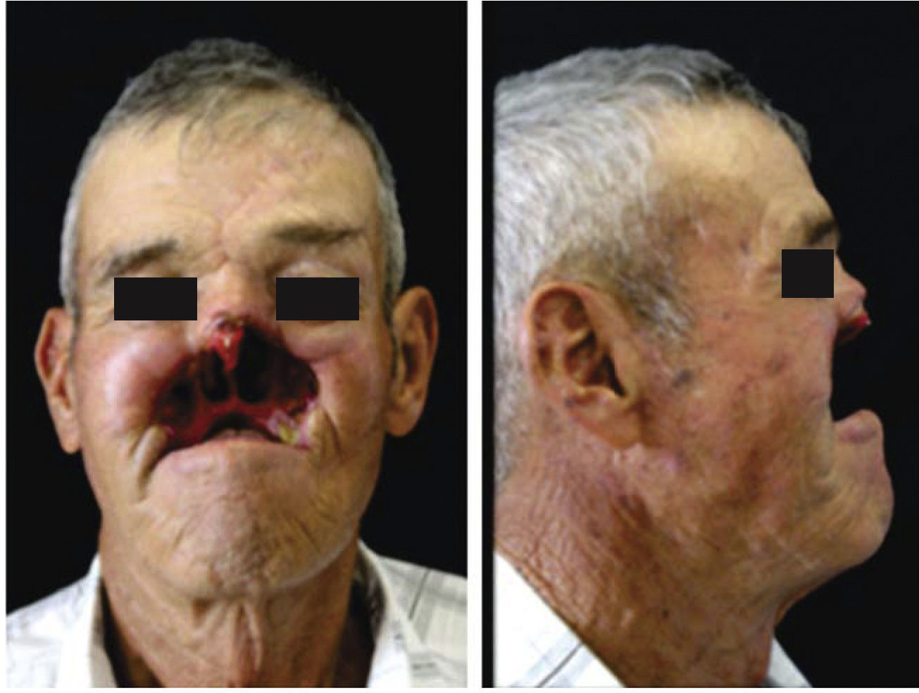
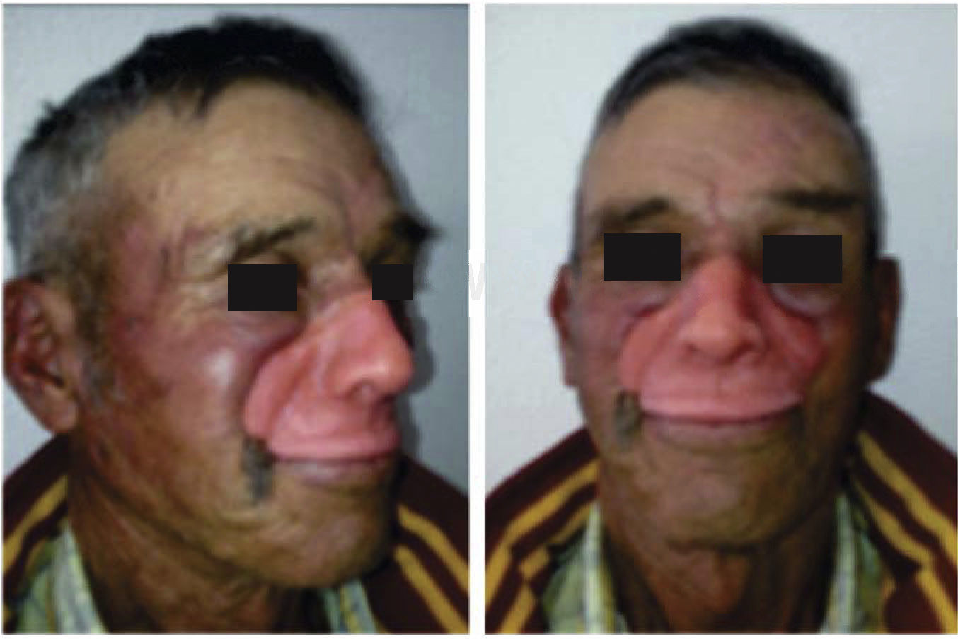
A 6 x 4cm lesion was described, with overall cartilage, left wing of the nose full destruction, ipsilaterally extending to skin of the cheek, partially infiltrating the mucosa of the upper lip in the mouth, additionally exhibiting adenomegalies in the neck. Basal cell carcinoma diagnosis was emitted after biopsy; this prompted treatment with 40Gy radiotherapy, according to evolution. Initial nodule persisted after completion radiation therapy, therefore it was suggested to increase dosage and request assessment of the oncological surgical team. The aforementioned team confirmed lesion recurrence, prompting thus undertaking partial maxillectomy and rhinectomy (Figure 1).

After six months recovery and lacking evidence of tumor activity the patient was remitted to Maxillofacial Prosthesis Service for assessment and rehabilitation. Intra oral examination revealed loss of upper lip, anterior portion of oral vestibule, and teeth 16, 17, and 27 in the remaining palate, as well as open communication to the nasal cavity. Extraoral examination revealed a defect limiting with proper nasal bones and floor of the orbit in the left region, irregular borders, absence of nasal cartilages and compromised left maxillary sinus. Due to the aforementioned situation, it was proposed to manufacture an filling intraoral prosthesis with lateral extension to support nasolabial structures.
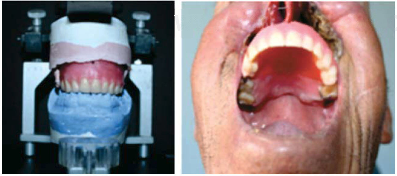
An intraoral impression with alginate was conventionally taken, placing gauzes to cover exposed naso-pharyngeal communication, in order to achieve a model in type III plaster (gypsum), so as to later manufacture retainers cast in chrome-cobalt, the base of the register (where prosthetic palate depth will be established, with physiological methods of deglutition and phonation)11 as well as adaptation and individualization of the wax cylinder. After conducting aesthetic, phonetic and prosthetic tests, the rest of craniomandibular relationships were established. To this effect, articulators are of the utmost importance in order to establish the correct relationship and function of the patient’ s working models according to fundamental bases of total prosthodontics (Figure 2).

Resin teeth were selected and articulated, placing them according to lower teeth. A final test was conducted in wax, taking special care in the structure's thickness, since it will directly influence the final prosthesis’ weight. Conventional laboratory procedures were undertaken, in order to obtain the intraoral prosthesis. In this prosthesis, an acrylic lateral extension was manufactured, which tripodized the structure thus improving its stability, additionally playing the role of a prosthetic device to house the magnet responsible for retention, location and insertion of the facial structure (Figure 3).12–15

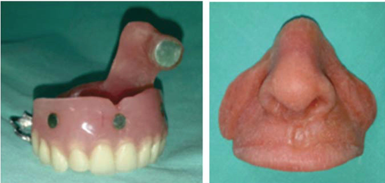
In order to manufacture the nasolabial prosthesis, an alginate impression was taken, with great care to locate the obturator in a suitable position to thus achieve necessary support of facial middle third and obtain the model in type IV plaster. With the final model in hand and the support of preoperative photographs of the patient, wax works of nose, lips and adjacent tissues were undertaken with all-season wax.
Once the final modelling was achieved and patient and his family granted consent, an analogous base was adapted for the magnets so they could match those found in the obturator, after which laboratory procedures were performed (Figure 4). Intrinsic characterization was achieved with medical grade silicon and pigments trying to replicate the different colors of the structures to be rehabilitated. The model was packed and subjected to three tonnes hydraulic pressure for 24hours (Figure 5).


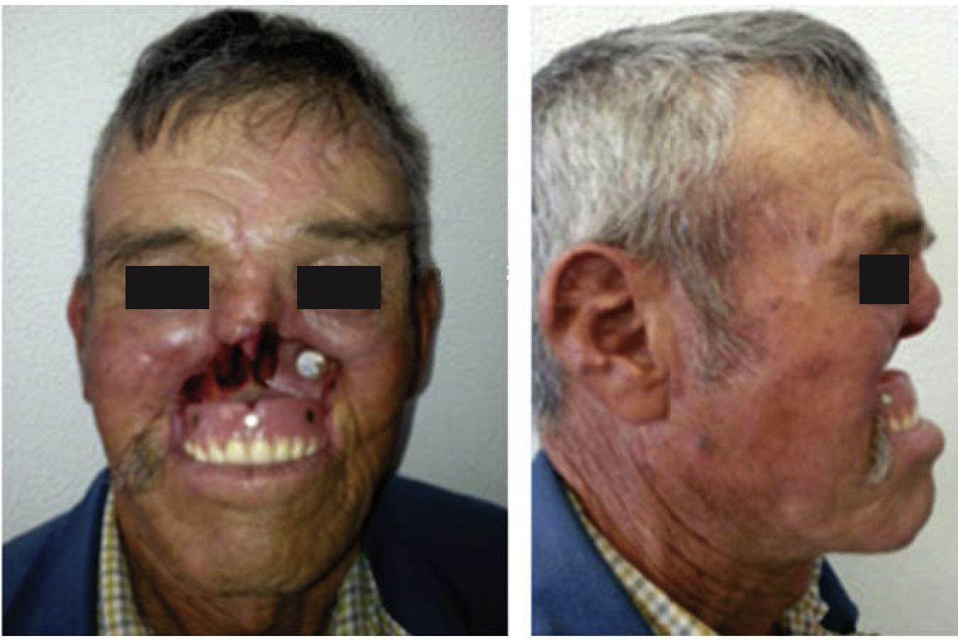
After this time, silicon full polymerization was assessed, it was withdrawn from the model and the prosthesis was tried on and adapted in the patient so as to continue with extrinsic characterization. In it, care was taken of specific details of traits of the lips, columella, nostrils, expression lines, and borders of the defect, which must be as thin and as diffuse as possible in order to blend in when in contact with the skin. Afterwards, and following manufacturers’ indications, the prosthesis was dried, sealed and dulled.
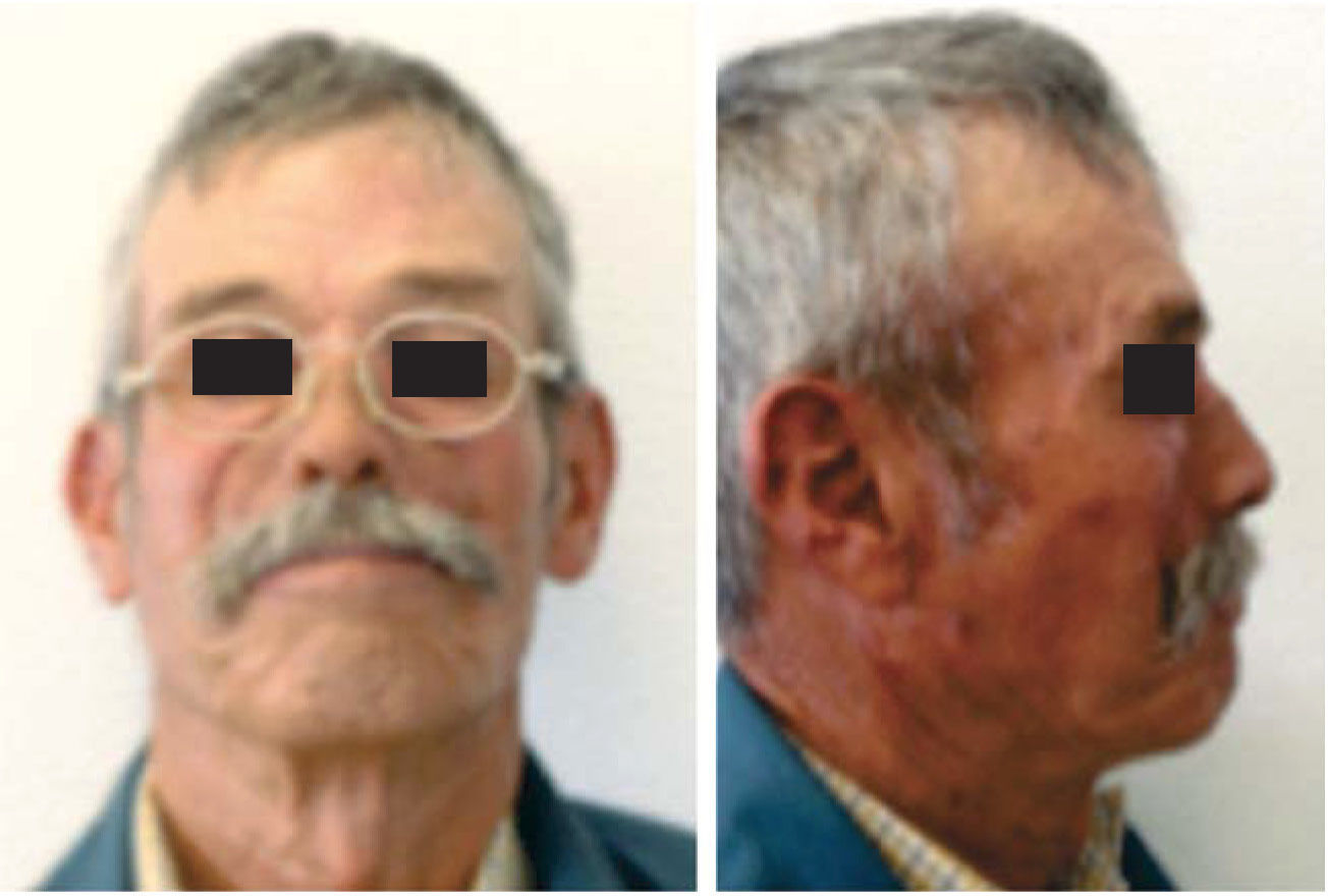
At the final stage, it was attempted to replicate the patient's own singular traits, such as the moustache and use of glasses. These elements were part of the day to day life of his post-operative environment; they will create suitable cosmetic effect which will represent a distraction from his final appearance. Finally, the obturator was positioned and the facial prosthesis was put in place, supplementing its retention with use of adhesives, this will help to join and mimic the borders of the prosthesis with the defect's margin. The patient was instructed on recommendations and indications for use and care of his prosthesis. In spite of the defect's complexity, rehabilitation was achieved, which would allow the patient to speak and communicate clearly, in addition to being able to chew and swallow, since the device created a barrier which would not allow food filtration into involved cavities. With respect to facial appearance, it could be said that cosmetic result was satisfactory, since the defect was dissimulated by distracting elements. The patient was satisfied with the result, and expressed he felt confident with his appearance (Figure 6).
DISCUSSION
Presently, in the medical fields, development of new technologies represent great advances, nevertheless, if one bears in mind social inequalities in most populations, health services will sustain limited access.16 For this reason, many ideal treatments reported in scientific literature cannot be a reality for many patients.
Extensive surgical resections in the face require non-conventional rehabilitating treatments. Yañez et al mention that for maxillary reconstruction one counts from use of obturator prostheses, local flaps, pedicle flaps, up to microsurgical flaps, all depending on type of defect and patient prognosis. We would like to point out that for patients undergoing maxillectomies, we consider microsurgical reconstruction the best alternative providing more suitable aesthetic and functional results.
Kornblith et al reported suitable physiological response when the psychological status of the patient with oncological surgery sequels was improved, with use of an obturator prosthesis as long as the soft palate is not involved or the defect extends into the orbital cavity.
Pigno presented adaptation of an extension for the space of the nasal defect in prosthetic treatment of patients which have been subjected to hemimaxillectomies. In the case here presented this would generate a point of support which would ease tension of masticatory movements exerted on the prosthetic ensemble as a whole.
On the other hand, Rogers Lowe et al compared results, prognosis and perception in quality of life indicators in a series of patients, out of which some were prosthetically and others surgically rehabilitated; in that study they did not find statistically significant differences in the results.17 This can be understood if we consider that patients using removable filling appliances, require adaptation when considering an external element for vital functions, whereas patients subjected to reconstructive surgery have sometimes very high expectations, hoping for circumstances almost identical to those they possessed preoperatively.
With respect to facial structure, silicon represents a very favorable option, due to its similarity in texture, shape and color (Beumer); nevertheless, this material is far from ideal since it presents drawbacks of durability, flexibility, biocompatibility and hygiene. Thus, new alternatives must be sought to achieve meeting requirements of patient as well as clinician.
CONCLUSIONSFrom the very beginning of treatment, importance of rehabilitation study, diagnosis and planning of intraoral and facial defects, as well as cancergenerated alterations and sequels must be assessed and transmitted to the patient; likewise, treatment must be conducted multi-disciplinarily.
Choice of placing palatal obturator or conducting surgical reconstruction must be based on well-defined criteria; both techniques are useful when a suitable treatment plan is devised taking into account individual requirements for each patient.
Comprehensive rehabilitation of patients with head and neck cancer is a process in which maxillofacial prosthesis allows as many designs and appliances as the specialist might develop, always having the target of patient reintegration into society and improvement of his quality of life.
This article can be read in its full version in the following page: http://www.medigraphic.com/facultadodontologiaunam
Resident of the Maxillofacial Prosthesis Specialty, National School of Dentistry, National University of Mexico (UNAM).