


Rheology is the science that studies deformation of objects when submitted to external forces. This science was associated to the scalpel blade, which is a cutting tool of therapeutic importance.
ObjectiveTo describe and compare deformation of the cutting surface of Bard Parker No. 15 scalpel blades (Elite® and Paramount® brands), performing one to four cuts in ex vivo pig gums.
Material and methodsComparative-descriptive study. Commercial use pig mandibles were used. Cuts with 20 scalpel blades per brand were performed. A texture analyzer with force to perform these cuts was used, keeping regulated speed and constant position. A stereo-microscope was used to photograph and compare the surface of the scalpel blades before and after being used. ImageJ software was used to measure deformed areas of the scalpel blades in pictures.
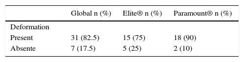
ResultsAverage cutting effort from both brands was 49.1 Newton (N), this effort did not statistically differ in each particular brand, Elite® reported an overall cutting effort of 48.1N [27.2-179.7], meanwhile Paramount® showed 49.1N [0.0- 60.3] (p = 0.776). The deformations of the scalpel blades were evaluated. For Elite®, 75% of the scalpel blades suffered some kind of deformation; for Paramount®, 90% did.
ConclusionThe cutting effort performed by the scalpel blades increases with each incision performed, which influenced directly the physical deformation of each blade.
La reología es la ciencia que estudia la deformación de los objetos sometidos a fuerzas externas. Se quiso relacionar esta ciencia con el bisturí, el cual es una herramienta de corte de importancia terapéutica.
ObjetivoDescribir y comparar la defor- mación de la superficie cortante de las hojas de bisturí de Bard Parker número 15 de las marcas comerciales Elite® y Paramount®, realizando de uno a cuatro incisiones en encía de cerdos ex vivo.
Material y métodosEstudio de tipo descriptivo comparativo. Se utilizaron mandíbulas de cerdos de uso comercial. Se efectuaron cortes con 20 hojas de bisturí por cada marca comercial. Se utilizó un texturómetro con fuerzas para realizar los cortes, con velocidad regulada, posición constante. Se empleó un estereomicroscopio para fotografiar y comparar la superficie de los escalpelos antes y después de ser utilizados. Se empleó el software ImageJ, para medir el área deformada de las hojas en fotos.
ResultadosEl es- fuerzo en promedio al corte de ambas marcas fue de 49.1 Newtons (N), este esfuerzo no difiere estadísticamente en cada marca en particular, Elite® reportó un esfuerzo general a los cortes de 48.1N [27.2-179.7], mientras que Paramount® mostró 49.1N [0.0-60.3] (p= 0.776). Se evaluó la deformación de las hojas de bisturí. Para Eli- te® se deformaron el 75% de las hojas; para la marca Paramount® fue del 90%.
Conclusión: El esfuerzo al corte realizado por las ho- jas del bisturí aumenta con cada incisión que se realiza influyendo esto directamente con la deformación física de la hoja.
Scalpel blades are an essential element in surgical treatments within the oral cavity, they have experienced a wide evolutionary process and great historical use in various surgical fields.1,2 Within the dental literature there are relatively few scientific studies that describe their behavior when applying the principles of rheology, such as suggested by Carter et al in 2005.3 Physical alterations that occur in its shape, caused by use, could be prevented to further avoid possible alterations that elicited by performing successive incisions in an individual during surgical procedures,4 caused by expected deformations in the cutting surface of the scalpel blade.5 This situation can eventually inflict larger damage in the tissue, with tissue alterations which will not be a subject of study in the present article. This eventually leads to longer scarring and recovery periods. It is fundamental to establish that in academic formation scenarios of surgical importance, the following is not taught, nor is it quantified in force units, the amount of pressure required or the degree of pressure that should be applied to a scalpel blade, in order to obtain a therapeutic result, without it deforming in any detectable way. It is equally important to mention that there is no exact or quantifiable parameter, thus it was decided to use rheology due to its investigative ability, what this science achieves is to study the effective link between exerted force on a material and the possible deformation that a material or tissue experiences.6
The present study aimed to describe deformation of the cutting surfaces and cutting effort of the No. 15 scalpel blades of two commercial brands, when performing one to four mucoperiosteal incisions in ex vivo pig mandibles by applying different degrees of strength expressed in Newton (N). This procedure, was carried out with a texture analyzer possessing suitable forces to perform the cuts while maintaining a regulated cutting speed and constant angulation. One of the major detected constraints, which are common on any study, was that not enough background information or references were found regarding description of the active part of a scalpel blade after being used.
The importance of this article, product of research, is that it makes a contribution that allows us to demonstrate and quantify the deformations or alterations that are produced on scalpel blades, which simultaneously decrease their cutting capacity when repeatedly used in surgical procedures within the oral cavity.
MATERIAL AND METHODSA comparative-descriptive study was performed.
As an experimentation model, commercially available ex vivo pig mandibles were used, no animal was harmed for this study.
As a sample, 20 No. 15 scalpel blades from the brands Elite® and Paramount®, for Bard Parker No. 3 scalpel handles were used, these were available in the country where the study was developed. The scalpel blades were divided into four groups for each brand and each group had assigned the names: A, B, C and D for the brand Elite® and A’, B’, C’ and D’ for Paramount®. Each group was made of five scalpel blades each.
Picture-taking was standardized by using a formwork, avoiding the movements of the scalpel blade. The photographs were taken with a Nikon d7000 and a stereo-microscope from the brand «D & D Implementos», each scalpel blade from the brands Elite® and Paramount® had one picture taken right after they were removed from their original packaging.
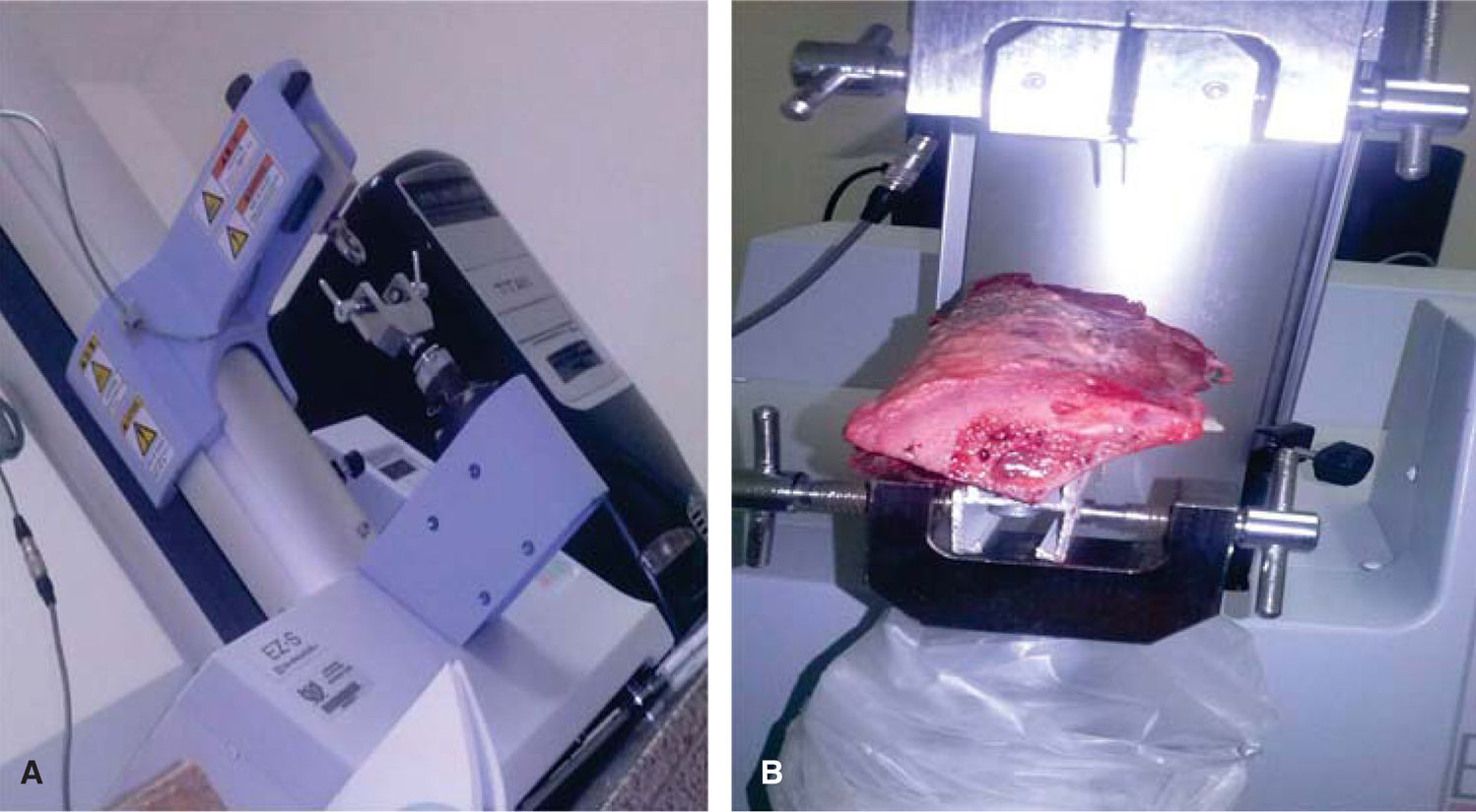
The scalpel blades were taken to the Shimadzu texture analyzer model EZ-S, serial number 346- 54909-33, 50-60Hz (Figure 1), this equipment reaches a maximum capacity of 500N of pressure for the performance of the tests.
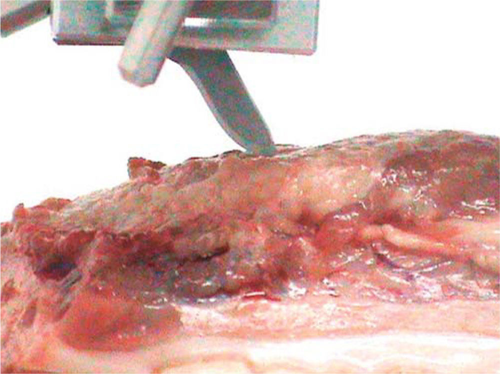
 Fragment of a pig mandible attached to the texture analyzer used for rheological tests.")
In the texture analyzer, the mucoperiosteal penetrating cuts were performed in a 45-degree angle in pig mandibles (Figure 2). The pig mandibles were selected because of their similitude with the human oral tissue. Also, they offer the possibility of being fitted and cut for an easy access of the cutting tools attached to the texture analyzer. The penetrating incision was made at a constant speed of 10mm per minute until reaching the bone.

Using the first group of the scalpel blades from both brands, only one cut was performed and had pictures taken afterwards. Then we proceeded with the second groups of the scalpel blades, and with a previous picture of the blades taken, a simulation of the cuts in the mandible was performed with the same standardization of cutting pressure and angle. The photographs were taken after two incisions were performed with each blade. Then we proceeded with the group C, performing three cuts per blade and in the group D, four cuts per blade.
Fracture of two Paramount® scalpel blades is reported in groups of three and four cuts. These blades were not replaced. Data collected from these fractured blades was used in the statistic report.
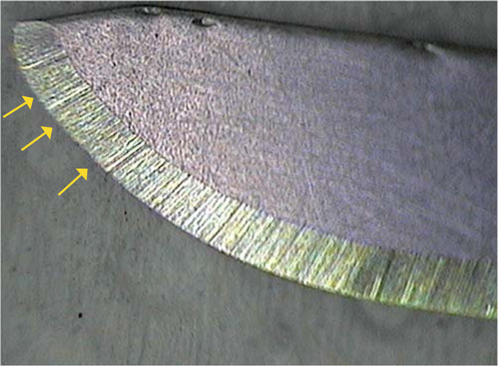
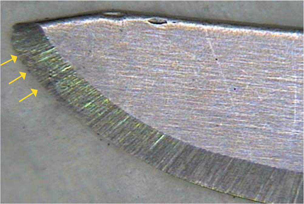
Photographic description was twofold. In the first instance, photographs were taken before and after the cuts and they were compared with each other to determine a qualitative variable that showed whether there was or wasn’t physical evidence of blade deformation, such as loss of continuity or alterations in the blade's active section. The second instance was carried out with help of ImageJ software7 where the area of the photographed active part of the scalpel blade was measured to then compare it by means of equivalence of pixels to millimeters, having previously calibrated software, to translate picture results from before and after the cuts provided by the program; this was conducted in a similar manner to that of Fukushima and Tomita in 2009, when they assessed changes in Guinea pigs’ conjunctive tissue (conjunctiva).8
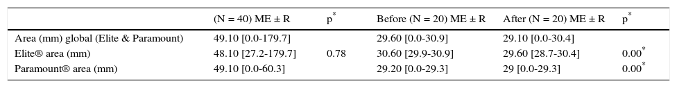
RESULTSPhysical deformation and cutting effort was evaluated in every No. 15 scalpel blade used as sample. Initially, scalpel blade area variability was assessed, measuring the cutting effort in Newton (N). In general, both brands reported a median of 49.1N [0.0-179.7]; however, this effort did not statistically differ in each particular brand (p > 0.05). Elite® brand reported a general cutting effort of 48.1N [27.2-179.7], meanwhile Paramount® showed 49.1N [0.0-60.3] (p = 0.776). When comparing variability showed in the area of the scalpel blades in millimeters before and after the cuts for each brand in particular, it was found that Elite® blades showed a median of 30.6mm [29.9-30.9], this area decreased significantly to 29.6mm [28.7-30.4] after performing all the different cuts. (p = 0.00). Likewise for Paramount® scalpel blades, the area before use was 29.2mm [0.0- 29.3] this significantly decreased to 29mm [0.0-29.3] (p= 0.00), thus, statistically significant differences for both brands were found (Table I). Global variability of scalpel blade area before and after cuts.
Variability of global scalpel blade area before and after cuts.
| (N = 40) ME ± R | p* | Before (N = 20) ME ± R | After (N = 20) ME ± R | p* | |
|---|---|---|---|---|---|
| Area (mm) global (Elite & Paramount) | 49.10 [0.0-179.7] | 29.60 [0.0-30.9] | 29.10 [0.0-30.4] | ||
| Elite® area (mm) | 48.10 [27.2-179.7] | 0.78 | 30.60 [29.9-30.9] | 29.60 [28.7-30.4] | 0.00* |
| Paramount® area (mm) | 49.10 [0.0-60.3] | 29.20 [0.0-29.3] | 29 [0.0-29.3] | 0.00* |
* Kruskal-Wallis (p < 0.05).
The variability of the area of the scalpel blade was also determined in millimeters, according to number of cuts, a significant decrease was reported in the observed area after cuts were performed, as shown in table II. When analyzing the area of the scalpel blades after number of cuts and according to brand, it was observed that Elite® brand blades did not show statistically signifi cant difference after performing the diverse cuts (p = 0.687). The opposite was observed in Paramount® scalpel blades, which showed a significant decrease in their area after performing the diverse cuts (p = 0.00) (Table II).
Variability of the area in mm according to the number of cuts performed.
| Global (mm) | Elite® (mm) | Paramount® (mm) | |||||||
|---|---|---|---|---|---|---|---|---|---|
| Before (N = 10) ME ± R | After (N = 10) ME ± R | p | After (N = 5) ME ± R | Before (N = 5) ME ± R | p | Before (N = 5) ME ± R | After (N = 5) ME ± R | p | |
| 1 Cut | 29.60 | 29.45 | 0.010* | 30.4 | 30.10 | 29.20 | 29.10 | ||
| [29.1-30.8] | [29.0-30.3] | [29.9-30.8] | [29.6-30.3] | [29.1-29.3] | [29.0-29.3] | ||||
| 2 Cuts | 29.75 | 29.15 | 30.8 | 29.7 | 29.20 | 29.00 | |||
| [29.1-30.9] | [28.9-30.1] | 0.002* | [30.2-30.9] | [29.3-30.1] | [29.1-29.3] | [28.9-29.0] | |||
| 3 Cuts | 29.80 | 29.05 | 0.004* | 30.50 | 29.60 | 0.697 | 29.20 | 28.90 | 0.00* |
| [0.0-30.9] | [0.0-30.4] | [30.4-30.9] | [29.1-30.4] | [0.0-29.2] | [0.0-29.0] | ||||
| 4 Cuts | 29.95 | 29.00 | 0.004* | 30.7 | 29.00 | 29.20 | 29.00 | ||
| [0.0-30.9] | [0.0-29.4] | [30.6-30.9] | [28.7-29.4] | [0.0-29.3] | [0.0-29.0] | ||||
* Kruskal-Wallis (p < 0.05).
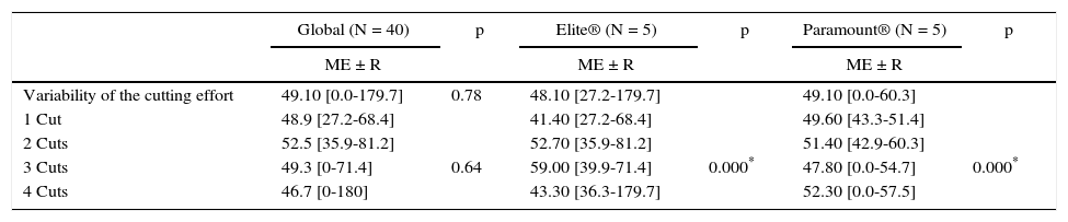
Likewise, the blade variability was evaluated according to number of cuts performed within each blade group. In general, no statistically significant differences in cutting effort of one, two, three and four cuts were observed; maximum reported effort was 52.05N [35.9-81.2] when performing two cuts (p = 0.64). When analyzing cutting effort according to brand, it was observed that Elite® scalpel blades exhibited greater effort when the number of cuts increased; thus, one cut needed an effort of 41.4 N; two cuts needed 52.7N and three cuts 59 N; showing a statistically significant difference (p = 0.00). In Paramount® blades, a significant increase in the required cutting effort was observed when performing the diverse cuts, a statistically signifi cant difference was thus revealed. (p = 0.00) (Table III).
Variability of the cutting effort (N) according to number of cuts.
| Global (N = 40) | p | Elite® (N = 5) | p | Paramount® (N = 5) | p | |
|---|---|---|---|---|---|---|
| ME ± R | ME ± R | ME ± R | ||||
| Variability of the cutting effort | 49.10 [0.0-179.7] | 0.78 | 48.10 [27.2-179.7] | 49.10 [0.0-60.3] | ||
| 1 Cut | 48.9 [27.2-68.4] | 41.40 [27.2-68.4] | 49.60 [43.3-51.4] | |||
| 2 Cuts | 52.5 [35.9-81.2] | 52.70 [35.9-81.2] | 51.40 [42.9-60.3] | |||
| 3 Cuts | 49.3 [0-71.4] | 0.64 | 59.00 [39.9-71.4] | 0.000* | 47.80 [0.0-54.7] | 0.000* |
| 4 Cuts | 46.7 [0-180] | 43.30 [36.3-179.7] | 52.30 [0.0-57.5] |
* Kruskal-Wallis (p < 0.05).
One of the most important evaluated conditions of the study allowed us to determine physical deformation of the scalpel blades from both brands; 75% of the Elite® blades were deformed regardless of the number of cuts; while deformation percentage of Paramount® scalpel blades was 90%. This shows the high deformation frequency of scalpel blades after use (Table IV). Nevertheless, in order to obtain better details of the generated deformation degree, scalpel blades areas were analyzed before and after performing the diverse cuts (Figures 3 and 4).


The Bard Parker No. 15 scalpel blade for a No. 3 scalpel handle is one of the most commonly used instruments in the field of oral surgery and it's still the gold standard for any kind of procedures that imply excision, incision or dissection of the soft tissue to further reach the surgical site or sites in the oral cavity, it is recognized for its capacity to achieve fine and clean cuts which, in the end, will favor the scarring process.
The object of this study allowed us to describe and compare the sharp edge deformation of Bard Parker scalpel blades from two different brands, using an experimental model and performing one to four incisions in ex vivo pig gums.
Various studies carried out to date, focus their interest in studying the effect of different instruments in the scarring process of the mucosa after diverse surgical procedures without determining or describing the deformities elicited in the scalpel blade. Authors such as Arshad et al compared the advantages and disadvantages of CO2 laser and the scalpel blade in the scarring of wounds after an oral exposition and surgical procedures in soft tissue. They observed the scarring process of the wound and they compared it, both clinically andhistologically, 24hours after surgery.9 They found significantly greater differences in the appearance of the wound, formation of scar tissue and thickness of the wound in the group that was intervened with a scalpel blade, however they did not evaluate the deformation or the effort that the instrument suffered during the procedure. Other authors who have done studies where they evaluated other types of primary outcomes were Sinha and Gallagher;10 they compared the performance of different instruments in the scarring process when a steel blade, ultrasonic blade, mono or bipolar electro-surgery instruments or CO2 laser were used while performing oral surgery in an animal model, the samples were collected for a histopathological study, traction resistance was measured, moreover they evaluated aspects such as hemostasis, coagulation, tissue adherence, wound scarring, reepithelialization and inflammation degree, and they found that the ultrasonic blade was the best tool in controlling the hemostasis and further coagulation. Greatest traction resistance was observed when using steel scalpel blade and ultrasonic blade. Most of these studies are directed to assess tissue scarring by evaluating different instruments, without assessing the variability in the instruments’ cutting effort. Ryu et al,11 compared wound scarring after performing incisions with a scalpel blade, CO2 laser or ER laser in the mucosa of Guinea pigs biopsy samples from each oral mucosa wound were examined and the expression of TNF-α and TGF-β1 by immunohistochemical staining were determined. The authors concluded that the ER laser achieved more benefits in the scarring process than the rest of the instruments. Chanthasopeephan et al,5 studied cuts from nonuniform thickness liver tissue with three different cutting speeds (0.1 cm/sec; 1.27 cm/sec; y 2.54 cm/ sec). They found that the depth of the cut plays a very important role in the force that needs to be applied while cutting. Using a stereo camera and an image processing system proved to be an effective method to measure the depth of the scalpel blades into the tissue, while clearly showing the cutting progress, it also showed similar results to the ones reported in this study, where a signifi cant increase of the effort required when performing the diverse cuts was reported in both evaluated brands (p = 0.00).
It's reported that when performing liver or intestinal surgeries, authors such as Leong, Coffey and Hill,12 are using methods of assisted radiofrequency to perform resections or hepatectomy, using a built prototype to carry out surgical simulations between radiofrequency ablation and liver resection. However, they reported a lack of literature studying the mechanical aspects, properties and interactions between the cutting tool, the tissue and the liver, with respect to resistance or fi rmness of the tissue considering the tool or the employed style of cut.
According to results found in the present study, it is valid to infer that scalpel blades suffer a noticeable process of deformation, which forces the operator to progressively use greater strength in every new cut to dissect the tissue, which in turn necessarily produces a decrease of the cutting speed which translates at the same time in an increase of the deformation resistance of the tissue.
From a scarring application point of view, this phenomenon is opposed to the right handling of the tissue, since to more modifications or alterations suffered by the tissue, more complex and slow will be its scarring process; as was proposed by Malik et al in 2011.9 At any rate, it can¿t be ascertained is at which exact moment deformation suffered by scalpel blades and increased cutting effort will interfere in the scarring process after surgery is performed, even if it is assumed beforehand.
CONCLUSIONSSimilarly, it is affirmed that after the first cut is performed, deformation of the scalpel blades increases, which can be expressed in qualitative terms; the moment we visually compare photographs from before and after.
After conducting photographic analysis using ImageJ software, where the area of the active part of the blade was measured using photographs from before and after, it is possible to conclude that decrease of scalpel blade's photographed area is directly related to the number of cuts performed with each blade.
This article can be read in its full version in the following page: http://www.medigraphic.com/facultadodontologiaunam
DDS, PhD in Biomedical Sciences and Professor, University of Cartagena. Specialist in Periodontics, Javeriana University. Degree of the Universidad del Norte. Director of GITOUC group.
Chemistry-Pharmaceutics Degree and Professor, University of Cartagena. Degree in Science and Technology of Biotechnologi- cal Processes, University of La Habana.
DDS and Specialist in Stomatology and Oral Surgery, University of Cartagena.
Food Engineer, Food Laboratory, University of Cartagena.
DDS, Javeriana University. Endodontics Specialist and Professor, University of Cartagena. Epidemiology Degree, National University. Professor at Corporación Universitaria Rafael Núñez.