


Lecanicillium fungicola causes dry bubble disease in Agaricus bisporus mushrooms leading to significant economic losses in commercial production.
AimsTo monitor the infection process of L. fungicola in Brazilian strains of A. bisporus.
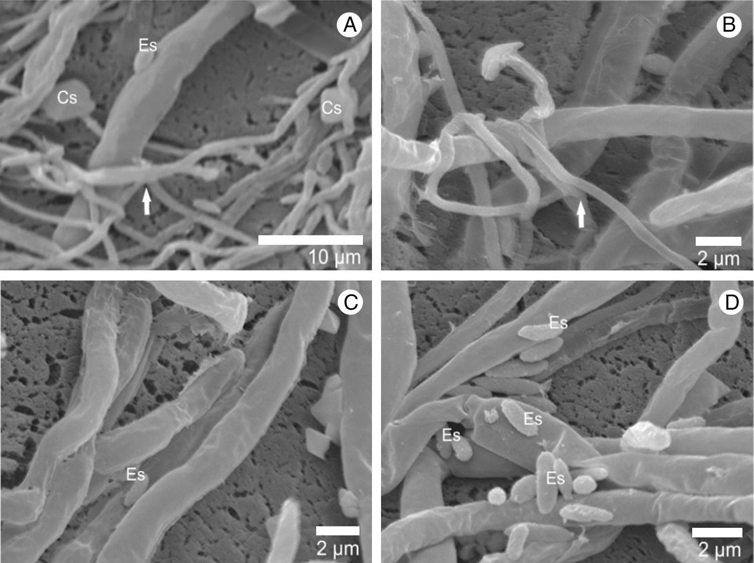
MethodsThe interaction between the mycelium of L. fungicola (LF.1) and three strains of A. bisporus (ABI 7, ABI 11/14 and ABI 11/21) was studied. Electron microscopy and X-ray microanalyses of vegetative growth and basidiocarp infection were evaluated.
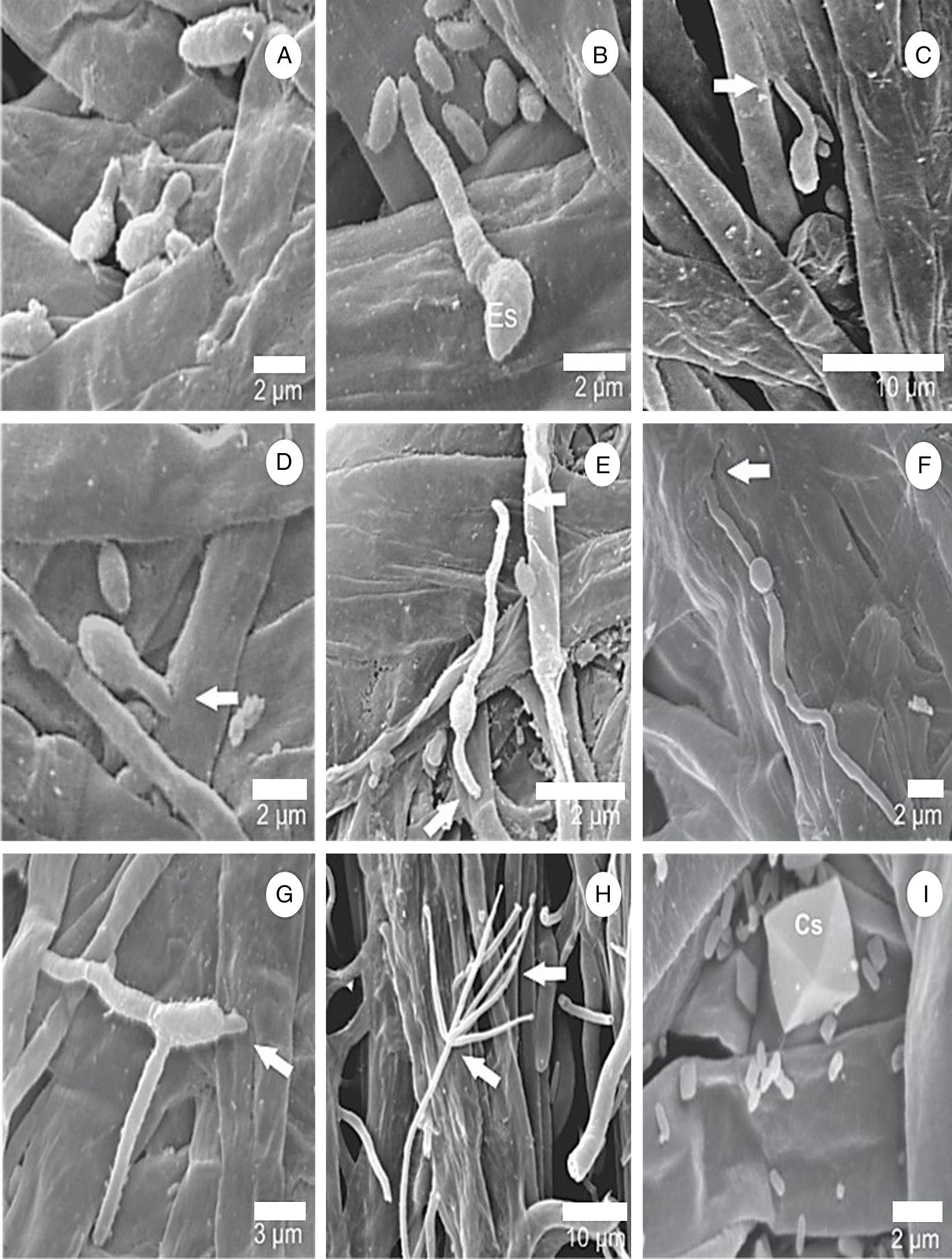
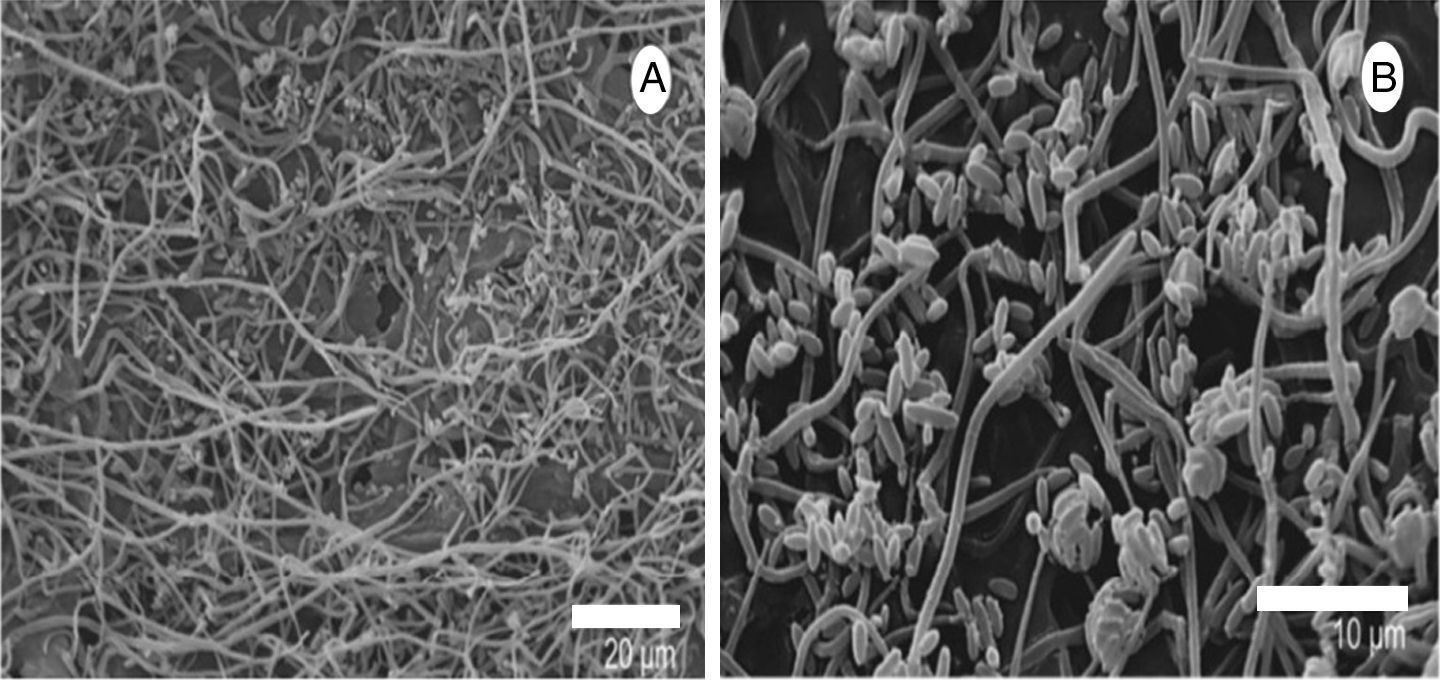
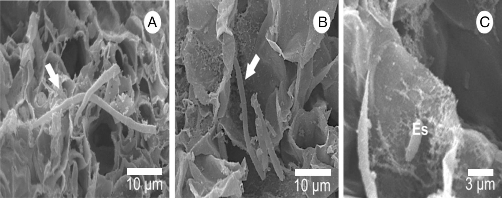
ResultsMicrographs show that the vegetative mycelium of the Brazilian strains of A. bisporus is not infected by the parasite. The images show that the pathogen can interlace the hyphae of A. bisporus without causing damage, which contributes to the presence of L. fungicola during the substrate colonization, allowing their presence during primordial formation of A. bisporus. In the basidiocarp, germ tubes form within 16h of infection with L. fungicola and the beginning of penetration takes place within 18h, both without the formation of specialized structures.
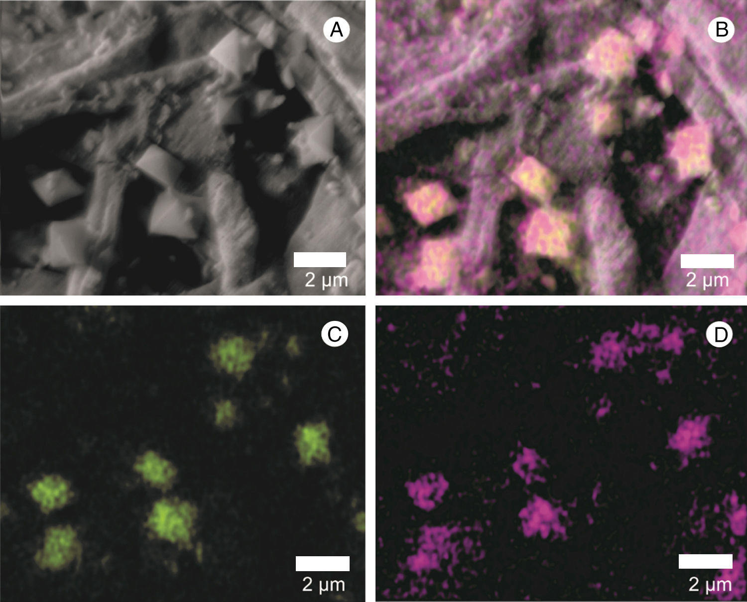
ConclusionsScanning electron microscopy enabled the process of colonization and reproduction to be observed within the formation of phialides, conidiophores and verticils of L. fungicola. The formation of calcium oxalate crystals by the pathogen was also visible using the X-ray microanalysis, both at the hyphae in the Petri plate and at basidiocarp infection site.
Lecanicillium fungicola es el agente causal de la enfermedad de la mole seca en Agaricus bisporus, responsable de importantes pérdidas económicas en la producción comercial de esta seta.
ObjetivosComprobar el proceso de infección de L. fungicola en cepas brasileñas de A. bisporus.
MétodosSe estudió la interacción entre el micelio de L. fungicola (LF.1) y tres cepas de A. bisporus (ABI 7, ABI 11/14 y ABI 11/21). Se evaluaron mediante microscopia electrónica y microanálisis de rayos X el crecimiento vegetativo y la infección de los basidiocarpos.
ResultadosLas micrografías muestran que el micelio vegetativo de las cepas brasileñas de A. bisporus no resultó afectado por la infección del parásito. Las imágenes muestran también cómo el agente patógeno puede entrelazar las hifas de A. bisporus sin causar daños, lo que contribuye a la perpetuación de L. fungicola durante la colonización del sustrato y durante la formación de los primordios de A. bisporus. En el basidiocarpo, los tubos germinales se forman después de 16h de la infección con L. fungicola y el comienzo de la penetración tiene lugar tras 18h, sin formación de estructuras especializadas.
ConclusionesLa microscopia electrónica permite observar el proceso de colonización y reproducción con la formación de fiálides, conidióforos y verticilos de L. fungicola. La formación de cristales de oxalato de calcio por parte del agente patógeno también fue visible mediante el microanálisis por rayos X, tanto en la infección de las hifas en placa de Petri como en la de los basidiocarpos.
Artículo
Comprando el artículo el PDF del mismo podrá ser descargado
Precio 19,34 €
Comprar ahora

