

The longitudinal arch of the human foot is a complex mechanical structure that must be compliant on uneven surfaces and also have sufficient stiffness to allow the foot to be an efficient propulsive organ during walking and running gait. To serve these functions, the longitudinal arch has a unique four-layer load-sharing system consisting of the plantar fascia, plantar intrinsic muscles, plantar arch extrinsic muscles and plantar ligaments. These four layers of tension load-bearing elements, working together with the osseous elements which serve as the framework of the longitudinal arch, work synergistically to increase longitudinal arch stiffness during weightbearing activities. The passive tension load-bearing elements of this load-sharing system, the plantar fascia and plantar ligaments, are not under direct central nervous system control and thus serve to stiffen the longitudinal arch with an automatic stiffening mechanism that is based on Achilles tendon tension and plantar forefoot loading. The active tension load-bearing elements, the plantar intrinsic and plantar extrinsic muscles, are under direct central nervous system control and serve to increase or decrease the stiffness of the medial and lateral longitudinal arches depending on the type and intensity of the prevailing weightbearing activity of the individual. Together, the elements of the longitudinal arch load-sharing system ensure that proper weightbearing function of the longitudinal arch, and the foot and lower extremity, can still occur even when a failure of one of these tension load-bearing elements occurs due to injury.
El arco longitudinal del pie humano es una estructura mecánica compleja que debe ser flexible en superficies irregulares y también tener suficiente rigidez para permitir al pie comportarse como un órgano propulsivo eficaz durante la marcha y carrera. Para realizar esas funciones, el arco longitudinal tiene un sistema único de 4 capas de reparto de cargas formado por la fascia plantar, músculos plantares intrínsecos, músculos plantares extrínsecos y ligamentos plantares. Estas 4 capas de elementos de soporte de carga tensional, trabajando conjuntamente con los elementos óseos que componen el arco longitudinal, trabajan sinérgicamente para aumentar la rigidez del arco longitudinal en situaciones de carga. Los elementos pasivos de soporte de carga tensional de este sistema son la fascia plantar y los ligamentos plantares que no están bajo el control directo del sistema nervioso central y sirven para aumentar la rigidez del arco longitudinal con un mecanismo de rigidez automático que se basa en la tensión del tendón de Aquiles y la carga plantar del antepié. Los elementos activos de soporte de carga tensional son los músculos plantares intrínsecos y extrínsecos que están bajo control directo del sistema nervioso central y sirven para aumentar o disminuir la rigidez de los arcos longitudinales medial y lateral dependiendo del tipo e intensidad de la actividad predominante de carga del individuo. Juntos, los elementos del sistema de reparto de cargas del arco longitudinal aseguran una función correcta del arco longitudinal en carga incluso cuando de uno de estos elementos falla por lesión.
Leonardo Da Vinci (1452–1519), the Renaissance painter, sculptor, architect and inventor called the human foot “a masterpiece of engineering and a work of art”.1 Within his notebooks are multiple drawing and descriptions of the form and shape of the human foot which clearly demonstrate Da Vinci's appreciation of the longitudinal arch of the foot and how it changes shape during weightbearing activities.2
The longitudinal arch of the foot has also been the focus of medical professionals for generations. Over a century ago, in 1896, a New York City orthopedic surgeon, Royal Whitman, described how the collapse of the longitudinal arch of the foot could create the condition known as “weak foot”, which was also known at the time as “flat-foot”, “splay-foot”, and “pronated foot”.3,4 Feet with high longitudinal arches, first called pes cavus by Little in 1853, was a condition also known at the time as “hollow foot”, “clawfoot”, “bolt foot”, “pes arcuatus”, and “anterior equinus”.5,6
Even in the modern era, podiatrists and clinicians still pay close attention to the longitudinal arch as one of the key structures of the human foot. The extremes in longitudinal arch height are thought to lead to pathology and dysfunction of the foot. Mahan and Flanigan noted that individuals with decreased longitudinal arch height, or pes planus, may suffer from pain, fatigue, joint degeneration and/or associated deformities such as hallux valgus, hammer toes and metatarsalgia.7 Individuals with increased longitudinal arch height, or pes cavus, were noted by Smith and Green to cause abnormal weightbearing stresses, instability of the foot and ankle, restricted foot mobility, and difficulty in fitting shoes.5
Since generations of health professionals have considered the longitudinal arch of the foot to be an important structural and functional component of the foot and lower extremity, it is important for the podiatrist and clinician to fully understand the biomechanical characteristics of the longitudinal arch during weightbearing activities. As such, a new concept in longitudinal arch biomechanics, the Longitudinal Arch Load-Sharing System (LALSS), which was first described by Kirby in 2012,8 will be detailed to help explain the intricate mechanical balance of forces acting on and within the longitudinal arch that allow the foot to possess an exquisite balance of both flexibility and stability throughout years upon years of repetitive use.
Engineering concept of load-sharing systemsLoad-sharing is a common concept in mechanical and electrical systems where redundancy of operation is vital to the reliability of the system as a whole. Examples of load-sharing systems include the use of multiple supporting cables in suspension bridges, multiple engines in aircraft, multiple electric generators in power-generation systems, multiple processors in computers and multiple servers in distributed computer systems. In a load-sharing system, the load on a system is equally or unequally shared by the components of the system. In that way, when one component fails, the system will remain operational, but the loads now acting on the remaining components of the load-sharing system will be increased. However, if all the components remain operational, the loads on each component of the load-sharing system will be reduced.9,10
One familiar type of load-sharing system that is mechanically analogous to the longitudinal arch of the foot is found within the rear suspension of trucks where both leaf-springs and shock absorbers are used to support and dampen vertical accelerations between the truck chassis and its rear axle. Both the leaf springs and the shock absorbers work together to stiffen the rear suspension and help prevent the truck chassis from bottoming-out onto the rear truck axle when driving while heavily loaded or when driving over bumpy roads. Both the leaf springs and shock absorbers will deform under increased loads and will recover to its less compressed shape with decreased load. If the shock absorbers fail in the truck's rear suspension load-sharing system, the leaf springs will have increased load. If the leaf springs fail, the shock absorbers will have increased load. However, when each component of the load-sharing system of a rear suspension is working properly, both leaf springs and shock absorbers will have decreased loading force acting on them during vehicle operation.
Recent technological advances now have made variable stiffness shock absorbers available for the suspensions of vehicles that may be automatically adjusted by microprocessors or may be manually adjusted by the driver in order to improve the comfort and handling characteristics of the vehicle.11,12 The usefulness of the mechanical analogy of a rear suspension of a truck to the longitudinal arch of the foot will become more apparent in the following review of the structure, function, and both passive and active controlling mechanisms of the LALSS of the foot during weightbearing activities.
Compression load-bearing elements of the longitudinal arch load-sharing systemSince the longitudinal arch of the foot is subjected to significant external loads from ground reaction force (GRF) during weightbearing activities, it must have a system by which it can not only resist these loads, but also respond to the loads with a variable degree of stiffness. Peak GRF loads during walking range from 1.1 to 1.5 times body weight (BW), whereas, during running, peak GRF loads are double that of walking.13 During jumping activities, GRF acting on the plantar foot can easily exceed over 4 times BW.14
These potentially large magnitudes of GRF acting on the plantar aspect of the foot will tend to temporarily flatten and lengthen the longitudinal arch. When GRF loads are reduced or removed from the plantar foot, the longitudinal arch will return back to its higher and shorter unloaded shape. Thus, much like the leaf spring of the truck rear suspension mentioned earlier that deforms under load and then recovers its shape once the load is reduced, the longitudinal arch deforms under the loading forces from GRF and then returns to its original shape upon unloading. This loading-unloading cycle of the longitudinal arch occurs thousands of times a day during an individual's daily weightbearing activities.
In order to function with this spring-like nature day after day, and year after year, the longitudinal arch of the foot has multiple components that allow both passive and active control of its function. First of all, the mechanical characteristics of the structural framework of the longitudinal arch, the bones of the longitudinal arch, need to be discussed. The osseous structures of the longitudinal arch are the compression load-bearing elements of the foot that allow the foot to be able to resist the compression loading forces that act on and within the foot during weightbearing activities. In addition, bone is not only excellent at resisting compression loading forces, but is also very good at resisting bending and torsional loads.15
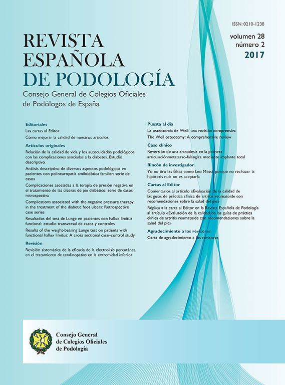
The rearfoot, consisting of the talus and calcaneus, plantarflexes relative to the ground with longitudinal arch flattening and then dorsiflexes relative to the ground with longitudinal arch elevation (Fig. 1). The forefoot, consisting of the navicular, cuboid, cuneiforms, and metatarsals, dorsiflexes relative to the rearfoot with longitudinal arch flattening and plantarflexes relative to the rearfoot with longitudinal arch elevation. Every osseous element of the longitudinal arch, like the wooden rafters within the roof of a house, can each individually resist compression, bending and torsion loads. However, since the bones of the longitudinal arch are linked together by ligaments, tendons and muscle, the longitudinal arch functions very differently than if the longitudinal arch was made of solid lengths of bone with no joints.
 acts on the plantar rearfoot and forefoot. Longitudinal arch elevation, involving rearfoot dorsiflexion and the forefoot plantarflexion, occurs when GRF is reduced on the plantar foot.")
The osseous compression load-bearing elements serve to provide the structural framework of the longitudinal arch. The talus and calcaneus form the rearfoot and the navicular, cuboid, cuneiform and metatarsals form the forefoot. Longitudinal arch flattening, involving the motions of rearfoot plantarflexion and forefoot dorsiflexion, occurs when ground reaction force (GRF) acts on the plantar rearfoot and forefoot. Longitudinal arch elevation, involving rearfoot dorsiflexion and the forefoot plantarflexion, occurs when GRF is reduced on the plantar foot.
The joints of the longitudinal arch allow necessary motion, while still allowing for longitudinal arch stability depending on the weightbearing demands of the individual. In other words, with the bones working together as a compression load-bearing framework, along with its ligaments, tendons and muscles, the longitudinal arch has both sufficient mobility and stability to allow it to function optimally for the individual in response to the variable magnitudes of external forces from GRF acting on the plantar foot during daily weightbearing activities.
Tension load-bearing elements of the longitudinal arch load-sharing systemEven though each bone of the longitudinal arch can individually resist compression, bending and torsion loads better than ligament, muscles and tendons, the bones of the longitudinal arch when functioning as a unit, with their multi-segmented arch structure, cannot resist total arch collapse without the important tension load-bearing elements of the plantar longitudinal arch: the plantar fascia, plantar intrinsic muscles, extrinsic muscles of the plantar longitudinal arch and plantar ligaments. These plantarly located tension load-bearing structures are the four elements of the LALSS which work synergistically with each other to prevent and regulate the flattening and elongation of the longitudinal arch during weightbearing activities.
The most superficial layer of the tension load-bearing elements of the LALSS is the plantar fascia, otherwise known as the central component of the plantar aponeurosis (Fig. 2). The plantar fascia originates from the plantar aspect of the medial calcaneal tubercle as a relatively thick band which then thins as it spreads distally to form five separate slips that each insert into the bases of the proximal phalanges of all five digits.16 The plantar fascia, like all fascial and ligamentous structures, is a passive structure, not being dependent on the central nervous system (CNS) to increase its tension forces.8
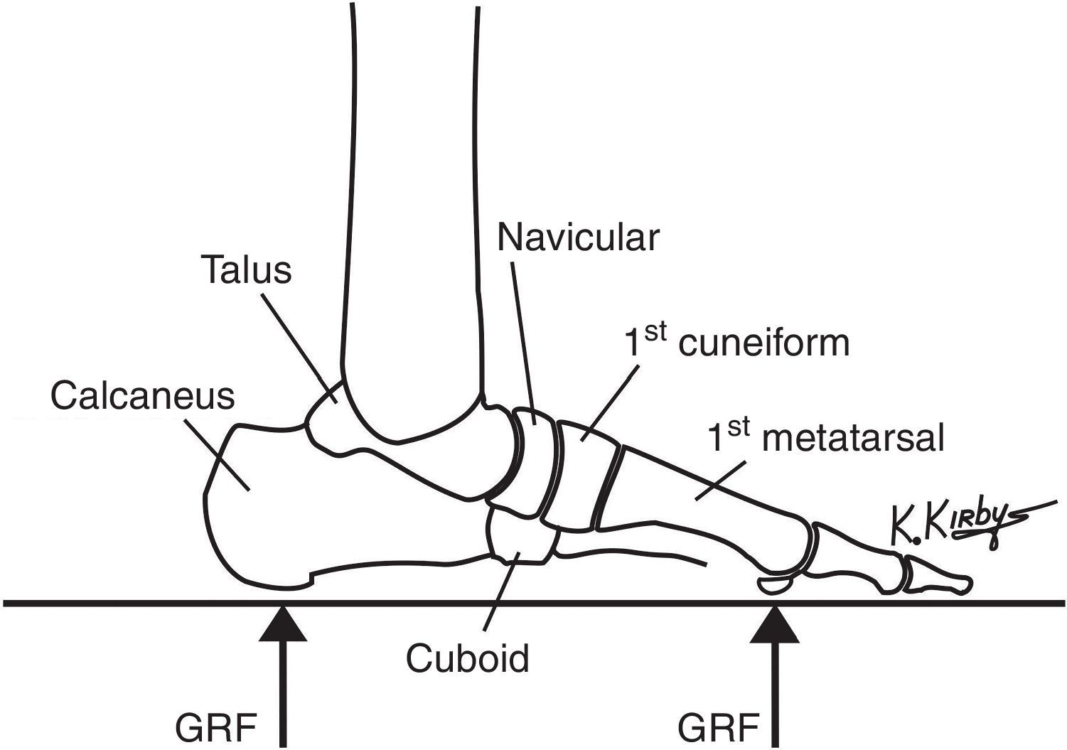
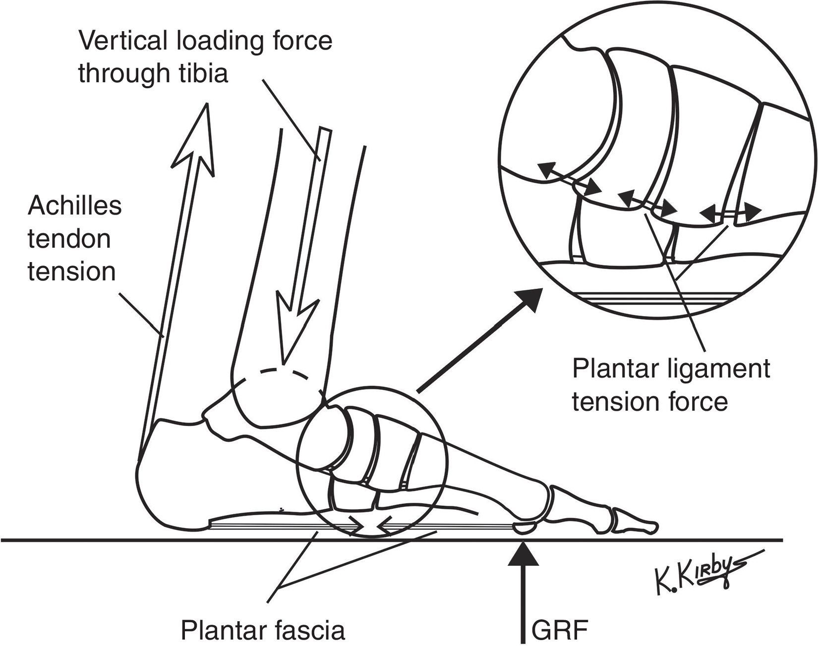
. Spanning the plantar foot from the medial calcaneal tubercle to the digital bases, the plantar fascia passively increases its tension force when GRF acts on the plantar forefoot, which, in turn, increases the stiffness of the longitudinal arch. The increase in plantar forefoot GRF is associated with an increase in force from the tibia onto the dorsal talar dome and an increase in Achilles tendon tension force during walking and running.")
The plantar fascia is a passive structure and forms the most superficial layer of tension load-bearing elements of the longitudinal arch load-sharing system (LALSS). Spanning the plantar foot from the medial calcaneal tubercle to the digital bases, the plantar fascia passively increases its tension force when GRF acts on the plantar forefoot, which, in turn, increases the stiffness of the longitudinal arch. The increase in plantar forefoot GRF is associated with an increase in force from the tibia onto the dorsal talar dome and an increase in Achilles tendon tension force during walking and running.
Hicks’ classic research showed that by increasing tension force within the plantar fascia in both live and cadaver feet with hallux dorsiflexion, the longitudinal arch could become more elevated by what he called the “windlass effect”.17 The plantar fascia is subjected to large tension forces which have been estimated to be 0.96 times body weight in simulated walking experiments using cadaver legs and feet.18 Other researchers have found that upon transection of the plantar fascia, the longitudinal arch of the foot significantly flattens and elongates.19,20 In addition, the plantar fascia has been clearly shown to be elastic in nature 21 and, when it is transected, the longitudinal arch of the foot becomes less stiff (i.e. more likely to flatten on plantar loading of the foot with GRF).22
Just deep to the plantar fascia is the next layer of the tension load-bearing elements of the LALSS, the plantar intrinsic muscles (Fig. 3). The abductor hallucis (AH), flexor digitorum brevis (FDB), abductor digiti quinti and quadratus plantae (QP) muscles are the plantar intrinsic muscles which are most important in preventing excessive flattening and elongation of the longitudinal arch, with the other plantar intrinsic muscles having a less important role.23
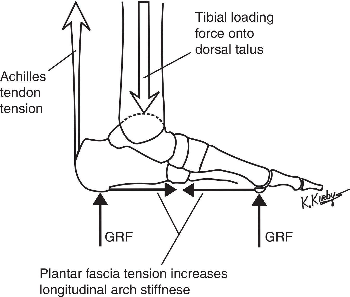
 and form the layer of tension load-bearing elements of the LALSS just deep to the plantar fascia. The abductor hallucis, and the other plantar intrinsic muscles which span the longitudinal arch, serve to actively stiffen the longitudinal arch when GRF loads increase on the plantar foot to prevent excessive longitudinal arch flattening during weightbearing activities.")
The plantar intrinsic muscles are actively controlled by the central nervous system (CNS) and form the layer of tension load-bearing elements of the LALSS just deep to the plantar fascia. The abductor hallucis, and the other plantar intrinsic muscles which span the longitudinal arch, serve to actively stiffen the longitudinal arch when GRF loads increase on the plantar foot to prevent excessive longitudinal arch flattening during weightbearing activities.
Excellent recent research on the function of the plantar intrinsics by Luke Kelly and coworkers, using bi-polar fine-wire electromyography inserted into the AH, FDB and QP muscles, show that the plantar intrinsic muscles are activated by the CNS to help stiffen the longitudinal arch of the foot to aid in both unipedal and bipedal balance.24 With increased loading of the longitudinal arch, the CNS increases the contractile activity of the plantar intrinsics and with electrical stimulation of these muscles, the plantar intrinsics have the ability to elevate the longitudinal arch.25 Plantar intrinsic EMG activity has also been shown to increase during the stance phase of walking and running, with more EMG activity occurring during faster running than during walking.26
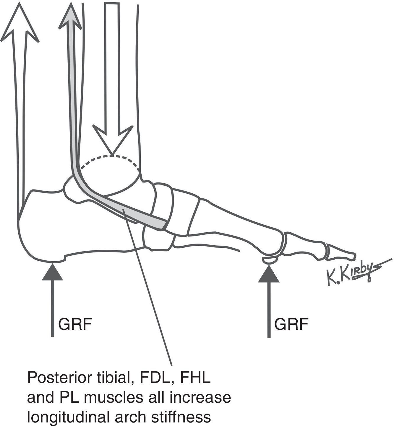
Deep to the plantar intrinsic muscles are the extrinsic muscles of the plantar longitudinal arch which form the next layer of tension load-bearing elements of the LALSS: the posterior tibial (PT), flexor digitorum longus (FDL), flexor hallucis longus (FHL) and peroneus longus (PL) muscles. The PT muscle, with its tendon inserting into the navicular tuberosity and also into the plantar aspects of the cuneiforms, cuboid and metatarsal bases, provides active support to the longitudinal arch by exerting a forefoot plantarflexion moment (i.e. a rotational force that resists longitudinal arch flattening) with its contractile activity (Fig. 4). The FDL, with its insertions into the lesser digit distal phalanges, and the FHL, with its insertion on the hallux distal phalanx, both cross nearly the full length of the plantar longitudinal arch. With contractile activity of the FDL and FHL, a forefoot plantarflexion moment is generated due to the increased proximally-directed compression force at the metatarsophalangeal joints that results. The PL muscle and its tendon, entering laterally into the plantar midfoot and then crossing the longitudinal arch to insert onto the first metatarsal base, generates a first ray plantarflexion moment and a forefoot plantarflexion moment during its contractile activity.
, flexor hallucis longus (FHL) and peroneus longus (PL) muscles are all actively controlled by the CNS and form the layer of the LALSS between the plantar intrinsic muscles and plantar ligaments. The posterior tibial muscle, illustrated above, along with the FDL, FHL and PL muscles, all cause a forefoot plantarflexion moment with their contractile activity, which increases the longitudinal arch stiffness and also acts to decelerate longitudinal arch flattening and/or accelerate longitudinal arch elevation during gait.")
The posterior tibial, flexor digitorum longus (FDL), flexor hallucis longus (FHL) and peroneus longus (PL) muscles are all actively controlled by the CNS and form the layer of the LALSS between the plantar intrinsic muscles and plantar ligaments. The posterior tibial muscle, illustrated above, along with the FDL, FHL and PL muscles, all cause a forefoot plantarflexion moment with their contractile activity, which increases the longitudinal arch stiffness and also acts to decelerate longitudinal arch flattening and/or accelerate longitudinal arch elevation during gait.
Like the plantar intrinsic muscles, the PT, FDL, FHL and PL muscles are all actively controlled by the CNS, which allows the body to regulate the stiffness of the both the medial and lateral longitudinal arches depending on the type and intensity of the individual's weightbearing activities.8 Much like the rear suspension of the truck that has microprocessor or manual control of its shock absorbers to allow variable rear suspension stiffness and variable vertical acceleration damping capabilities for different driving conditions and vehicle loads, the CNS can alter the magnitudes and temporal patterns of efferent motor activity to any or all of the plantar intrinsic or plantar extrinsic muscles to optimize the stiffness and vertical acceleration damping capabilities of the medial and lateral longitudinal arches of the foot for any given weightbearing activity.
The deepest layer of the tension load-bearing elements of the LALSS, the plantar ligaments, are, like the plantar fascia, passive structures which will only develop tension forces within their fibers when they are elongated (Fig. 5). Upon weightbearing, the forefoot dorsiflexes on the rearfoot, causing a flattening and lengthening of the longitudinal arch, which, in turn, causes all the plantar ligaments and plantar fascia to elongate and generate increased tension force on the osseous elements of the longitudinal arch which they attach to.

The plantar ligaments are the passive tension load-bearing elements that form the deepest layer of the LALSS. As weightbearing loads from GRF increase, the longitudinal arch flattens which increases the plantar ligaments tension force. The plantar ligaments and plantar fascia work together, without direct CNS control, to increase the longitudinal arch stiffness when longitudinal arch flattening motions increase their passive tension force. Together with the actively-controlled plantar intrinsic and plantar extrinsic muscles, the four layers of the tension load-bearing elements of the LALSS work synergistically with each other to regulate longitudinal arch stiffness during weightbearing activities.
The increased plantar ligament tension seen with longitudinal arch flattening motion helps stiffen the longitudinal arch since both layers of the passive structures of the LALSS, the plantar fascia and plantar ligaments, working synergistically to increase the longitudinal arch stiffness. In the eleven cadaver feet loaded plantarly with 920N of force in their research, Crary and coworkers found that the average strain in the spring ligament increased by 52% and average strain in long plantar ligament increased by 94% after plantar fasciotomy.27 In other words, with plantar fasciotomy, the longitudinal arch elongated and flattened which increased the strain in both the spring ligament and long plantar ligament, indicating that all passive tension load-bearing elements within the LALSS work together to prevent longitudinal arch flattening.
Functional synergy of the longitudinal arch load-sharing systemDuring weightbearing activities, the longitudinal arch must flatten slightly to help dampen or attenuate the impact forces of GRF acting on the plantar foot. The medial and lateral longitudinal arches may also need to change shape to adapt to uneven terrain. In addition, the longitudinal arch must be stiff enough so that muscular forces from the gastrocnemius and soleus muscles are transmitted efficiently through the foot through and to the plantar forefoot to become a mechanically effective propulsive force during walking, running, jumping and other activities. Alterations in longitudinal arch stiffness must be regulated continuously by the CNS to optimize the location and magnitude of the plantar loads acting on the foot so that the foot may become a more efficient weightbearing organ during all weightbearing activities.8
In order to accomplish the biomechanical goal of being compliant enough to allow normal deformation during the first half of stance phase and stiff enough to allow effective push-off force during the second half of stance of walking, the LALSS uses the passive plantar fascia and plantar ligaments along with the actively-controlled plantar intrinsic and plantar extrinsic muscles to continuously regulate the stiffness of the longitudinal arch. In other words, the four layers of the LALSS work synergistically, both passively and actively, to form a load-sharing system that maintains the integrity and optimizes the function of the longitudinal arch during weightbearing activities.8
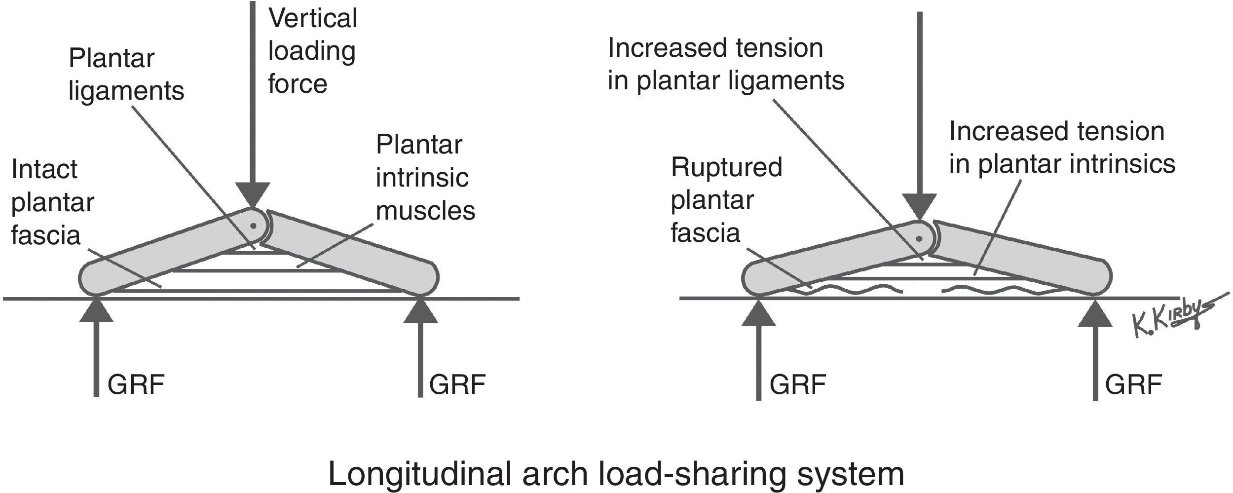
A key mechanical characteristic of each of the four tension load-sharing elements of the LALSS is that they each work to perform similar functions for the longitudinal arch of the foot so that when one element fails, the remaining elements still will allow proper longitudinal arch function. This also means, however, that when one structure of the LALSS fails (e.g. plantar fascia or plantar ligament rupture), the other tension load-bearing structures will be subjected to increased tension loads to allow proper longitudinal arch function (Fig. 6). Therefore, the four tension load-bearing elements of the LALSS, work to not only reduce the tension loads on all the elements of the LALSS, but guarantee that when one element fails, the longitudinal arch will still have enough strength and stiffness to maintain its shape and function properly during weightbearing activities.8
. With a rupture of the plantar fascia, the loss of plantar tension force from the plantar fascia will cause increased flattening of the longitudinal arch (right). The increased arch flattening will passively increase the tension within the plantar ligaments and will also cause the CNS to increase its efferent output to the plantar intrinsic muscles. The CNS may also respond by increasing the contractile activity of the extrinsic muscles of the plantar longitudinal arch (extrinsic muscles not illustrated in this model). In this manner, the unique load-sharing arrangement of the tension load-bearing elements of the LALSS will still allow proper longitudinal arch function even though one of its elements has ceased to function.")
In this simplified model of the longitudinal of the foot, three of the four layers of the tension load-bearing elements of the LALSS are illustrated from superficial to deep: the plantar fascia, plantar intrinsic muscles and plantar ligaments. When vertical loading forces are applied to the dorsal aspect of the longitudinal arch and GRF increases plantar to the rearfoot and forefoot, the longitudinal arch starts to flatten which passively increases the tension within the plantar fascia and plantar ligaments. The CNS will also respond to arch flattening by increasing the contractile activity of the plantar intrinsics (left). With a rupture of the plantar fascia, the loss of plantar tension force from the plantar fascia will cause increased flattening of the longitudinal arch (right). The increased arch flattening will passively increase the tension within the plantar ligaments and will also cause the CNS to increase its efferent output to the plantar intrinsic muscles. The CNS may also respond by increasing the contractile activity of the extrinsic muscles of the plantar longitudinal arch (extrinsic muscles not illustrated in this model). In this manner, the unique load-sharing arrangement of the tension load-bearing elements of the LALSS will still allow proper longitudinal arch function even though one of its elements has ceased to function.
One of the most significant design factors of the LALSS is the mechanical integration of passive elements, the plantar fascia and plantar ligaments, with the CNS controlled elements, the plantar intrinsic and plantar extrinsic muscles of the foot. As noted earlier, since tension force within the plantar fascia and plantar ligaments are not controlled directly by the CNS, the plantar fascia and plantar ligaments can only exert tension forces on their origins and insertions when the forefoot dorsiflexes on the rearfoot, or, in other words, when the longitudinal arch flattens and lengthens.
For example, during sitting and lying activities, since the forefoot becomes relatively plantarflexed on the rearfoot, the plantar fascia and plantar ligaments exert only negligible tension forces on the osseous elements of the longitudinal arch. The passive plantarflexion of the forefoot relative to the rearfoot during sitting and lying shortens the distance between the origins and insertions of the plantar fascia and the plantar ligaments so that these tension load-bearing structures are not elongated and, thus, have negligible tension forces during these types of non-weightbearing activities.
If, however, GRF acts on the plantar forefoot, or if the ankle joint dorsiflexors exert contractile activity on the forefoot, a forefoot dorsiflexion moment is created which tends to make the forefoot dorsiflex on the rearfoot. This forefoot dorsiflexion moment will cause the longitudinal arch to flatten and elongate, which will, in turn, elongate the plantar fascia and plantar ligaments until they exert a given magnitude of tension force. Once the tension force within the plantar fascia and plantar ligaments cause sufficient internal forefoot plantarflexion moments to counterbalance the forefoot dorsiflexion moments, then longitudinal arch flattening and elongation will cease and the longitudinal arch will become stable.
In addition, any dorsiflexion forces acting on the forefoot either from GRF and/or muscular contractile activities will also cause a simultaneous ankle joint dorsiflexion moment that will place the Achilles tendon and gastrocnemius and soleus muscles under increased tension loads. In other words, when GRF is increased on the plantar forefoot, the tension force within the plantar fascia, plantar ligaments and the Achilles tendon is increased. Conversely, when GRF is decreased on the plantar forefoot, the tension within the plantar fascia, plantar ligaments and Achilles tendon is decreased.12
Therefore, Achilles tendon tension, plantar fascia tension and plantar ligament tension forces are mechanically linked so that when the forefoot is dorsiflexed on the rearfoot during weightbearing activities, the plantar fascia, plantar ligaments and the Achilles tendon will all elongate and experience a passive increase in tension force.28 The direct mechanical effect of this increase in plantar fascia and plantar ligament tension force is that the longitudinal arch is passively, and automatically, stiffened during walking and running gait to help limit longitudinal arch flattening and elongation during late midstance and propulsion.8
As GRF is increased on the plantar forefoot, and longitudinal arch stiffness increases until it is of sufficient magnitude to prevent further forefoot dorsiflexion on the rearfoot (i.e. longitudinal arch flattening). Once the longitudinal arch becomes stiff enough (i.e. rigid enough) then Achilles tendon tension forces can more efficiently increase GRF to the plantar forefoot for propulsive activities. Thus, the increased stiffness of the midtarsal and midfoot joints that occur due to increased plantar ligament and plantar fascia tension forces effectively makes walking, running or jumping activities more efficient and metabolically economical activities.8
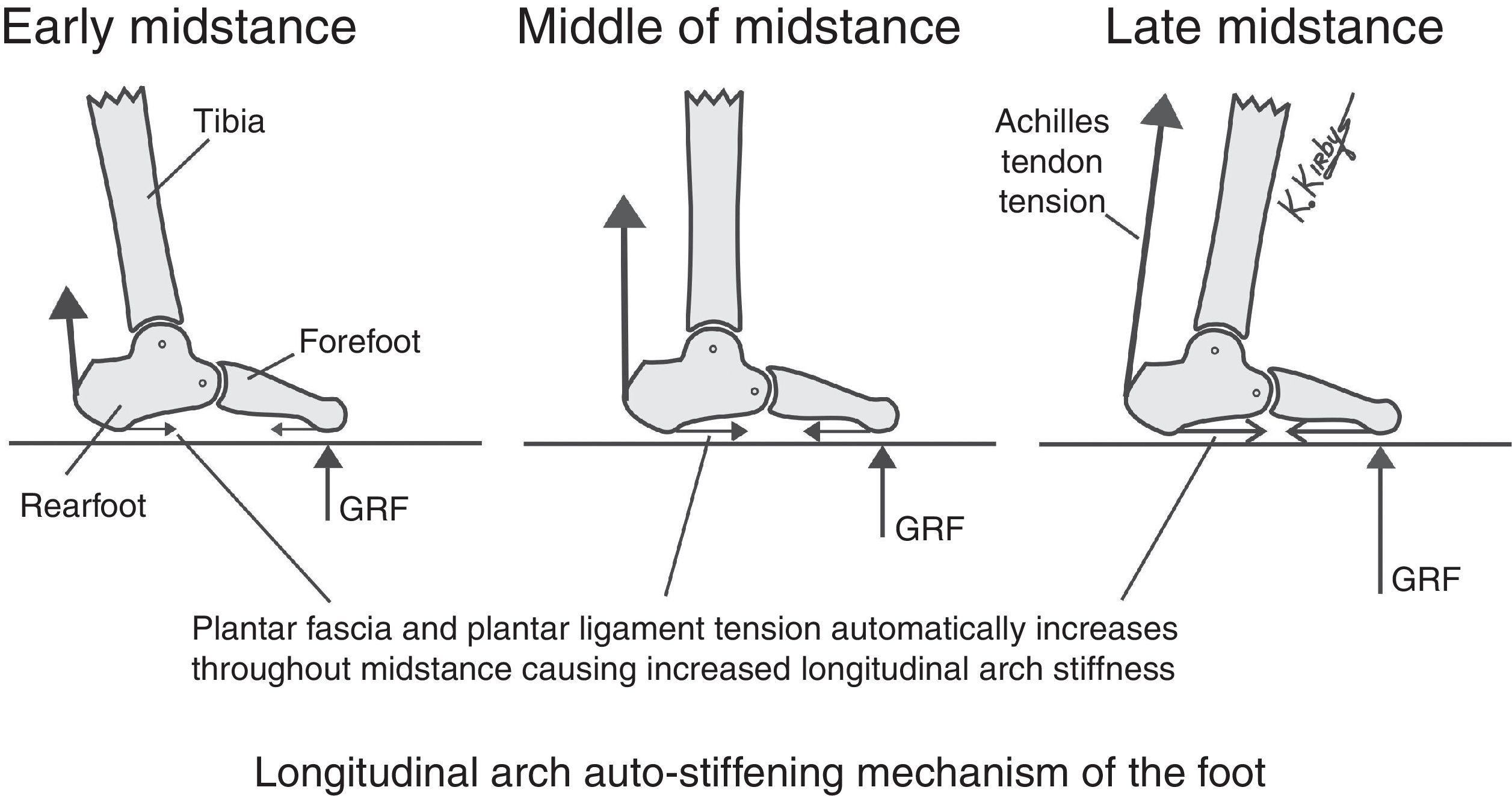
Longitudinal arch auto-stiffening mechanismIn 1993, Dananberg had noted three distinct “autosupport” mechanisms, calcaneocuboid joint “locking” secondary to plantar fascia “tightening”, the “locking wedge and truss effect”, and the windlass effect, which were thought to permit the foot to support the stresses applied to it during gait.29 More recently, in 2012, Kirby noted another automatic function of the foot, the Longitudinal Arch Auto-Stiffening Mechanism,30 which allows automatic stiffening of the longitudinal arch as the foot progresses from the beginning to the end of midstance during gait (Fig. 7).
. By the middle of midstance, as Achilles tendon force and forefoot GRF increase, the longitudinal arch automatically stiffens due to the increase in passive tension force within the plantar fascia and plantar ligaments (middle). Just before heel-off, when Achilles tendon tension and GRF plantar to the forefoot are greatest, the longitudinal arch stiffness also automatically further increases in magnitude due to the increased passive plantar fascia and plantar ligament tension forces (right). The Longitudinal Arch Auto-Stiffening Mechanism greatly decreases the metabolic demand of walking and running activities since the automatic increase in longitudinal arch stiffness at the initiation of propulsion allows the power from the gastrocnemius and soleus muscles to be more efficiently transferred to the plantar forefoot through the Achilles tendon. As a result, the Longitudinal Arch Auto-Stiffening Mechanism results in less longitudinal arch flattening motion during late midstance and propulsion which improves the mechanical efficiency of gait.")
The longitudinal arch auto-stiffening mechanism is dependent on the unique anatomical arrangement of the Achilles tendon, plantar fascia and plantar ligaments within the human foot. In early midstance, the GRF plantar to the forefoot and Achilles tendon tension force is of relatively low magnitude which causes a relatively small amount of plantar fascia tension and plantar ligament tension which, in turn, causes little increase in longitudinal arch stiffness (left). By the middle of midstance, as Achilles tendon force and forefoot GRF increase, the longitudinal arch automatically stiffens due to the increase in passive tension force within the plantar fascia and plantar ligaments (middle). Just before heel-off, when Achilles tendon tension and GRF plantar to the forefoot are greatest, the longitudinal arch stiffness also automatically further increases in magnitude due to the increased passive plantar fascia and plantar ligament tension forces (right). The Longitudinal Arch Auto-Stiffening Mechanism greatly decreases the metabolic demand of walking and running activities since the automatic increase in longitudinal arch stiffness at the initiation of propulsion allows the power from the gastrocnemius and soleus muscles to be more efficiently transferred to the plantar forefoot through the Achilles tendon. As a result, the Longitudinal Arch Auto-Stiffening Mechanism results in less longitudinal arch flattening motion during late midstance and propulsion which improves the mechanical efficiency of gait.
The Longitudinal Arch Auto-Stiffening Mechanism is directly due to the (1) unique arched structure of the osseous elements of the longitudinal arch, (2) the posterior location of the Achilles tendon insertion which causes not only an ankle joint plantarflexion moment but also a rearfoot plantarflexion moment which tends to flatten the longitudinal arch, and (3) the plantar locations of the plantar fascia and plantar ligaments which span the longitudinal arch and passively resist longitudinal arch flattening as GRF increases on the plantar forefoot during midstance.30
As a result of this unique morphologic configuration of the bones, muscles and ligaments of the human foot and leg, the automatic increase in plantar fascia and plantar ligament tension that mechanically results due to increasing tension within the Achilles tendon also automatically stiffens the longitudinal arch as midstance progresses which likely reduces the metabolic cost of walking and running. More importantly, these automatic mechanically-linked actions which increase the stiffness of the longitudinal arch as midstance progresses do not require direct CNS control. In other words, even a lifeless cadaver foot with no muscle activity and no CNS control will demonstrate “automatic” longitudinal arch stiffening as long as Achilles tendon tension increases and plantar forefoot GRF loads also increase in response to the increase in Achilles tendon tension force. Therefore, the Longitudinal Arch Auto-Stiffening Mechanism is a totally passive mechanism of the foot, requiring no extra metabolic energy to perform its longitudinal arch stiffening function, and only requires the Achilles tendon generate sufficient tension force to resist the external ankle joint dorsiflexion moment caused by GRF acting on the plantar forefoot.30
Active control of the longitudinal arch load-sharing systemThe CNS-controlled plantar intrinsic muscles and the PT, FDL, FHL and PL muscles all work together as active elements of the LALSS which allow the foot to be a dynamic weightbearing organ for the body. These active LALSS tension load-bearing elements can adjust medial and/or lateral longitudinal arch stiffness so that the magnitude and plantar location of GRF can be altered at any instant during gait. The CNS may modulate longitudinal arch stiffness by increasing or decreasing the contractile activity to any combination of the plantar intrinsic muscles or the PT, FDL, FHL and/or PL muscles which can make, as necessary, the medial or lateral longitudinal arch become either a more stiff spring or a more compliant spring during weightbearing activities.8
For example, situations which require the foot to conform to an uneven surface or require the forefoot to remain plantigrade in a fast side-to-side movement may require the CNS to simultaneously reduce efferent motor signals to decrease medial longitudinal arch stiffness and to increase efferent motor signals to increase lateral longitudinal arch stiffness. Much like the microprocessor-controlled shock absorbers of an advanced vehicle rear suspension system that optimizes comfort, efficiency and safety over a variety of surfaces and loading forces during driving, the CNS control of the contractile activities of the plantar intrinsics and plantar extrinsic muscles regulates the finely-tuned LALSS for the individual so that their weightbearing activities can be performed smoothly, efficiently and with reduced risk of injury.8
ConclusionAs Leonardo Da Vinci noted over six centuries ago, the human foot is a masterpiece of engineering and a work of art. One of these engineering marvels of the human foot is the longitudinal arch and its unique and elegant load-sharing system, the LALSS. With its passive elements that automatically increase the stiffness of the longitudinal arch with increasing Achilles tendon tension and increasing plantar forefoot loads, and its active elements that allow the CNS to instantly modify its stiffness and the plantar locations and magnitudes of GRF acting on the plantar foot, the LALSS allows for more effective and metabolically efficient weightbearing activities. The LALSS allows the human foot to function as an actively-controlled variable-stiffness spring, with the CNS continually optimizing the stiffness of both the medial and lateral longitudinal arches to improve the biomechanical function of the weightbearing individual. The podiatrist and clinician must fully appreciate these remarkable synergistic mechanisms within the longitudinal arch in order to better understand the normal function of the foot and lower extremity and to allow them to choose the most mechanically efficient and therapeutically effective conservative and surgical treatments for their patients with mechanically-based pathologies of their foot and lower extremity.